



Orellanine

Based on 1 Customer Validation
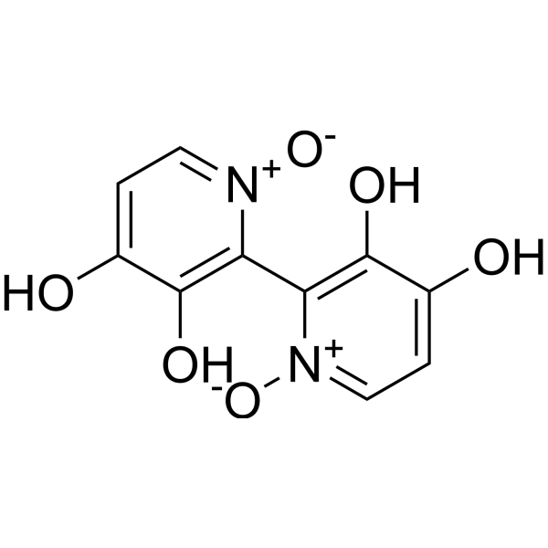
Orellanine, a nephrotoxic alkaloid found in Cortinarius orellanus, is an orally active and selective non-competitive inhibitor of alkaline phosphatase. Orellanine chelates iron, generates reactive oxygen species (ROS), induces DNA scission, forms ortho-semiquinone radicals, downregulates antioxidant defenses, and inhibits mitochondrial function. Orellanine induces caspase 8/9-mediated apoptosis. Orellanine inhibits synthesis of proteins, RNA, DNA, and mitochondrial protein synthesis, with metabolic activation required for cell-free protein synthesis inhibition. Orellanine can be used for the research of metastatic clear cell renal cell carcinoma, acute renal failure, chronic renal insufficiency, and kidney damage.
For research use only. We do not sell to patients.
- Purity : 95.0%
- CAS No.: 37338-80-0
- Formula: C10H8N2O6
- Molecular Weight:252.18
-
Storage:Powder -20°C, 3 years ; In solvent -80°C, 6 months , -20°C, 1 month

To place orders, for customer services and technical support, please contact: MedChemExpress USA
Tel: 609-228-6898 E-mail: [email protected] [email protected]


-
 Biological Activity
-
Chemical Information
-
Solvent & Solubility
- Protocols
- Purity & Documentation
- References
-
 Help & FAQs


All Caspase Isoforms
More

Biological Activity
Description
IC50 & Target
[1]|
Caspase 8 |
Caspase 9 |
In Vitro
Orellanine inhibits local clear cell renal cell carcinoma cell cultures, while showing no major toxicity to HUVEC, HEPG2, or MDA cell lines at equivalent concentrations[1].
Orellanine induces oxidative stress, mitochondrial dysfunction, and caspase 8/9-mediated apoptosis in SKRC-17, SKRC-52, and patient-derived metastatic clear cell renal cell carcinoma cell lines[1].
Orellanine inhibits pinocytosis in Amoeba proteus and growth of Dictyostelium discoideum and Escherichia coli cells[2].
Orellanine inhibits the DNA-dependent activity of both rat liver RNA polymerase B and Escherichia coli RNA polymerase[2].
Orellanine noncompetitively inhibits alkaline phosphatase, γ-glutamyl transpeptidase, and leucine aminopeptidase, and interrupts adenosine triphosphatase production at the proximal tubular brush border[2].
Orellanine (0.18 mM; 4-24 h) time-dependently inhibits protein, RNA, and DNA synthesis in Madin-Darby canine kidney (MDCK) cells without altering precursor uptake, reaching maximum inhibition of 73%, 80%, and 85% respectively after 24 h[3].
Orellanine (0.1-0.6 mM; 90 min pre-incubation, 30 min synthesis) inhibits protein synthesis in purified rat liver mitochondria with an IC50 of 0.3 mM, reaching a maximum inhibition of ~90% without altering leucine uptake into mitochondria[3].
MedChemExpress (MCE) has not independently confirmed the accuracy of these methods. They are for reference only.
In Vivo
Orellanine exhibits LD50 values of 15 mg/kg (p.o.) in cats[2].
Orellanine exhibits LD50 values of 8 mg/kg (i.p.) in Guinea pig[2].
MedChemExpress (MCE) has not independently confirmed the accuracy of these methods. They are for reference only.
- Nephrotoxicity Study
- Mitochondrial membrane-potential fluorescent assay
- Detection of Apoptosis
- TUNEL staining for apoptotic DNA fragmentation
- Annexin V plus membrane-impermeant dye apoptosis staining
- ROS/oxidative-stress fluorescent staining
- Mitochondrial membrane-potential and mitochondrial mass staining
- Apoptosis Solutions
Clinical Trial
| NCT Number | Sponsor | Condition | Start Date |
Phase

|
|---|---|---|---|---|
| NCT01329991 | 2011-05 | PHASE1 |


Chemical Information
-
CAS No. 37338-80-0
-
Appearance Solid
-
Molecular Weight 252.18
-
Formula C10H8N2O6
-
Color Light yellow to yellow
-
SMILES
[O-][N+]1=CC=C(O)C(O)=C1C2=C(O)C(O)=CC=[N+]2[O-]
-
Structure Classification
-
Initial Source
Cortinarius orellanus
-
Shipping
Room temperature in continental US; may vary elsewhere.
-
Storage
Powder -20°C 3 years In solvent -80°C 6 months -20°C 1 month


Solvent & Solubility
In Vitro:
DMSO : 20 mg/mL (79.31 mM; ultrasonic and warming and heat to 60°C; Hygroscopic DMSO has a significant impact on the solubility of product, please use newly opened DMSO)
Please refer to the solubility information to select the appropriate solvent. Once prepared, please aliquot and store the solution to prevent product inactivation from repeated freeze-thaw cycles.
Storage method and period of stock solution: -80°C, 6 months; -20°C, 1 month. When stored at -80°C, please use it within 6 months. When stored at -20°C, please use it within 1 month.
Please refer to the solubility information to select the appropriate solvent. Once prepared, please aliquot and store the solution to prevent product inactivation from repeated freeze-thaw cycles.
Storage method and period of stock solution: -80°C, 6 months; -20°C, 1 month. When stored at -80°C, please use it within 6 months. When stored at -20°C, please use it within 1 month.
Concentration (start) × Volume (start) = Concentration (final) × Volume (final)


Purity & Documentation
-
Data Sheet (276 KB)
-
SDS (394 KB)
- English - EN (394 KB)
- Français - FR (394 KB)
- Deutsch - DE (394 KB)
- Norwegian - NO (394 KB)
- Español - ES (394 KB)
- Swedish - SV (394 KB)
- Italian - IT (394 KB)
- Korean - KR (394 KB)
- Portuguese - PT (394 KB)
-
Handling Instructions (2659 KB)
References
[1]. Lyons MJ, et al. Orellanine: From Fungal Origin to a Potential Future Cancer Treatment. J Nat Prod. 2023;86(6):1620-1631. [Content Brief]
[2]. Dinis-Oliveira RJ, et al. Human and experimental toxicology of orellanine. Hum Exp Toxicol. 2016;35(9):1016-1029. [Content Brief]
[3]. Richard JM, et al. Orellanine inhibits protein synthesis in Madin-Darby canine kidney cells, in rat liver mitochondria, and in vitro: indication for its activation prior to in vitro inhibition. Toxicology. 1991;67(1):53-62. [Content Brief]


Complete Stock Solution Preparation Table
Please refer to the solubility information to select the appropriate solvent. Once prepared, please aliquot and store the solution to prevent product inactivation from repeated freeze-thaw cycles.
Storage method and period of stock solution: -80°C, 6 months; -20°C, 1 month. When stored at -80°C, please use it within 6 months. When stored at -20°C, please use it within 1 month.
| Optional Solvent | Concentration Solvent Mass | 1 mg | 5 mg | 10 mg | 25 mg |
|---|---|---|---|---|---|
| DMSO | 1 mM | 3.9654 mL | 19.8271 mL | 39.6542 mL | 99.1355 mL |
| 5 mM | 0.7931 mL | 3.9654 mL | 7.9308 mL | 19.8271 mL | |
| 10 mM | 0.3965 mL | 1.9827 mL | 3.9654 mL | 9.9136 mL | |
| 15 mM | 0.2644 mL | 1.3218 mL | 2.6436 mL | 6.6090 mL | |
| 20 mM | 0.1983 mL | 0.9914 mL | 1.9827 mL | 4.9568 mL | |
| 25 mM | 0.1586 mL | 0.7931 mL | 1.5862 mL | 3.9654 mL | |
| 30 mM | 0.1322 mL | 0.6609 mL | 1.3218 mL | 3.3045 mL | |
| 40 mM | 0.0991 mL | 0.4957 mL | 0.9914 mL | 2.4784 mL | |
| 50 mM | 0.0793 mL | 0.3965 mL | 0.7931 mL | 1.9827 mL | |
| 60 mM | 0.0661 mL | 0.3305 mL | 0.6609 mL | 1.6523 mL |


Orellanine Related Classifications
HY-W661499 Related Classifications
 Powered by Bioz
Keywords
- Orellanine
- 37338-80-0
- Phosphatase
- Apoptosis
- Reactive Oxygen Species (ROS)
- Caspase
- alkaline phosphatase
- kidney proximal tubular cells
- reactive oxygen species
- HUVEC
- Madin-Darby canine kidney (MDCK) cells
- caspase 8/9
- metastatic clear cell renal cell carcinoma
- HEPG2
- Cortinarius orellanus
- MDA cell lines
- Inhibitor
- inhibitor
- inhibit