



Phosphocreatine
Based on 3 publication(s) in Google Scholar
Phosphocreatine (creatine phosphate) is an organic compound found in vertebrate skeletal muscles. Phosphocreatine enhances antioxidant activity, and activates the TAK1 pathway to protect the heart. Phosphocreatine normalizing mitochondrial function and reducing oxidative stress via Akt mediated Nrf2/HO-1 pathway. Phosphocreatine provides renal protection by suppressing Apoptosis and ROS (Reactive Oxygen Species) generation through ERK mediated mediated Nrf-2/HO-1 pathway..
For research use only. We do not sell to patients.
- Purity : 99.60%
- CAS No.: 67-07-2
- Formula: C4H10N3O5P
- Molecular Weight:211.11
-
Storage:
4°C, sealed storage, away from moisture
* In solvent : -80°C, 6 months; -20°C, 1 month (sealed storage, away from moisture)

To place orders, for customer services and technical support, please contact: MedChemExpress USA
Tel: 609-228-6898 E-mail: [email protected] [email protected]


-
 Biological Activity
-
Chemical Information
-
Solvent & Solubility
- Protocols
- Purity & Documentation
- References
-
 Help & FAQs


-
HY-B2176
ATPEndogenous Metabolite -

HY-P2799
Creatine phosphokinase, Rabbit muscleATPase -
HY-W010388
CreatineEndogenous Metabolite -
HY-15066
CNQXAMPA Antagonist -
HY-B1777
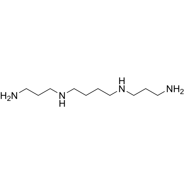
SpermineEndogenous Metabolite -
HY-D0861
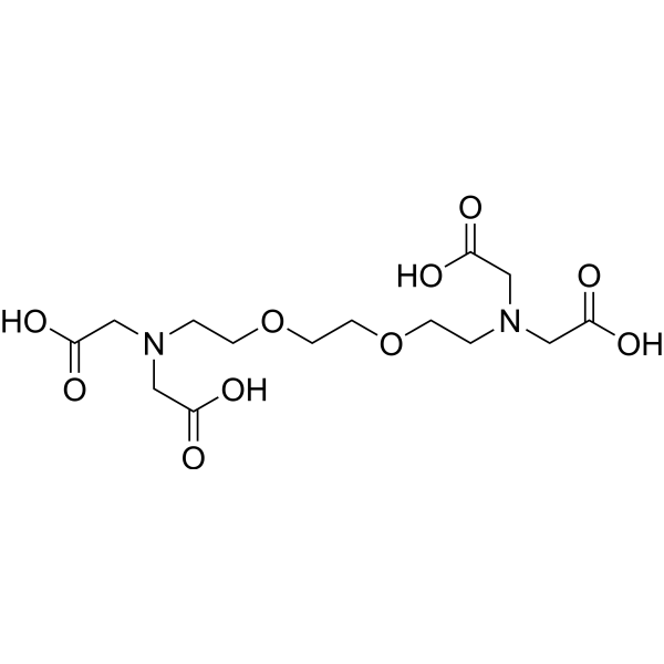
EGTACalcium Ion Chelator -

HY-15371
ForskolinAdenylate Cyclase Activator -
HY-B1776
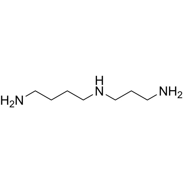
SpermidinePolyamine Compound -
HY-108505
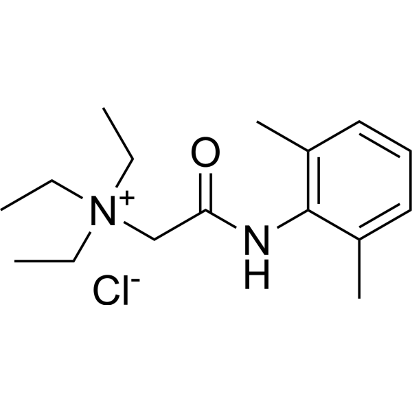
QX-314 chlorideSodium Channel Blocker -
HY-100714A
D-AP5NMDA Antagonist -
HY-B0228
AdenosineEndogenous Autacoid -

HY-P2799A
Creatine Kinase (CPK/CK), Bovine HeartCreatine Kinase
Publications Citing Use of MedChemExpress (MCE) Phosphocreatine
MoreAll Endogenous Metabolite Isoforms
MoreAll MAP3K Isoforms
More

Biological Activity
Description
IC50 & Target
|
Human Endogenous Metabolite |
In Vitro
Phosphocreatine (0-1 mM, 24 h) reveals the effect of anti-oxidant, anti-apoptosis and anti-necroptosis to protect agaist DOX (Doxorubicin) (HY-15142A)-induced cardiomyocytes injury in H9c2 cells by targeting TAK1[2].
Phosphocreatine (0-1 mM, 24 h) alleviates oxidative stress by increasing antioxidant activity, subsequently recovers expression level of TAK1 to baseline and reduces apoptosis and necroptosis in DOX-induced myocardial injury[2].
Phosphocreatine (5-20 mM, 24 h) attenuates cell injury and inhibits apoptosis induced by MGO (Methylglyoxal) (HY-106634) in PC12 cell[3].
Phosphocreatine (5-20 mM, 24 h) prevents loss of mitochondrial membrane permeability of MGO (Methylglyoxal) injured PC-12 cells[3].
Phosphocreatine (5-20 mM, 2 h) exhibits the neuroprotective effects in PC-12 cells relying on normalizing mitochondrial function and reducing oxidative stress via Akt mediated Nrf2/HO-1 pathway[3].
Phosphocreatine (5-40 mM, 24 h) at different concentrations might contribute to protection of the NRK-52E cells against MGO-induced kidney injury[4].
Phosphocreatine (10-40 mM, 4 h) suppresses kidney oxidative stress metabolites[4].
MedChemExpress (MCE) has not independently confirmed the accuracy of these methods. They are for reference only. Further protocols information, click here.
-
Cell Line:PC-12, NRK-52E cells
-
Concentration:0, 5, 10, 20, 40 mM
-
Incubation Time:24 h
-
Result:Was not toxic to PC-12 cells under the treatment conditions.
Significantly increased PC-12 cell viability at the concentrations of 5, 10 and 20 mM compared with MGO (Methylglyoxal) (HY-106634) groups.
Contributed to protection of the NRK-52E cells against MGO-induced kidney injury.
-
Cell Line:PC-12, NRK-52E cells
-
Concentration:5, 10, 20, 40 mM
-
Incubation Time:2 h
-
Result:Significantly suppressed the enhanced early apoptosis in PC-12 cells in a dose-dependent manner.
Increased the expression of Bcl-2, procaspase-3 and procaspase-9 in NRK-52E cells.
Decreased the expression of Bax and cleaved caspase-3 in NRK-52E cells..
Suppressed karyorrhexis and karyopyknosis in NRK-52E cells..
Decreased the apoptotic rate compared with MGO-treated cells in NRK-52E cells..
Prevented the losing of MMP (mitochondrial membrane potential) in NRK-52E cells.
-
Cell Line:PC-12, NRK-52E cells
-
Concentration:20, 40 mM
-
Incubation Time:24 h
-
Result:Increased the expression levels of Akt, Nrf2 (nuclear factor (erythroid-derived-2)-like 2 (Nrf2)) and HO-1 (Hemeoxygenase-1) in PC12 cells.
Increased the expression of nuclear Nrf2 levels, and decreased Nrf2 level in PC12 cells cytoplasm.
Increased the expression of p-Akt, HO-1 and Nrf2 with compared with pre-treatment for 2 h with LY294002 (a PI3K inhibitor) in PC12 cells.
Significantly increased Bcl-2 and procaspase-9 levels and decreased Bax, cleaved caspase-9 and cleaved caspase-3 C level in NRK-52E cells.
Decreased the expression of p-ERK and increased the Nrf2 and HO-1 expressions in NRK-52E cells.
In Vivo
Phosphocreatine (20-40 mg/kg, i.v., daily, 6 weeks) has a protective effect on the kidney tissues against diabetic nephropathy in SD (Sprague Dawley) rats[4].
MedChemExpress (MCE) has not independently confirmed the accuracy of these methods. They are for reference only.
-
Animal Model:Male Sprague Dawley (SD) rats, i.p., normal saline, 3 times; i.p., DOX 2 mg/kg, 7 times; i.p., normal saline, 3 times [2].
-
Dosage:200 mg/kg
-
Administration:i.p., once every other day, 7 weeks
-
Result:Improved the heart function abnormality.
Lowered myocardial apoptosis.
Recovered the expression of Nrf2, SOD, FoxO3a and diminished C-Casp3, Bax/Bcl2 in the myocardial tissue of rats.
Markedly improved myocardial necroptosis, as indicated by decreasing expression of RIP3 and CaMKII.
Increased expression level of TAK1.
-
Animal Model:Male Sprague Dawley (SD) rats, i.p., 70 mg/kg (STZ(Streptozotocin) (HY-13753)), daily, 6 weeks [4].
-
Dosage:20, 40 mg/kg
-
Administration:i.p., daily, 6 weeks
-
Result:Reduced hyperglycemia compared with STZ (Streptozotocin) (HY-13753) -treated rats.
Increased the weight of rats gradually compared with STZ (Streptozotocin) (HY-13753) group.
Decreased kidney weight index (kidney weight/body weight).
Decreased MDA level and increased of GSH and SOD levels compared with STZ group.
Decreased the apoptotic rate compared with MGO-treated groups.


Chemical Information
-
CAS No. 67-07-2
-
Appearance Solid
-
Molecular Weight 211.11
-
Formula C4H10N3O5P
-
Color White to off-white
-
SMILES
O=C(O)CN(C(NP(O)(O)=O)=N)C
-
Synonyms
Creatine phosphate; Creatinephosphoric acid
-
Structure Classification
-
Initial Source
-
Shipping
Room temperature in continental US; may vary elsewhere.
-
Storage
4°C, sealed storage, away from moisture
* In solvent : -80°C, 6 months; -20°C, 1 month (sealed storage, away from moisture)


Publications (3)
-
Journal Impact Factor
-
Most Recent
-
Autophagy
ESCRT III-mediated lysosomal repair improve renal tubular cell injury in cisplatin-induced AKI. [Abstract]2025 Sep;21(9):1927-1944. PMID: 40152606 -
-
Toxicol Appl Pharmacol
2022 Apr 15:441:115971. PMID: 35276125


Solvent & Solubility
In Vitro:
H2O : 175 mg/mL (828.95 mM; Need ultrasonic)
Please refer to the solubility information to select the appropriate solvent. Once prepared, please aliquot and store the solution to prevent product inactivation from repeated freeze-thaw cycles.
Storage method and period of stock solution: -80°C, 6 months; -20°C, 1 month (sealed storage, away from moisture). When stored at -80°C, please use it within 6 months. When stored at -20°C, please use it within 1 month.
* Note: If you choose water as the stock solution, please dilute it to the working solution, then filter and sterilize it with a 0.22 μm filter before use.
Please refer to the solubility information to select the appropriate solvent. Once prepared, please aliquot and store the solution to prevent product inactivation from repeated freeze-thaw cycles.
Storage method and period of stock solution: -80°C, 6 months; -20°C, 1 month (sealed storage, away from moisture). When stored at -80°C, please use it within 6 months. When stored at -20°C, please use it within 1 month.
* Note: If you choose water as the stock solution, please dilute it to the working solution, then filter and sterilize it with a 0.22 μm filter before use.
Concentration (start) × Volume (start) = Concentration (final) × Volume (final)

Protocols
-
Mitochondrial membrane-potential fluorescent assay
Mitochondrial membrane potential fluorescent assays estimate ΔΨm in living cells using lipophilic cationic dyes such as TMRM, TMRE, rhodamine 123, and JC-1, which accumulate in mitochondria according to membrane polarization; loss of signal after FCCP or CCCP treatment is interpreted as mitochondrial depolarization. TMRM/TMRE and rhodamine 123 are commonly used for semi-quantitative live-cell microscopy or flow cytometry, while JC-1 can report a shift from red aggregate fluorescence to green monomer fluorescence during depolarization; interpretation requires controls because dye concentration, quenching mode, cell type, dye efflux, and mitochondrial mass can affect fluorescence independently of ΔΨm.
-
Apoptosis
Apoptosis, also called programmed cell death, is generally characterized by distinct morphological characteristics.
-
TUNEL staining for apoptotic DNA fragmentation
TUNEL staining detects DNA strand breaks by using terminal deoxynucleotidyl transferase to add labeled nucleotides to exposed 3′-OH DNA termini, generating either microscopic staining in fixed cells or tissue sections, or fluorescence/cytometric signal in cell suspensions. TUNEL positivity reflects DNA fragmentation but should not be interpreted alone as definitive apoptosis, because TUNEL can also label necrotic, autolytic, mechanically damaged, or DNA-repair-associated DNA breaks.
-
Annexin V plus membrane-impermeant dye apoptosis staining
Annexin V-based apoptosis assays rely on the detection of phosphatidylserine (PS) externalization from the inner leaflet of the plasma membrane to the outer leaflet, an early biochemical hallmark of apoptosis. Fluorescently labeled Annexin V binds PS in a calcium-dependent manner, enabling identification of early apoptotic cells by flow cytometry or fluorescence microscopy. When combined with a membrane-impermeant DNA-binding dye (e. g. , propidium iodide), this approach allows discrimination between viable (Annexin V−/dye−), early apoptotic (Annexin V+/dye−), and late apoptotic or necrotic (Annexin V+/dye+) cell populations by assessing membrane integrity and PS exposure.
-
ROS/oxidative-stress fluorescent staining
ROS/oxidative-stress fluorescent staining uses cell-permeant fluorogenic probes that become fluorescent after oxidation inside cells or tissues; commonly used examples include DCFH-DA/DCFDA for broad cellular oxidant detection, DHE for superoxide-related signal detection, MitoSOX for mitochondrial superoxide-related signal detection, and CellROX probes for oxidative-stress-associated fluorescence readouts. The assay detects probe oxidation rather than a single ROS species unless the probe and analysis method have been chemically validated for that species. DCFH-DA enters cells, is deacetylated by intracellular esterases to DCFH, and produces fluorescent DCF after oxidation, so the readout is used as an operational measure of total cellular oxidative stress rather than a species-specific ROS measurement. DHE and MitoSOX can report superoxide-related oxidation, but red fluorescence alone can include non-specific ethidium-like oxidation products; HPLC or optimized spectral approaches are
-
Mitochondrial membrane-potential and mitochondrial mass staining
Mitochondrial membrane potential staining measures the electrochemical polarization across the mitochondrial inner membrane in live cells using lipophilic cationic fluorescent probes; early rhodamine-based work showed that selective mitochondrial dye accumulation is lost when the mitochondrial transmembrane potential is dissipated. JC-1 reports mitochondrial polarization by shifting from green monomer fluorescence to red J-aggregate fluorescence as dye concentration increases within energized mitochondria; therefore, the red/green fluorescence ratio is used as a relative readout of mitochondrial membrane potential. TMRE or TMRM staining provides a single-channel relative readout because these cationic rhodamine esters accumulate in polarized mitochondria, and lower fluorescence indicates reduced mitochondrial polarization when acquisition and dye-loading conditions are controlled. Mitochondrial mass staining is commonly performed with MitoTracker Green FM or related MitoTracker dyes as
-
Apoptosis Solutions
Apoptosis is a regulated, generally non-lytic cell-death pathway that removes unwanted, damaged, infected, or abnormal cells through coordinated morphological changes, caspase activation, DNA fragmentation, and membrane remodeling. The intrinsic apoptosis pathway is controlled mainly by mitochondrial outer membrane permeabilization, BCL-2 family proteins, cytochrome c release, apoptosome formation, caspase-9 activation, and downstream executioner caspase-3/7 activation. The extrinsic apoptosis pathway is initiated by death receptors such as Fas, TNFR, and TRAIL receptors, which recruit adaptor proteins and activate caspase-8 before engaging executioner caspases or mitochondrial amplification through BID cleavage. Apoptosis is linked to many phenotypes, including cancer cell killing, tissue homeostasis, immune regulation, neurodegeneration, infection response, and treatment-induced cytotoxicity; unresolved questions include how apoptosis interacts with necroptosis, pyroptosis, ferroptos


Purity & Documentation
-
Data Sheet (281 KB)
-
SDS (393 KB)
- English - EN (393 KB)
- Français - FR (393 KB)
- Deutsch - DE (393 KB)
- Norwegian - NO (393 KB)
- Español - ES (393 KB)
- Swedish - SV (393 KB)
- Italian - IT (393 KB)
- Korean - KR (393 KB)
- Portuguese - PT (393 KB)
-
Handling Instructions (2659 KB)
References
[1]. Feldman EB, et al. Creatine: a dietary supplement and ergogenic aid. Nutr Rev. 1999 Feb;57(2):45-50. [Content Brief]
[2]. Wang C, et al. Phosphocreatine attenuates doxorubicin-induced cardiotoxicity by inhibiting oxidative stress and activating TAK1 to promote myocardial survival in vivo and in vitro. Toxicology. 2021 Aug;460:152881. [Content Brief]
[3]. Li H, et al. Neuroprotective effect of phosphocreatine on oxidative stress and mitochondrial dysfunction induced apoptosis in vitro and in vivo: Involvement of dual PI3K/Akt and Nrf2/HO-1 pathways. Free Radic Biol Med. 2018 May 20;120:228-238. [Content Brief]
[4]. Shopit A, et al. Protection of diabetes-induced kidney injury by phosphocreatine via the regulation of ERK/Nrf2/HO-1 signaling pathway. Life Sci. 2020 Feb 1;242:117248. [Content Brief]


Complete Stock Solution Preparation Table
Please refer to the solubility information to select the appropriate solvent. Once prepared, please aliquot and store the solution to prevent product inactivation from repeated freeze-thaw cycles.
Storage method and period of stock solution: -80°C, 6 months; -20°C, 1 month (sealed storage, away from moisture). When stored at -80°C, please use it within 6 months. When stored at -20°C, please use it within 1 month.
| Optional Solvent | Concentration Solvent Mass | 1 mg | 5 mg | 10 mg | 25 mg |
|---|---|---|---|---|---|
| H2O | 1 mM | 4.7369 mL | 23.6843 mL | 47.3687 mL | 118.4217 mL |
| 5 mM | 0.9474 mL | 4.7369 mL | 9.4737 mL | 23.6843 mL | |
| 10 mM | 0.4737 mL | 2.3684 mL | 4.7369 mL | 11.8422 mL | |
| 15 mM | 0.3158 mL | 1.5790 mL | 3.1579 mL | 7.8948 mL | |
| 20 mM | 0.2368 mL | 1.1842 mL | 2.3684 mL | 5.9211 mL | |
| 25 mM | 0.1895 mL | 0.9474 mL | 1.8947 mL | 4.7369 mL | |
| 30 mM | 0.1579 mL | 0.7895 mL | 1.5790 mL | 3.9474 mL | |
| 40 mM | 0.1184 mL | 0.5921 mL | 1.1842 mL | 2.9605 mL | |
| 50 mM | 0.0947 mL | 0.4737 mL | 0.9474 mL | 2.3684 mL | |
| 60 mM | 0.0789 mL | 0.3947 mL | 0.7895 mL | 1.9737 mL | |
| 80 mM | 0.0592 mL | 0.2961 mL | 0.5921 mL | 1.4803 mL | |
| 100 mM | 0.0474 mL | 0.2368 mL | 0.4737 mL | 1.1842 mL |
* Note: If you choose water as the stock solution, please dilute it to the working solution, then filter and sterilize it with a 0.22 μm filter before use.


Phosphocreatine Related Classifications
HY-D0885 Related Classifications
 Powered by Bioz