



Rhoifolin
Based on 1 publication(s) in Google Scholar
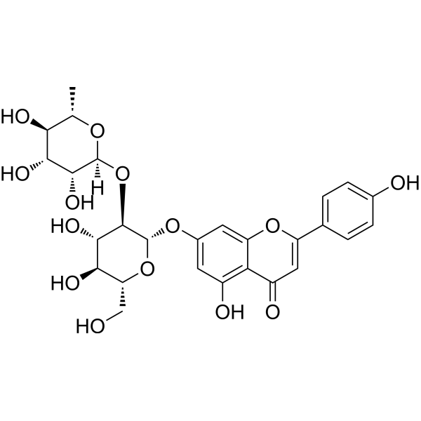
Rhoifolin is a flavone glycoside can be isolated from Rhus succedanea. Rhoifolin has anti-diabetic effect acting through enhanced adiponectin secretion, tyrosine phosphorylation of insulin receptor-β and glucose transporter 4 (GLUT 4) translocation. Rhoifolin has an anti-inflammatory action via multi-level regulation of inflammatory mediators. Rhoifolin ameliorates titanium particle-stimulated osteolysis and attenuates osteoclastogenesis via RANKL-induced NF-κB and MAPK pathways. Rhoifolin also has cytotoxic activity against different cancer cell lines.
Nur für Forschungszwecke. Wir verkaufen nicht an Patienten.
- Reinheit : 99.25%
- CAS. Nr.: 17306-46-6
- Formel: C27H30O14
- Molecular Weight:578.52
-
Speicherung:
4°C, protect from light, stored under nitrogen
* In solvent : -80°C, 6 months; -20°C, 1 month (protect from light, stored under nitrogen)

To place orders, for customer services and technical support, please contact: MedChemExpress USA
Tel: 609-228-6898 E-mail: [email protected] [email protected]


-
 Biologische Aktivität
-
Chemical Information
-
Lösungsmittel & Löslichkeit
- Protokoll
- Reinheit & Dokumentation
- Verweise
-
 Help & FAQs


-
HY-B0141
EstradiolHuman Endogenous Metabolite -
HY-N2258
PoncirinAnti-Hyperalgesia Agent -
HY-N4122
NeodiosminFlavone Glycoside -

HY-N0755R
Rhoifolin (Standard)Flavone Glycoside -
HY-N0628
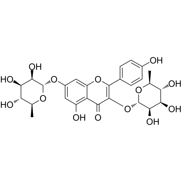
KaempferitrinInsulin Receptor Activator -
HY-144873
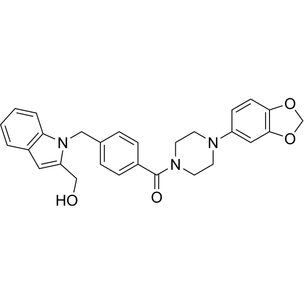
AVJ16IGF2BP1 Inhibitor -
HY-P2542
GIP (3-42), humanGIP Receptor Agonist -
HY-W040127
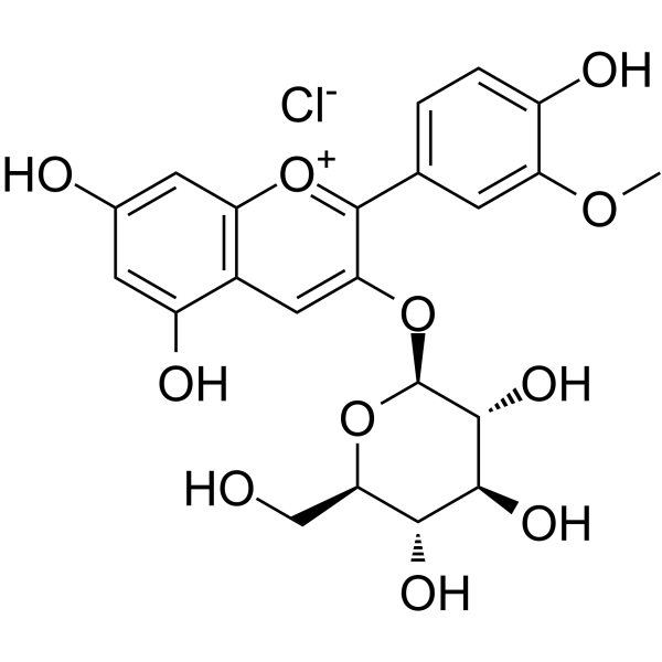
Peonidin 3-O-glucoside chlorideInsulin Secretagogue -
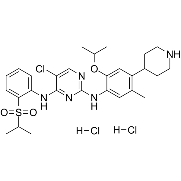
HY-15656A
Ceritinib dihydrochlorideALK Inhibitor -
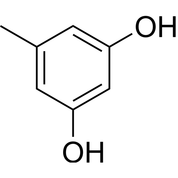
HY-D0168
OrcinolMelanogenesis Inhibitor -
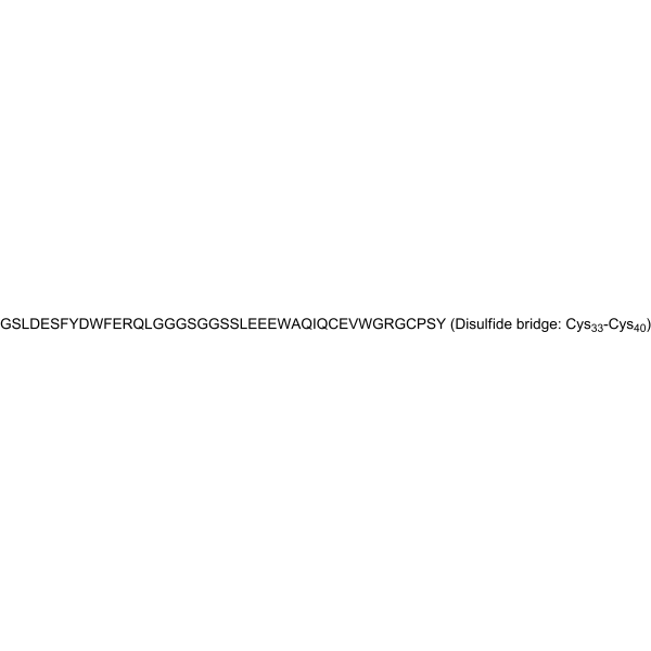
HY-P2093
S961Insulin Receptor Antagonist -
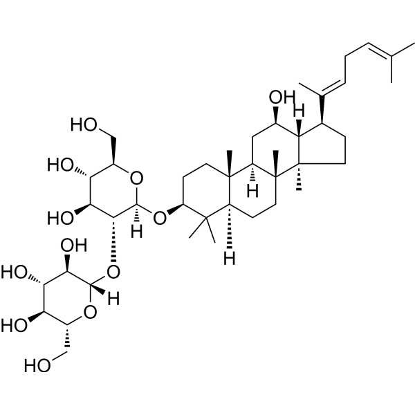
HY-N0908
Ginsenoside Rg5IGF-1R Agonist
Publications Citing Use of MedChemExpress (MCE) Rhoifolin
More

Biologische Aktivität
Beschreibung
|
insulin receptor-β |
GLUT4 |
NF-κB |
MAPK |
Cellular Effect
|
Cell Line

|
Type | Value | Description | References |
|---|---|---|---|---|
| Erythrocyte | IC50 |
95.9 μM
Compound: 13
|
Antioxidant activity assessed as inhibition of AAPH-induced hemolysis in rat RBC after 3 hrs
Antioxidant activity assessed as inhibition of AAPH-induced hemolysis in rat RBC after 3 hrs
|
[PMID: 12828473] |
In Vitro
In Vivo
MedChemExpress (MCE) has not independently confirmed the accuracy of these methods. They are for reference only.
-
Animal Model:Swiss albino mice (10 Gy irradiation induced certain haematological and cardiac biochemical abnormalities)[4]
-
Dosage:12, 24, 30, 36, and 40 mg/kg
-
Administration:PO, for 7 days
-
Result:Diminished the toxic effect of radiation by decreasing the level of lipid peroxides measured as malondialdehyde, ameliorating the alterations in nitric oxide, lactate dehydrogenase, creatine kinase in plasma and tissue and the plasma lipid profile.
- Kinase activity and phosphorylation assays
- Western Blot
- Cell Cytotoxicity Assay
- Research Protocol for Endocrine Diseases
- Protocol for Kinase activity and phosphorylation assays
- Research Protocol for Inflammation-related Diseases
- Osteoclast differentiation from monocyte/macrophage precursors
- Cell Viability Determination by MTT Colorimetric Assay


Chemical Information
-
CAS. Nr. 17306-46-6
-
Appearance Solid
-
Molecular Weight 578.52
-
Formel C27H30O14
-
Color Light yellow to yellow
-
SMILES
O=C(C=C(C1=CC=C(O)C=C1)OC2=CC(O[C@H](O[C@H](CO)[C@@H](O)[C@@H]3O)[C@@H]3O[C@@](O[C@@H](C)[C@H](O)[C@H]4O)([H])[C@@H]4O)=C5)C2=C5O
-
Structure Classification
-
Initial Source
-
Versand
Room temperature in continental US; may vary elsewhere.
-
Speicherung
4°C, protect from light, stored under nitrogen
* In solvent : -80°C, 6 months; -20°C, 1 month (protect from light, stored under nitrogen)


Publications (1)
-
Journal Impact Factor
-
Most Recent
-
Metabolism
2026 May 20:182:156651. PMID: 42162901


Lösungsmittel & Löslichkeit
In Vitro:
DMSO : 62.5 mg/mL (108.03 mM; Need ultrasonic; Hygroscopic DMSO has a significant impact on the solubility of product, please use newly opened DMSO)
Please refer to the solubility information to select the appropriate solvent. Once prepared, please aliquot and store the solution to prevent product inactivation from repeated freeze-thaw cycles.
Storage method and period of stock solution: -80°C, 6 months; -20°C, 1 month (protect from light, stored under nitrogen). When stored at -80°C, please use it within 6 months. When stored at -20°C, please use it within 1 month.
Please refer to the solubility information to select the appropriate solvent. Once prepared, please aliquot and store the solution to prevent product inactivation from repeated freeze-thaw cycles.
Storage method and period of stock solution: -80°C, 6 months; -20°C, 1 month (protect from light, stored under nitrogen). When stored at -80°C, please use it within 6 months. When stored at -20°C, please use it within 1 month.
Konzentration (Stammlösung) × Volumen (Stammlösung) = Konzentration (Ziellösung) × Volumen (Ziellösung)
In Vivo:
Select the appropriate dissolution method based on your experimental animal and administration route.
- For the following dissolution methods, please ensure to first prepare a clear stock solution using an In Vitro approach and then sequentially add co-solvents:
- To ensure reliable experimental results, the clarified stock solution can be appropriately stored based on storage conditions. As for the working solution for In Vivo experiments, it is recommended to prepare freshly and use it on the same day.
- The percentages shown for the solvents indicate their volumetric ratio in the final prepared solution. If precipitation or phase separation occurs during preparation, heat and/or sonication can be used to aid dissolution.
Add each solvent one by one: 10% DMSO 40% PEG300 5% Tween-80 45% Saline
Solubility: ≥ 2.08 mg/mL (3.60 mM); Clear solution
This protocol yields a clear solution of ≥ 2.08 mg/mL (saturation unknown).
Taking 1 mL working solution as an example, add 100 μL DMSO stock solution (20.8 mg/mL) to 400 μL PEG300, and mix evenly; then add 50 μL Tween-80 and mix evenly; then add 450 μL Saline to adjust the volume to 1 mL.
Preparation of Saline: Dissolve 0.9 g sodium chloride in ddH₂O and dilute to 100 mL to obtain a clear Saline solution.
Add each solvent one by one: 10% DMSO 90% (20% SBE-β-CD in Saline)
Solubility: ≥ 2.08 mg/mL (3.60 mM); Clear solution
This protocol yields a clear solution of ≥ 2.08 mg/mL (saturation unknown).
Taking 1 mL working solution as an example, add 100 μL DMSO stock solution (20.8 mg/mL) to 900 μL 20% SBE-β-CD in Saline, and mix evenly.
Preparation of 20% SBE-β-CD in Saline (4°C, storage for one week): 2 g SBE-β-CD powder is dissolved in 10 mL Saline, completely dissolve until clear.
In Vivo Dissolution Calculator
Please enter the basic information of animal experiments:
-
-
-
-
Recommended: Prepare an additional quantity of animals to account for potential losses during experiments.
Please enter your animal formula composition:
-
%DMSO +
Recommended: Keep the proportion of DMSO in working solution below 2% if your animal is weak.
-
%+
-
+%Tween-80 + +
-
%Saline +
The co-solvents required include: DMSO, . All of co-solvents are available by MedChemExpress (MCE). , Tween 80. All of co-solvents are available by MedChemExpress (MCE).
Working solution concentration: 0.22 mg/mL
Method for preparing stock solution: mg drug dissolved in μL DMSO. Stock solution concentration: mg/mL. * In solvent : -80°C, 6 months; -20°C, 1 month (protect from light, stored under nitrogen)
1. Take μL DMSO stock solution;
2. Add μL .
μL , mix evenly;
3. Then add μL Tween 80, mix evenly;
4. Then add μL
Please ensure that the stock solution in the first step is dissolved to a clear state, and add co-solvents in sequence. You can use ultrasonic heating (ultrasonic cleaner, recommended frequency 20-40 kHz), vortexing, etc. to assist dissolution.


Reinheit & Dokumentation
-
Data Sheet (277 KB)
-
SDS (393 KB)
- English - EN (393 KB)
- Français - FR (393 KB)
- Deutsch - DE (393 KB)
- Norwegian - NO (393 KB)
- Español - ES (393 KB)
- Swedish - SV (393 KB)
- Italian - IT (393 KB)
- Korean - KR (393 KB)
- Portuguese - PT (393 KB)
-
Handling Instructions (2659 KB)
Verweise
[1]. Rao YK, et al. Insulin-Mimetic Action of Rhoifolin and Cosmosiin Isolated from Citrus grandis (L.) Osbeck Leaves: Enhanced Adiponectin Secretion and Insulin Receptor Phosphorylation in 3T3-L1 Cells. Evid Based Complement Alternat Med. 2011;2011:624375. [Content Brief]
[2]. Liao S, et al. Rhoifolin ameliorates titanium particle-stimulated osteolysis and attenuates osteoclastogenesis via RANKL-induced NF-κB and MAPK pathways. J Cell Physiol. 2019 Aug;234(10):17600-17611. [Content Brief]


Complete Stock Solution Preparation Table
Please refer to the solubility information to select the appropriate solvent. Once prepared, please aliquot and store the solution to prevent product inactivation from repeated freeze-thaw cycles.
Storage method and period of stock solution: -80°C, 6 months; -20°C, 1 month (protect from light, stored under nitrogen). When stored at -80°C, please use it within 6 months. When stored at -20°C, please use it within 1 month.
| Optional Solvent | Concentration Solvent Mass | 1 mg | 5 mg | 10 mg | 25 mg |
|---|---|---|---|---|---|
| DMSO | 1 mM | 1.7285 mL | 8.6427 mL | 17.2855 mL | 43.2137 mL |
| 5 mM | 0.3457 mL | 1.7285 mL | 3.4571 mL | 8.6427 mL | |
| 10 mM | 0.1729 mL | 0.8643 mL | 1.7285 mL | 4.3214 mL | |
| 15 mM | 0.1152 mL | 0.5762 mL | 1.1524 mL | 2.8809 mL | |
| 20 mM | 0.0864 mL | 0.4321 mL | 0.8643 mL | 2.1607 mL | |
| 25 mM | 0.0691 mL | 0.3457 mL | 0.6914 mL | 1.7285 mL | |
| 30 mM | 0.0576 mL | 0.2881 mL | 0.5762 mL | 1.4405 mL | |
| 40 mM | 0.0432 mL | 0.2161 mL | 0.4321 mL | 1.0803 mL | |
| 50 mM | 0.0346 mL | 0.1729 mL | 0.3457 mL | 0.8643 mL | |
| 60 mM | 0.0288 mL | 0.1440 mL | 0.2881 mL | 0.7202 mL | |
| 80 mM | 0.0216 mL | 0.1080 mL | 0.2161 mL | 0.5402 mL | |
| 100 mM | 0.0173 mL | 0.0864 mL | 0.1729 mL | 0.4321 mL |


Rhoifolin Related Classifications
HY-N0755 Related Classifications
 Powered by Bioz