



Rubrofusarin gentiobioside

Based on 1 Customer Validation
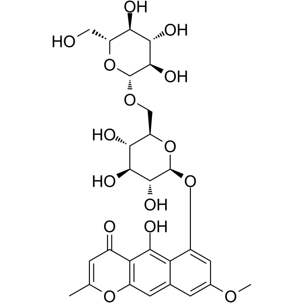
Rubrofusarin gentiobioside (Rrubrofusarin-6-β-gentiobioside) is an orally active weak inhibitor of PTP1B and MAO-A (IC50 >100 μM), and its glycosylation modification results in lower biological activity than its aglycone Rubrofusarin (HY-130307). Rubrofusarin gentiobioside promotes AMPK phosphorylation in an LKB1-independent manner and inhibits the mTOR signaling pathway, thereby downregulating the expressions of PPARγ, C/EBPα, as well as FAS, LPL and aP2. Rubrofusarin gentiobioside inhibits lipid accumulation, reduces body weight and epididymal white adipose tissue volume, improves fatty liver, and shows no cytotoxicity to hepatocytes. Rubrofusarin gentiobioside is widely used in obesity-related studies.
For research use only. We do not sell to patients.
- Purity : 99.07%
- CAS No.: 24577-90-0
- Formula: C27H32O15
- Molecular Weight:596.53
-
Storage:
4°C, sealed storage, away from moisture and light
* In solvent : -80°C, 6 months; -20°C, 1 month (sealed storage, away from moisture and light)

To place orders, for customer services and technical support, please contact: MedChemExpress USA
Tel: 609-228-6898 E-mail: [email protected] [email protected]


-
 Biological Activity
-
Chemical Information
-
Solvent & Solubility
- Protocols
- Purity & Documentation
- References
-
 Help & FAQs


All AMPK Isoforms
More

Biological Activity
Description
IC50 & Target
[1]|
MAO-A |
PPAR-γ |
In Vitro
Rubrofusarin gentiobioside (200, 400 μM; 48 h) is non-cytotoxic to 3T3-L1 murine pre-adipocytes[1].
Rubrofusarin gentiobioside (50, 100, 200 μM) activates AMPKα in an LKB1-independent manner, suppresses mTOR phosphorylation, and does not regulate the PI3K/AKT pathway in differentiated 3T3-L1 murine pre-adipocytes at concentrations of 50, 100, and 200 μM[1].
Rubrofusarin gentiobioside (400 μM; 48 h) is non-cytotoxic to human adipose mesenchymal stem cells at concentrations up to 400 μM when incubated for 48 h[1].
Rubrofusarin gentiobioside (25-100 μM) is non-toxic to HepG2 human hepatocarcinoma cells at concentrations up to 100 μM[2].
Rubrofusarin gentiobioside has limited druglike properties per Lipinski’s rule and poor predicted ADME characteristics including low intestinal absorption and blood-brain barrier penetration[2].
MedChemExpress (MCE) has not independently confirmed the accuracy of these methods. They are for reference only. Further protocols information, click here.
-
Cell Line:3T3-L1 murine pre-adipocytes
-
Concentration:0, 50, 100, 200, 400 μM
-
Incubation Time:48 h
-
Result:Did not exert cytotoxicity at concentrations up to 200 μM.
Caused a statistically significant reduction in cell viability (p < 0.05) relative to the untreated control at 400 μM.
-
Cell Line:human adipose mesenchymal stem cells (hAMSCs)
-
Concentration:0, 50, 100, 200, 400 μM
-
Incubation Time:48 h
-
Result:Did not exhibit cytotoxicity at any tested concentration up to 400 μM.
In Vivo
MedChemExpress (MCE) has not independently confirmed the accuracy of these methods. They are for reference only.
-
Animal Model:C57BL/6 J mice with Obesity (male, 4 weeks old, 16-18 g, HFD-induced obesity)[1]
-
Dosage:50 mg/kg/day; 100 mg/kg/day
-
Administration:p.o.; daily; 10 weeks
-
Result:Reduced weight gain by 19.7% (50 mg/kg/day) and 30.9% (100 mg/kg/day) relative to HFD-only group.
Reduced epididymal white adipose tissue weight relative to HFD-only group.
Prevented liver weight increase relative to HFD-only group.
Decreased serum total cholesterol, LDL-cholesterol, free fatty acid, glucose, and leptin levels, and increased adiponectin levels relative to HFD-only group.
Reduced adipocyte size to 4079 μM2 (50 mg/kg/day) and 2813 μM2 (100 mg/kg/day) relative to 5944 μM2 in HFD-only group.
Reduced hepatic lipid accumulation relative to HFD-only group.
Decreased protein and mRNA expression of PPARγ and C/EBPα in epididymal white adipose tissue and liver relative to HFD-only group.
Increased phosphorylation of AMPK and decreased phosphorylation of mTOR in epididymal white adipose tissue and liver relative to HFD-only group.
Downregulated mRNA expression of FAS, LPL, and aP2 in epididymal white adipose tissue and liver relative to HFD-only group.


Chemical Information
-
CAS No. 24577-90-0
-
Appearance Solid
-
Molecular Weight 596.53
-
Formula C27H32O15
-
Color Light yellow to yellow
-
SMILES
O[C@H]1[C@H](OC2=CC(OC)=CC3=C2C(O)=C4C(C=C(C)OC4=C3)=O)O[C@H](CO[C@H]5[C@@H]([C@H]([C@@H]([C@H](O5)CO)O)O)O)[C@@H](O)[C@@H]1O
-
Synonyms
Rubrofusarin-6-β-gentiobioside
-
Structure Classification
-
Shipping
Room temperature in continental US; may vary elsewhere.
-
Storage
4°C, sealed storage, away from moisture and light
* In solvent : -80°C, 6 months; -20°C, 1 month (sealed storage, away from moisture and light)


Solvent & Solubility
In Vitro:
DMSO : 100 mg/mL (167.64 mM; Need ultrasonic; Hygroscopic DMSO has a significant impact on the solubility of product, please use newly opened DMSO)
Please refer to the solubility information to select the appropriate solvent. Once prepared, please aliquot and store the solution to prevent product inactivation from repeated freeze-thaw cycles.
Storage method and period of stock solution: -80°C, 6 months; -20°C, 1 month (sealed storage, away from moisture and light). When stored at -80°C, please use it within 6 months. When stored at -20°C, please use it within 1 month.
Please refer to the solubility information to select the appropriate solvent. Once prepared, please aliquot and store the solution to prevent product inactivation from repeated freeze-thaw cycles.
Storage method and period of stock solution: -80°C, 6 months; -20°C, 1 month (sealed storage, away from moisture and light). When stored at -80°C, please use it within 6 months. When stored at -20°C, please use it within 1 month.
Concentration (start) × Volume (start) = Concentration (final) × Volume (final)
In Vivo:
Select the appropriate dissolution method based on your experimental animal and administration route.
- For the following dissolution methods, please ensure to first prepare a clear stock solution using an In Vitro approach and then sequentially add co-solvents:
- To ensure reliable experimental results, the clarified stock solution can be appropriately stored based on storage conditions. As for the working solution for In Vivo experiments, it is recommended to prepare freshly and use it on the same day.
- The percentages shown for the solvents indicate their volumetric ratio in the final prepared solution. If precipitation or phase separation occurs during preparation, heat and/or sonication can be used to aid dissolution.
Add each solvent one by one: 10% DMSO 40% PEG300 5% Tween-80 45% Saline
Solubility: ≥ 2.5 mg/mL (4.19 mM); Clear solution
This protocol yields a clear solution of ≥ 2.5 mg/mL (saturation unknown).
Taking 1 mL working solution as an example, add 100 μL DMSO stock solution (25.0 mg/mL) to 400 μL PEG300, and mix evenly; then add 50 μL Tween-80 and mix evenly; then add 450 μL Saline to adjust the volume to 1 mL.
Preparation of Saline: Dissolve 0.9 g sodium chloride in ddH₂O and dilute to 100 mL to obtain a clear Saline solution.
Add each solvent one by one: 10% DMSO 90% (20% SBE-β-CD in Saline)
Solubility: ≥ 2.5 mg/mL (4.19 mM); Clear solution
This protocol yields a clear solution of ≥ 2.5 mg/mL (saturation unknown).
Taking 1 mL working solution as an example, add 100 μL DMSO stock solution (25.0 mg/mL) to 900 μL 20% SBE-β-CD in Saline, and mix evenly.
Preparation of 20% SBE-β-CD in Saline (4°C, storage for one week): 2 g SBE-β-CD powder is dissolved in 10 mL Saline, completely dissolve until clear.
In Vivo Dissolution Calculator
Please enter the basic information of animal experiments:
-
-
-
-
Recommended: Prepare an additional quantity of animals to account for potential losses during experiments.
Please enter your animal formula composition:
-
%DMSO +
Recommended: Keep the proportion of DMSO in working solution below 2% if your animal is weak.
-
%+
-
+%Tween-80 + +
-
%Saline +
The co-solvents required include: DMSO, . All of co-solvents are available by MedChemExpress (MCE). , Tween 80. All of co-solvents are available by MedChemExpress (MCE).
Working solution concentration: 0.22 mg/mL
Method for preparing stock solution: mg drug dissolved in μL DMSO. Stock solution concentration: mg/mL. * In solvent : -80°C, 6 months; -20°C, 1 month (sealed storage, away from moisture and light)
1. Take μL DMSO stock solution;
2. Add μL .
μL , mix evenly;
3. Then add μL Tween 80, mix evenly;
4. Then add μL
Please ensure that the stock solution in the first step is dissolved to a clear state, and add co-solvents in sequence. You can use ultrasonic heating (ultrasonic cleaner, recommended frequency 20-40 kHz), vortexing, etc. to assist dissolution.

Protocols
-
Kinase activity and phosphorylation assays
Kinase activity assays measure the ability of kinases to transfer phosphate groups from ATP to specific substrates, while phosphorylation assays detect the presence and levels of phosphorylated proteins. Common methods include radiolabeled ATP incorporation (e. g. ,), ADP release detection via bioluminescence (e. g. ,[3]), enzyme-linked immunosorbent assays (ELISA) for phospho-specific epitopes (e. g. ,[6]), and microtiter-based formats for high-throughput screening (e. g. ,[8]). The ADP-Glo assay quantifies kinase activity by measuring ADP produced during phosphorylation using a luciferase-based system. Radiometric assays involve autoradiography or scintillation counting after incorporation of 32P-labeled ATP into substrate proteins. ELISA-based approaches rely on phospho-specific antibodies to detect activated kinases in cell lysates or purified samples.
-
Western Blot
Western blotting (WB) is a commonly used experimental method in molecular biology, biochemistry, and immunogenetics for identifying and quantifying target proteins. It combines gel electrophoresis with immunoassay, enabling researchers to analyze protein expression, post-translational modifications, and molecular weight.
-
3T3-L1 preadipocyte-to-adipocyte differentiation
3T3-L1 preadipocytes are induced to differentiate after growth arrest using adipogenic media containing insulin, dexamethasone, and IBMX; differentiation is assessed by lipid-droplet accumulation, triglyceride increase, Oil Red O staining, and adipocyte-marker induction such as PPARγ and C/EBPα.
-
Cell Cytotoxicity Assay
Cytotoxicity assays are usually based on the assessment of cell membrane damage, which can also be indirectly detected by measuring cell viability. Detection methods include MTT assay, CKK-8 assay, LDH assay and ATP assay, etc.
-
Mammalian live/dead viability and cytotoxicity staining
Live/dead viability and cytotoxicity staining assays are based on the simultaneous detection of intracellular esterase activity in metabolically active (viable) cells and membrane integrity loss in non-viable cells. In commonly used dual-staining approaches, membrane-permeant fluorogenic substrates are converted by intracellular esterases into fluorescent products in live cells, while impermeant DNA-binding dyes selectively enter cells with compromised plasma membranes and label nucleic acids in dead or dying cells, enabling discrimination between viable and non-viable populations by fluorescence microscopy or flow cytometry.
-
Large-size fat particle sorting
Large-size fat particle sorting is widely used to isolate cells up to 200 μm in diameter. Single-cell flow sorting will allow greater insight into adipocyte heterogeneity by identifying gene expression, protein composition, and metabolic signatures at the single-cell level.
-
Protocol for Kinase activity and phosphorylation assays
Kinase activity assays measure transfer of phosphate from ATP to a protein or peptide substrate, generating phosphorylated substrate, ADP, or incorporated radiolabeled phosphate as the readout; phosphorylation assays measure site-specific phosphorylation in cells or tissues as a proxy for kinase-pathway activation, inhibition, or substrate regulation. Phosphorylation can be detected by phospho-specific Western blot, immunoprecipitation kinase assay, phospho-immunofluorescence, phospho-flow cytometry, luminescent ADP detection, radiolabeled ATP incorporation, or reporter-based pathway assays, and these readouts can be applied to cancer cells, primary neurons, mouse tumors, organoids, inflammatory macrophages, ferroptosis studies, and mitophagy studies when the kinase target is biologically relevant.
-
Research Protocol for Metabolic Diseases
AMP-activated protein kinase, AMPK, is a conserved cellular energy sensor that responds to reduced cellular energy status and coordinates metabolism by increasing ATP-generating catabolic pathways while suppressing ATP-consuming anabolic processes. In metabolic disease research, the AMPK pathway is experimentally relevant because it regulates hepatic lipid synthesis, fatty acid oxidation, glucose production, skeletal-muscle glucose disposal, mTORC1-linked biosynthesis, autophagy, mitochondrial homeostasis, and whole-body energy balance. The central pathway logic is that energy stress, metformin, exercise-like stimulation, or direct AMPK activators increase AMPKα Thr172 phosphorylation and downstream substrate phosphorylation, including ACC and RAPTOR. Phosphorylation of ACC suppresses lipogenesis and supports fatty acid oxidation, whereas phosphorylation of RAPTOR suppresses mTORC1 signaling and links cellular energy status to growth and protein synthesis control. The pathway is linked
-
Lipid Droplets: Oil Red O/Sudan Dye Lipid Staining
Lipid droplets are intracellular organelles with a neutral-lipid core that stores triacylglycerols and sterol esters, and Oil Red O or Sudan dyes detect these hydrophobic lipid deposits by partitioning into retained lipids in fresh or frozen specimens. Oil Red O stains neutral triglycerides and lipids in frozen tissue sections or air-dried cytologic preparations, while Sudan Black B has also been used as a histochemical fat stain for lipid-rich tissue structures.
-
Cell Viability Determination by MTT Colorimetric Assay
The following protocol uses the MTT colorimetric assay as a classic literature-established method for assessing cell viability/metabolic activity in cultured mammalian cells. MTT[3-(4,5-dimethylthiazol-2-yl)-2,5-diphenyltetrazolium bromide] is reduced by metabolically active cells to a colored formazan product; the amount of formazan is quantified spectrophotometrically and provides an indirect measure of metabolically active viable cells. Importantly, MTT reduction reflects cellular oxidoreductase/metabolic activity rather than an absolute direct count of living cells, so changes in cellular metabolism can alter the signal independently of cell number.


Purity & Documentation
-
Data Sheet (281 KB)
-
SDS (393 KB)
- English - EN (393 KB)
- Français - FR (393 KB)
- Deutsch - DE (393 KB)
- Norwegian - NO (393 KB)
- Español - ES (393 KB)
- Swedish - SV (393 KB)
- Italian - IT (393 KB)
- Korean - KR (393 KB)
- Portuguese - PT (393 KB)
-
Handling Instructions (2659 KB)
References
[1]. Han YH, et al. Rubrofusarin-6-β-gentiobioside inhibits lipid accumulation and weight gain by regulating AMPK/mTOR signaling. Phytomedicine. 2019;62:152952. [Content Brief]
[2]. Paudel P, et al. Rubrofusarin as a Dual Protein Tyrosine Phosphate 1B and Human Monoamine Oxidase-A Inhibitor: An in Vitro and in Silico Study. ACS Omega. 2019;4(7):11621-11630. Published 2019 Jul 3. [Content Brief]


Complete Stock Solution Preparation Table
Please refer to the solubility information to select the appropriate solvent. Once prepared, please aliquot and store the solution to prevent product inactivation from repeated freeze-thaw cycles.
Storage method and period of stock solution: -80°C, 6 months; -20°C, 1 month (sealed storage, away from moisture and light). When stored at -80°C, please use it within 6 months. When stored at -20°C, please use it within 1 month.
| Optional Solvent | Concentration Solvent Mass | 1 mg | 5 mg | 10 mg | 25 mg |
|---|---|---|---|---|---|
| DMSO | 1 mM | 1.6764 mL | 8.3818 mL | 16.7636 mL | 41.9090 mL |
| 5 mM | 0.3353 mL | 1.6764 mL | 3.3527 mL | 8.3818 mL | |
| 10 mM | 0.1676 mL | 0.8382 mL | 1.6764 mL | 4.1909 mL | |
| 15 mM | 0.1118 mL | 0.5588 mL | 1.1176 mL | 2.7939 mL | |
| 20 mM | 0.0838 mL | 0.4191 mL | 0.8382 mL | 2.0955 mL | |
| 25 mM | 0.0671 mL | 0.3353 mL | 0.6705 mL | 1.6764 mL | |
| 30 mM | 0.0559 mL | 0.2794 mL | 0.5588 mL | 1.3970 mL | |
| 40 mM | 0.0419 mL | 0.2095 mL | 0.4191 mL | 1.0477 mL | |
| 50 mM | 0.0335 mL | 0.1676 mL | 0.3353 mL | 0.8382 mL | |
| 60 mM | 0.0279 mL | 0.1397 mL | 0.2794 mL | 0.6985 mL | |
| 80 mM | 0.0210 mL | 0.1048 mL | 0.2095 mL | 0.5239 mL | |
| 100 mM | 0.0168 mL | 0.0838 mL | 0.1676 mL | 0.4191 mL |


Rubrofusarin gentiobioside Related Classifications
HY-N4177 Related Classifications
 Powered by Bioz