



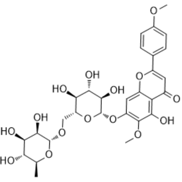
Pectolinarin
Based on 3 publication(s) in Google Scholar
Pectolinarin possesses anti-inflammatory activity. Pectolinarin inhibits secretion of IL-6 and IL-8, as well as the production of PGE2 and NO. Pectolinarin suppresses cell proliferation and inflammatory response and induces apoptosis via inactivation of the PI3K/Akt pathway.
For research use only. We do not sell to patients.
- Purity : 99.82%
- CAS No.: 28978-02-1
- Formula: C29H34O15
- Molecular Weight:622.57
-
Storage:
4°C, protect from light
* In solvent : -80°C, 6 months; -20°C, 1 month (protect from light)

To place orders, for customer services and technical support, please contact: MedChemExpress USA
Tel: 609-228-6898 E-mail: [email protected] [email protected]


-
 Biological Activity
-
Chemical Information
-
Solvent & Solubility
- Protocols
- Purity & Documentation
- References
-
 Help & FAQs


-

HY-N0314R
Pectolinarin (Standard)IL-6/8 Inhibitor -
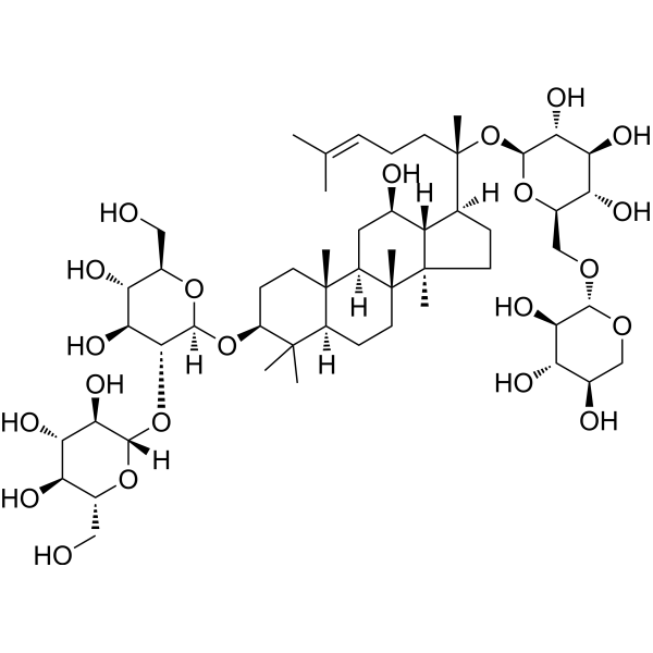
HY-N0041
Ginsenoside Rb3COX Inhibitor -
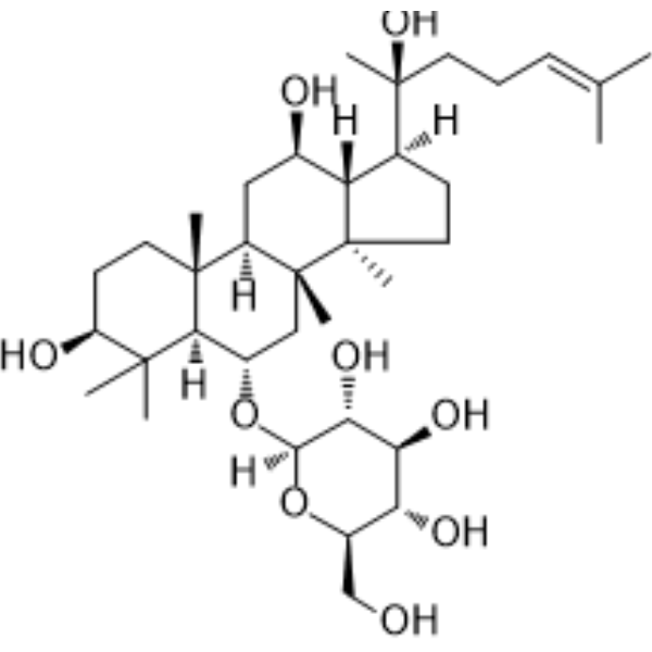
HY-N0604
Ginsenoside Rh1Endogenous Metabolite -
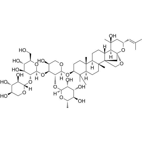
HY-N0660
Jujuboside BSaponin -
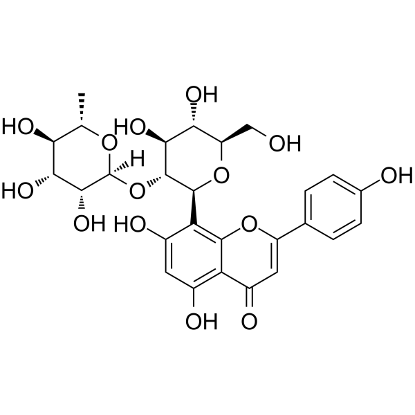
HY-N0534
Vitexin-2"-O-rhamnosideFlavonoid Glycoside -
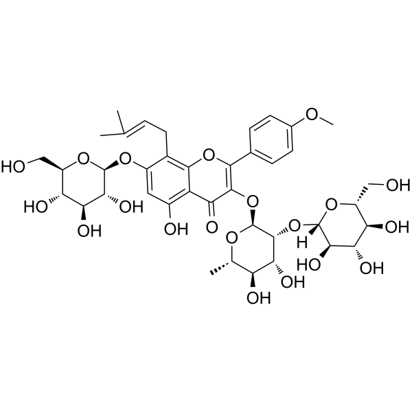
HY-N0257
Epimedin ANatural Product -
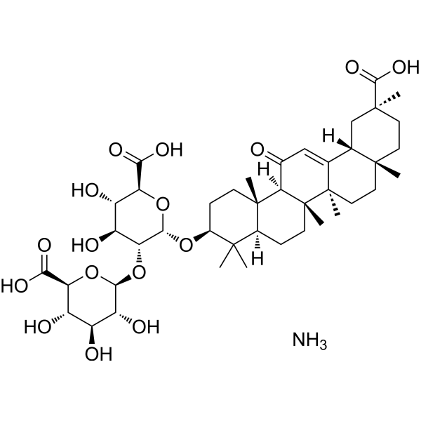
HY-76225
Ammonium glycyrrhizinateAnti-tumor Agent -
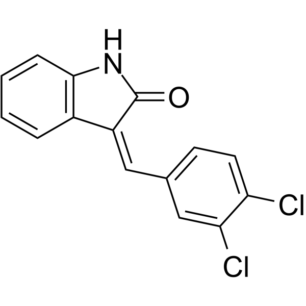
HY-21293
SU5201IL-2 Inhibitor -
HY-N0041R
Ginsenoside Rb3 (Standard) -

HY-N0660R
Jujuboside B (Standard)Platelet Aggregation Inhibitor -
HY-N0604R
Ginsenoside Rh1 (Standard)Endogenous Metabolite -

HY-N0534R
Vitexin-2"-O-rhamnoside (Standard)Standard of Vitexin-2"-O-rhamnoside
Publications Citing Use of MedChemExpress (MCE) Pectolinarin
More Customer Validation & Images

-
Cell Proliferation/Viability Assay

-
WB
-
IF
-
ELISA
-
RT-PCR


Biological Activity
Description
IC50 & Target
[2]|
IL-6 |
EP |
IL-8 |
Cellular Effect
|
Cell Line

|
Type | Value | Description | References |
|---|---|---|---|---|
| A-375 | IC50 |
32.9 μM
Compound: 1
|
Antiproliferative activity against human A375 cells after 48 hrs by sulforhodamine B assay
Antiproliferative activity against human A375 cells after 48 hrs by sulforhodamine B assay
|
[PMID: 18818071] |
| A549 | IC50 |
38.8 μM
Compound: 1
|
Antiproliferative activity against human A549 cells after 48 hrs by sulforhodamine B assay
Antiproliferative activity against human A549 cells after 48 hrs by sulforhodamine B assay
|
[PMID: 18818071] |
| ACHN | IC50 |
17.2 μM
Compound: 1
|
Antiproliferative activity against human ACHN cells after 48 hrs by sulforhodamine B assay
Antiproliferative activity against human ACHN cells after 48 hrs by sulforhodamine B assay
|
[PMID: 18818071] |
| ACHN | IC50 |
17.22 μM
Compound: Pectolinarin
|
In vitro inhibitory concentration against renal adenocarcinoma cell line (ACHN) in the presence of taxol
In vitro inhibitory concentration against renal adenocarcinoma cell line (ACHN) in the presence of taxol
|
[PMID: 16125932] |
| C32 | IC50 |
7.2 μM
Compound: 1
|
Antiproliferative activity against human C32 cells after 48 hrs by sulforhodamine B assay
Antiproliferative activity against human C32 cells after 48 hrs by sulforhodamine B assay
|
[PMID: 18818071] |
| Caco-2 | IC50 |
6.18 μM
Compound: Pectolinarin
|
In vitro inhibitory concentration against human colorectal adenocarcinoma Caco-2 cells in the presence of vinblastine sulfate salt
In vitro inhibitory concentration against human colorectal adenocarcinoma Caco-2 cells in the presence of vinblastine sulfate salt
|
[PMID: 16125932] |
| Caco-2 | IC50 |
6.2 μM
Compound: 1
|
Antiproliferative activity against human Caco-2 cells after 48 hrs by sulforhodamine B assay
Antiproliferative activity against human Caco-2 cells after 48 hrs by sulforhodamine B assay
|
[PMID: 18818071] |
| COR-L23 | IC50 |
5 μM
Compound: 1
|
Antiproliferative activity against human COR-L23 cells after 48 hrs by sulforhodamine B assay
Antiproliferative activity against human COR-L23 cells after 48 hrs by sulforhodamine B assay
|
[PMID: 18818071] |
| COR-L23 | IC50 |
5.03 μM
Compound: Pectolinarin
|
In vitro inhibitory concentration against human carcinoma COR-L23 cells in the presence of vinblastine sulfate salt
In vitro inhibitory concentration against human carcinoma COR-L23 cells in the presence of vinblastine sulfate salt
|
[PMID: 16125932] |
| HEK293 | EC50 |
>100 μM
Compound: 18
|
Agonist activity at mouse PPARgamma expressed in HEK293 cells co-expressing with Gal4 reporter vector after 24 hrs by dual-luciferase reporter assay
Agonist activity at mouse PPARgamma expressed in HEK293 cells co-expressing with Gal4 reporter vector after 24 hrs by dual-luciferase reporter assay
|
[PMID: 24955889] |
| HepG2 | IC50 |
14.12 μM
Compound: Pectolinarin
|
In vitro inhibitory concentration against human hepatocellular carcinoma HepG-2 cells in the presence of vinblastine sulfate salt
In vitro inhibitory concentration against human hepatocellular carcinoma HepG-2 cells in the presence of vinblastine sulfate salt
|
[PMID: 16125932] |
| MRC5 | IC50 |
>80.3 μM
Compound: Pectolinarin
|
In vitro inhibitory concentration against human fetal lung MRC-5 cell line in the presence of vinblastine sulfate salt
In vitro inhibitory concentration against human fetal lung MRC-5 cell line in the presence of vinblastine sulfate salt
|
[PMID: 16125932] |
In Vitro
Pectolinarin increases Bax level, and decreases Bcl-2 level in rheumatoid arthritis fibroblast-like synoviocytes (RA-FLSs). Pectolinarin inactivates the PI3K/Akt pathway in RA-FLSs[2].
MedChemExpress (MCE) has not independently confirmed the accuracy of these methods. They are for reference only.
- Annexin V plus membrane-impermeant dye apoptosis staining
- BrdU Incorporation Assay
- Somatic Cell Culture
- CFSE Dye Dilution Proliferation Assay
- Dye-dilution cell tracking and proliferation staining
- CCK-8/WST-8 Cell Proliferation Assay
- Protocol for Cell Cycle
- Research Protocol for Inflammation-related Diseases
- Cell Counting-Based Growth Curve Assay
- Colony Formation (Clonogenic) Assay
- Cell Viability Determination by MTT Colorimetric Assay
- EdU Incorporation Assay (Click Chemistry-Based DNA Synthesis Measurement)
- Ki-67 Immunostaining Proliferation Assay
- Detection of Apoptosis
- TUNEL staining for apoptotic DNA fragmentation
- PCNA Immunodetection Proliferation Assay
- Cotton Pellet Granuloma
- Protocol for Cell Counting and Cell Density Analysis
- Carrageenan-Induced Paw Edema
- Apoptosis Solutions
- MTT Cell Proliferation Assay


Chemical Information
-
CAS No. 28978-02-1
-
Appearance Solid
-
Molecular Weight 622.57
-
Formula C29H34O15
-
Color Off-white to light yellow
-
SMILES
O=C1C=C(C2=CC=C(OC)C=C2)OC3=CC(O[C@H]4[C@@H]([C@H]([C@@H]([C@@H](CO[C@H]5[C@@H]([C@@H]([C@H]([C@H](C)O5)O)O)O)O4)O)O)O)=C(OC)C(O)=C13
-
Structure Classification
-
Shipping
Room temperature in continental US; may vary elsewhere.
-
Storage
4°C, protect from light
* In solvent : -80°C, 6 months; -20°C, 1 month (protect from light)


Publications (3)
-
Journal Impact Factor
-
Most Recent
-
Autophagy
XIAP-ULK1-Mediated mitophagy modulates carnitine metabolism to mitigate diabetic kidney disease. [Abstract]2025 Oct 25. PMID: 41139215 -
Mol Neurobiol
Pectolinarin Promotes Functional Recovery after Spinal Cord Injury by Regulating Microglia Polarization Through the PI3K/AKT Signaling Pathway. [Abstract]2025 Jul;62(7):8587-8602. PMID: 40014266
Pectolinarin purchased from MedChemExpress. Usage Cited in: Mol Neurobiol. 2025 Jul;62(7):8587-8602. [Abstract]
Cell viability of microglia treated with Pectolinarin (PTR) (0, 10, 20, 40, 80 μM) at diferent concentrations assessed by CCK-8 assay.
Pectolinarin purchased from MedChemExpress. Usage Cited in: Mol Neurobiol. 2025 Jul;62(7):8587-8602. [Abstract]
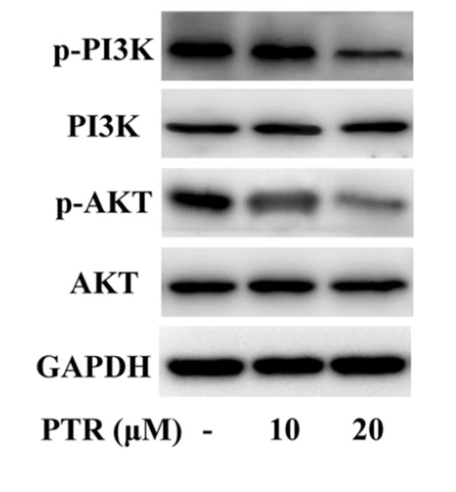
Western blot results of p-PI3K and p-AKT in microglia treated with Pectolinarin (PTR) (10, 20 μM).
Pectolinarin purchased from MedChemExpress. Usage Cited in: Mol Neurobiol. 2025 Jul;62(7):8587-8602. [Abstract]
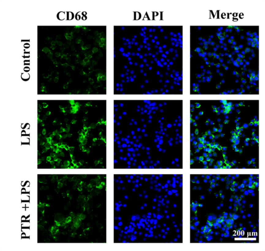
Immunofuorescence staining for CD68 in microglia treated with Pectolinarin (PTR) (20 μM).
Pectolinarin purchased from MedChemExpress. Usage Cited in: Mol Neurobiol. 2025 Jul;62(7):8587-8602. [Abstract]
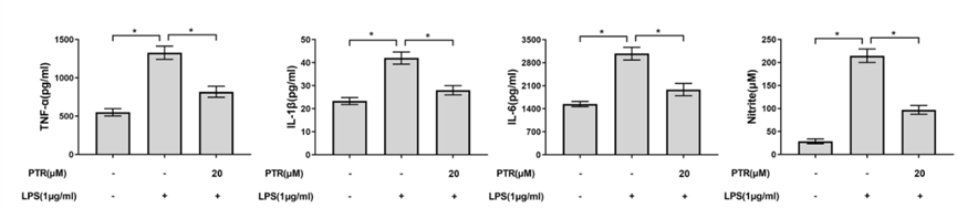
The levels of pro-infammatory mediators (TNF-α, IL-1β, IL-6, and nitrite) after Pectolinarin (PTR) (20 μM) treatment in microglia.
Pectolinarin purchased from MedChemExpress. Usage Cited in: Mol Neurobiol. 2025 Jul;62(7):8587-8602. [Abstract]
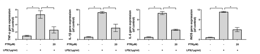
Expression levels of infammation-related genes (TNF-α, IL-1β, IL-6, and iNOS) in microglia after Pectolinarin (PTR) (20 μM) treatment.
-
J Cell Mol Med
2024 Feb;28(4):e18130. PMID: 38332511


Solvent & Solubility
In Vitro:
DMSO : 100 mg/mL (160.62 mM; ultrasonic and warming and heat to 60°C; Hygroscopic DMSO has a significant impact on the solubility of product, please use newly opened DMSO)
Please refer to the solubility information to select the appropriate solvent. Once prepared, please aliquot and store the solution to prevent product inactivation from repeated freeze-thaw cycles.
Storage method and period of stock solution: -80°C, 6 months; -20°C, 1 month (protect from light). When stored at -80°C, please use it within 6 months. When stored at -20°C, please use it within 1 month.
Please refer to the solubility information to select the appropriate solvent. Once prepared, please aliquot and store the solution to prevent product inactivation from repeated freeze-thaw cycles.
Storage method and period of stock solution: -80°C, 6 months; -20°C, 1 month (protect from light). When stored at -80°C, please use it within 6 months. When stored at -20°C, please use it within 1 month.
Concentration (start) × Volume (start) = Concentration (final) × Volume (final)
In Vivo:
Select the appropriate dissolution method based on your experimental animal and administration route.
- For the following dissolution methods, please ensure to first prepare a clear stock solution using an In Vitro approach and then sequentially add co-solvents:
- To ensure reliable experimental results, the clarified stock solution can be appropriately stored based on storage conditions. As for the working solution for In Vivo experiments, it is recommended to prepare freshly and use it on the same day.
- The percentages shown for the solvents indicate their volumetric ratio in the final prepared solution. If precipitation or phase separation occurs during preparation, heat and/or sonication can be used to aid dissolution.
Add each solvent one by one: 10% DMSO 40% PEG300 5% Tween-80 45% Saline
Solubility: 2.08 mg/mL (3.34 mM); Suspended solution; Need ultrasonic
This protocol yields a suspended solution of 2.08 mg/mL. Suspended solution can be used for oral and intraperitoneal injection.
Taking 1 mL working solution as an example, add 100 μL DMSO stock solution (20.8 mg/mL) to 400 μL PEG300, and mix evenly; then add 50 μL Tween-80 and mix evenly; then add 450 μL Saline to adjust the volume to 1 mL.
Preparation of Saline: Dissolve 0.9 g sodium chloride in ddH₂O and dilute to 100 mL to obtain a clear Saline solution.
Add each solvent one by one: 10% DMSO 90% (20% SBE-β-CD in Saline)
Solubility: ≥ 2.08 mg/mL (3.34 mM); Clear solution
This protocol yields a clear solution of ≥ 2.08 mg/mL (saturation unknown).
Taking 1 mL working solution as an example, add 100 μL DMSO stock solution (20.8 mg/mL) to 900 μL 20% SBE-β-CD in Saline, and mix evenly.
Preparation of 20% SBE-β-CD in Saline (4°C, storage for one week): 2 g SBE-β-CD powder is dissolved in 10 mL Saline, completely dissolve until clear.
In Vivo Dissolution Calculator
Please enter the basic information of animal experiments:
-
-
-
-
Recommended: Prepare an additional quantity of animals to account for potential losses during experiments.
Please enter your animal formula composition:
-
%DMSO +
Recommended: Keep the proportion of DMSO in working solution below 2% if your animal is weak.
-
%+
-
+%Tween-80 + +
-
%Saline +
The co-solvents required include: DMSO, . All of co-solvents are available by MedChemExpress (MCE). , Tween 80. All of co-solvents are available by MedChemExpress (MCE).
Working solution concentration: 0.22 mg/mL
Method for preparing stock solution: mg drug dissolved in μL DMSO. Stock solution concentration: mg/mL. * In solvent : -80°C, 6 months; -20°C, 1 month (protect from light)
1. Take μL DMSO stock solution;
2. Add μL .
μL , mix evenly;
3. Then add μL Tween 80, mix evenly;
4. Then add μL
Please ensure that the stock solution in the first step is dissolved to a clear state, and add co-solvents in sequence. You can use ultrasonic heating (ultrasonic cleaner, recommended frequency 20-40 kHz), vortexing, etc. to assist dissolution.


Purity & Documentation
-
Data Sheet (281 KB)
-
SDS (394 KB)
- English - EN (394 KB)
- Français - FR (394 KB)
- Deutsch - DE (394 KB)
- Norwegian - NO (394 KB)
- Español - ES (394 KB)
- Swedish - SV (394 KB)
- Italian - IT (394 KB)
- Korean - KR (394 KB)
- Portuguese - PT (394 KB)
-
Handling Instructions (2659 KB)
References
[1]. Lim H, et al. Anti-inflammatory activity of pectolinarigenin and pectolinarin isolated from Cirsium chanroenicum. Biol Pharm Bull. 2008 Nov;31(11):2063-7. [Content Brief]
[2]. Wang L, et al. Pectolinarin inhibits proliferation, induces apoptosis, and suppresses inflammation in rheumatoid arthritis fibroblast-like synoviocytes by inactivating the phosphatidylinositol 3 kinase/protein kinase B pathway.J Cell Biochem. 2019 Sep;120(9):15202-15210. [Content Brief]


Complete Stock Solution Preparation Table
Please refer to the solubility information to select the appropriate solvent. Once prepared, please aliquot and store the solution to prevent product inactivation from repeated freeze-thaw cycles.
Storage method and period of stock solution: -80°C, 6 months; -20°C, 1 month (protect from light). When stored at -80°C, please use it within 6 months. When stored at -20°C, please use it within 1 month.
| Optional Solvent | Concentration Solvent Mass | 1 mg | 5 mg | 10 mg | 25 mg |
|---|---|---|---|---|---|
| DMSO | 1 mM | 1.6062 mL | 8.0312 mL | 16.0625 mL | 40.1561 mL |
| 5 mM | 0.3212 mL | 1.6062 mL | 3.2125 mL | 8.0312 mL | |
| 10 mM | 0.1606 mL | 0.8031 mL | 1.6062 mL | 4.0156 mL | |
| 15 mM | 0.1071 mL | 0.5354 mL | 1.0708 mL | 2.6771 mL | |
| 20 mM | 0.0803 mL | 0.4016 mL | 0.8031 mL | 2.0078 mL | |
| 25 mM | 0.0642 mL | 0.3212 mL | 0.6425 mL | 1.6062 mL | |
| 30 mM | 0.0535 mL | 0.2677 mL | 0.5354 mL | 1.3385 mL | |
| 40 mM | 0.0402 mL | 0.2008 mL | 0.4016 mL | 1.0039 mL | |
| 50 mM | 0.0321 mL | 0.1606 mL | 0.3212 mL | 0.8031 mL | |
| 60 mM | 0.0268 mL | 0.1339 mL | 0.2677 mL | 0.6693 mL | |
| 80 mM | 0.0201 mL | 0.1004 mL | 0.2008 mL | 0.5020 mL | |
| 100 mM | 0.0161 mL | 0.0803 mL | 0.1606 mL | 0.4016 mL |


Pectolinarin Related Classifications
HY-N0314 Related Classifications
 Powered by Bioz