



Setidegrasib

Based on 1 Customer Validation
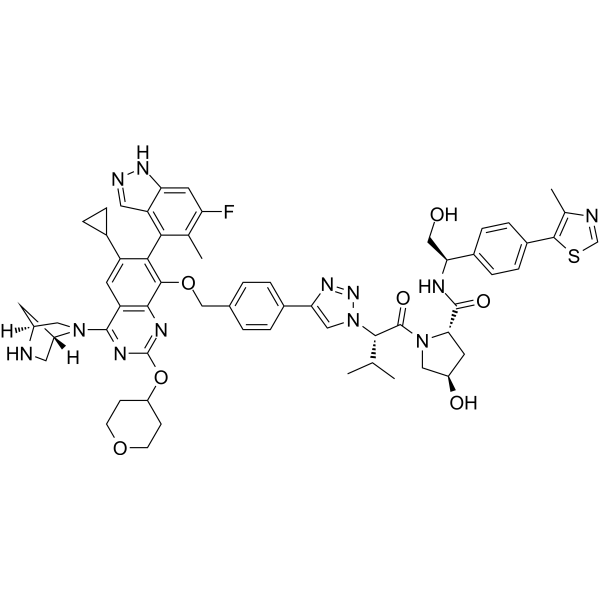
Setidegrasib (ASP-3082) is a PROTAC KRAS degrader (DC50: 37 nM). Setidegrasib induces the degradation of G12D-mutation KRAS protein. Setidegrasib suppresses p-ERK, p-AKT, p-S6 levels in AsPC-1 cells. Setidegrasib exhibits anti-tumor activity in various cancer xenograft models in mice. Setidegrasib can be used in studies involving KRAS(G12D)-mutated solid tumors, such as non-small cell lung cancer.
(Pink: K-RAS ligand (HY-168700); Blue: VHL ligand (HY-168699); Black: linker (HY-168698)).
For research use only. We do not sell to patients.
- Purity : 99.42%
- CAS No.: 2821793-99-9
- Formula: C60H65FN12O7S
- Molecular Weight:1117.30
-
Storage:
-20°C, protect from light
* In solvent : -80°C, 6 months; -20°C, 1 month (protect from light)

To place orders, for customer services and technical support, please contact: MedChemExpress USA
Tel: 609-228-6898 E-mail: [email protected] [email protected]


-
 Biological Activity
-
Chemical Information
-
Solvent & Solubility
- Protocols
- Purity & Documentation
- References
-
 Help & FAQs


-
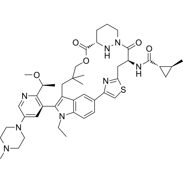
HY-148439
DaraxonrasibRAS(ON) Inhibitor -
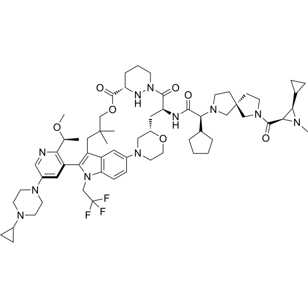
HY-156819
ZoldonrasibKRAS G12D Inhibitor -
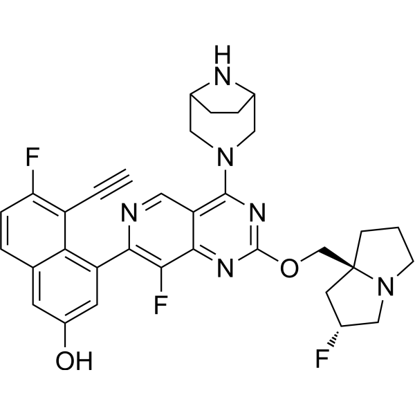
HY-134813
MRTX1133KRAS G12D Inhibitor -
HY-157228
ACBI3KRAS degrader -
HY-161176
PROTAC KRAS G12D degrader 1PROTAC KRAS G12D Degrader -
HY-15531
VenetoclaxBCL-2 Inhibitor -

HY-159127
HRS-4642KRAS G12D Inhibitor -

HY-173629
RMC-5127GTP-bound KRAS^{G12V} Inhibitor -

HY-176786
MCB-36KRAS PROTAC Degrader -

HY-168054
PROTAC K-Ras Degrader-3K-Ras PROTAC Degrader
All PROTACs Isoforms
More

Biological Activity
Description
IC50 & Target
[1][2]|
KRAS(G12D) 37 nM (DC50) |
VHL |
In Vitro
Setidegrasib (Example 8) (24 h) inhibits ERK phosphorylation in G12D-mutant KRAS-positive pancreatic cancer strain AsPC-1, with an IC50 of 15 nM[1].
Setidegrasib (6 days) inhibits anchorage-independent cell growth against G12D-mutant KRAS-positive pancreatic cancer strain AsPC-1, with an IC50 of 23 nM[1].
Setidegrasib (6 days) potently inhibits proliferation of multiple KRAS(G12D)-mutated cancer cell lines (PK-59, HPAC, GP2d, GP5d) in 3D assay, while showing weak activity (IC50 > 10 μM) against KRAS(WT) cells (A375, HT-29) and other KRAS mutants (G12V, G12C, G13D) via CellTiter-Glo assay[2].
Setidegrasib (1 μM, 4-24 h) selectively reduces KRAS(G12D) and DUSP4 (a MAPK-dependent gene) levels in AsPC-1 cells[2].
Setidegrasib (30-300 nM, 6-72 h) sustains KRAS(G12D) degradation (including GTP-bound form) and suppresses p-ERK, p-AKT, p-S6 levels in AsPC-1 cells[2].
MedChemExpress (MCE) has not independently confirmed the accuracy of these methods. They are for reference only.
-
Cell Line:AsPC-1 cells
-
Concentration:30, 100, 300 nM
-
Incubation Time:6, 24, 48, 72 h
-
Result:Sustained KRAS(G12D) degradation (including GTP-bound form).
Suppressed p-ERK, p-AKT, p-S6 levels in AsPC-1 cells.
In Vivo
MedChemExpress (MCE) has not independently confirmed the accuracy of these methods. They are for reference only.
-
Animal Model:PK-59, AsPC-1, HPAC, PK-1, GP2d, GP5d, A375, KP-4, and HT-29 cells were subcutaneously inoculated into the flank of 4-8 week-old male nude mice (Balb/c nu/nu) at 1-6 × 10⁶ cells per 0.1–0.2 ml(Matrigel (Corning):PBS = 1:1)/mouse[2]
-
Dosage:0.3, 1, 3, 10, 30 mg/kg
-
Administration:i.v., once/twice weekly, 14, 21, 22 days
-
Result:Achieved tumor growth inhibition in PK-59, LXFA 1125 NSCLC PDX, AsPC-1 (pancreatic) and GP2d (colorectal) KRAS(G12D)-mutated xenograft models of Balb/c nu/nu nude mice.
Showed no significant changes in body weight.
Showed no significant tumor growth inhibition in A375 (melanoma) and HT-29 (colorectal) KRAS(WT) xenograft models.
Clinical Trial
| NCT Number | Sponsor | Condition | Start Date |
Phase

|
|---|---|---|---|---|
| NCT01329991 | Plexxikon| | 2011-05 | PHASE1 |


Chemical Information
-
CAS No. 2821793-99-9
-
Appearance Solid
-
Molecular Weight 1117.30
-
Formula C60H65FN12O7S
-
Color White to off-white
-
SMILES
CC1=C(SC=N1)C2=CC=C(C=C2)[C@@H](NC([C@@H]3C[C@H](CN3C([C@H](C(C)C)N4C=C(N=N4)C5=CC=C(C=C5)COC6=[C@]([C@]7=C(C(F)=CC8=C7C=NN8)C)C(C9CC9)=CC%10=C6N=C(N=C%10N%11C[C@@H]%12C[C@H]%11CN%12)OC%13CCOCC%13)=O)O)=O)CO
-
Synonyms
ASP-3082; KRAS G12D inhibitor 17
-
Shipping
Room temperature in continental US; may vary elsewhere.
-
Storage
-20°C, protect from light
* In solvent : -80°C, 6 months; -20°C, 1 month (protect from light)


Solvent & Solubility
In Vitro:
DMSO : 100 mg/mL (89.50 mM; Need ultrasonic; Hygroscopic DMSO has a significant impact on the solubility of product, please use newly opened DMSO)
Please refer to the solubility information to select the appropriate solvent. Once prepared, please aliquot and store the solution to prevent product inactivation from repeated freeze-thaw cycles.
Storage method and period of stock solution: -80°C, 6 months; -20°C, 1 month (protect from light). When stored at -80°C, please use it within 6 months. When stored at -20°C, please use it within 1 month.
Please refer to the solubility information to select the appropriate solvent. Once prepared, please aliquot and store the solution to prevent product inactivation from repeated freeze-thaw cycles.
Storage method and period of stock solution: -80°C, 6 months; -20°C, 1 month (protect from light). When stored at -80°C, please use it within 6 months. When stored at -20°C, please use it within 1 month.
Concentration (start) × Volume (start) = Concentration (final) × Volume (final)
In Vivo:
Select the appropriate dissolution method based on your experimental animal and administration route.
- For the following dissolution methods, please ensure to first prepare a clear stock solution using an In Vitro approach and then sequentially add co-solvents:
- To ensure reliable experimental results, the clarified stock solution can be appropriately stored based on storage conditions. As for the working solution for In Vivo experiments, it is recommended to prepare freshly and use it on the same day.
- The percentages shown for the solvents indicate their volumetric ratio in the final prepared solution. If precipitation or phase separation occurs during preparation, heat and/or sonication can be used to aid dissolution.
Add each solvent one by one: 10% DMSO 40% PEG300 5% Tween-80 45% Saline
Solubility: ≥ 2.5 mg/mL (2.24 mM); Clear solution
This protocol yields a clear solution of ≥ 2.5 mg/mL (saturation unknown).
Taking 1 mL working solution as an example, add 100 μL DMSO stock solution (25.0 mg/mL) to 400 μL PEG300, and mix evenly; then add 50 μL Tween-80 and mix evenly; then add 450 μL Saline to adjust the volume to 1 mL.
Preparation of Saline: Dissolve 0.9 g sodium chloride in ddH₂O and dilute to 100 mL to obtain a clear Saline solution.
In Vivo Dissolution Calculator
Please enter the basic information of animal experiments:
-
-
-
-
Recommended: Prepare an additional quantity of animals to account for potential losses during experiments.
Please enter your animal formula composition:
-
%DMSO +
Recommended: Keep the proportion of DMSO in working solution below 2% if your animal is weak.
-
%+
-
+%Tween-80 + +
-
%Saline +
The co-solvents required include: DMSO, . All of co-solvents are available by MedChemExpress (MCE). , Tween 80. All of co-solvents are available by MedChemExpress (MCE).
Working solution concentration: 0.22 mg/mL
Method for preparing stock solution: mg drug dissolved in μL DMSO. Stock solution concentration: mg/mL. * In solvent : -80°C, 6 months; -20°C, 1 month (protect from light)
1. Take μL DMSO stock solution;
2. Add μL .
μL , mix evenly;
3. Then add μL Tween 80, mix evenly;
4. Then add μL
Please ensure that the stock solution in the first step is dissolved to a clear state, and add co-solvents in sequence. You can use ultrasonic heating (ultrasonic cleaner, recommended frequency 20-40 kHz), vortexing, etc. to assist dissolution.


Purity & Documentation
-
Data Sheet (284 KB)
-
SDS (252 KB)
- English - EN (252 KB)
- Français - FR (252 KB)
- Deutsch - DE (252 KB)
- Norwegian - NO (252 KB)
- Español - ES (252 KB)
- Swedish - SV (252 KB)
- Italian - IT (252 KB)
- Korean - KR (252 KB)
- Portuguese - PT (252 KB)
-
Handling Instructions (2659 KB)
References


Complete Stock Solution Preparation Table
Please refer to the solubility information to select the appropriate solvent. Once prepared, please aliquot and store the solution to prevent product inactivation from repeated freeze-thaw cycles.
Storage method and period of stock solution: -80°C, 6 months; -20°C, 1 month (protect from light). When stored at -80°C, please use it within 6 months. When stored at -20°C, please use it within 1 month.
| Optional Solvent | Concentration Solvent Mass | 1 mg | 5 mg | 10 mg | 25 mg |
|---|---|---|---|---|---|
| DMSO | 1 mM | 0.8950 mL | 4.4751 mL | 8.9501 mL | 22.3754 mL |
| 5 mM | 0.1790 mL | 0.8950 mL | 1.7900 mL | 4.4751 mL | |
| 10 mM | 0.0895 mL | 0.4475 mL | 0.8950 mL | 2.2375 mL | |
| 15 mM | 0.0597 mL | 0.2983 mL | 0.5967 mL | 1.4917 mL | |
| 20 mM | 0.0448 mL | 0.2238 mL | 0.4475 mL | 1.1188 mL | |
| 25 mM | 0.0358 mL | 0.1790 mL | 0.3580 mL | 0.8950 mL | |
| 30 mM | 0.0298 mL | 0.1492 mL | 0.2983 mL | 0.7458 mL | |
| 40 mM | 0.0224 mL | 0.1119 mL | 0.2238 mL | 0.5594 mL | |
| 50 mM | 0.0179 mL | 0.0895 mL | 0.1790 mL | 0.4475 mL | |
| 60 mM | 0.0149 mL | 0.0746 mL | 0.1492 mL | 0.3729 mL | |
| 80 mM | 0.0112 mL | 0.0559 mL | 0.1119 mL | 0.2797 mL |


Setidegrasib Related Classifications
HY-148273 Related Classifications
 Powered by Bioz