



SHP2/HDAC-IN-1
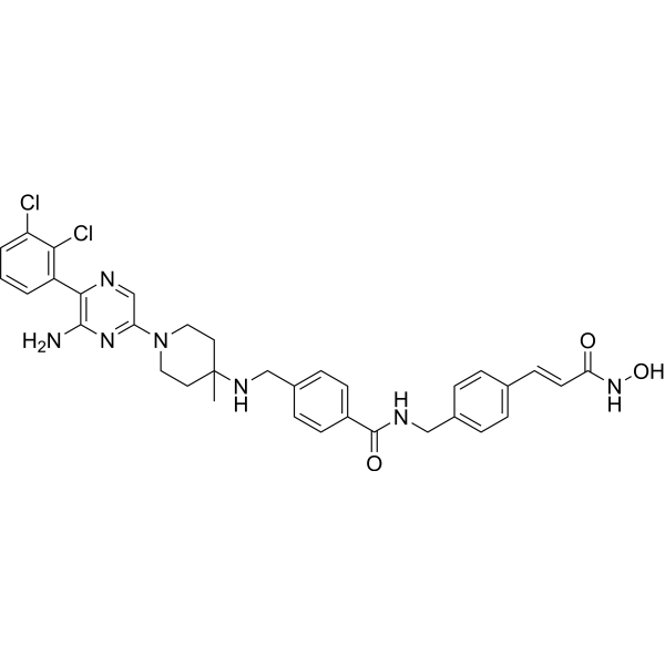
SHP2/HDAC-IN-1 is a dual allosteric SHP2/HDAC inhibitor with IC50 values of 20.4 nM (SHP2) and 25.3 nM (HDAC1) respectively. SHP2/HDAC-IN-1 triggers efficient antitumor immunity by activating T cells, enhancing the antigen presentation function and promoting cytokine secretion. SHP2/HDAC-IN-1 can be used in the research of cancer immunoresearch.
For research use only. We do not sell to patients.
- CAS No.: 2831230-38-5
- Formula: C34H35Cl2N7O3
- Molecular Weight:660.59
-
Storage:
Please store the product under the recommended conditions in the Certificate of Analysis.

To place orders, for customer services and technical support, please contact: MedChemExpress USA
Tel: 609-228-6898 E-mail: [email protected] [email protected]


-
 Biological Activity
-
Chemical Information
- Protocols
- Purity & Documentation
- References
-
 Help & FAQs




Biological Activity
Description
IC50 & Target
[1]|
HDAC1 25 nM (IC50) |
HDAC2 79 nM (IC50) |
HDAC3 233 nM (IC50) |
HDAC6 27 nM (IC50) |
SHP2 20.4 nM (IC50) |
Cellular Effect
|
Cell Line

|
Type | Value | Description | References |
|---|---|---|---|---|
| 4T1 | IC50 |
5.43 μM
Compound: 8t
|
Antiproliferative activity against mouse 4T1 cells assessed as cell viability incubated for 72 hrs by CCK-8 assay
Antiproliferative activity against mouse 4T1 cells assessed as cell viability incubated for 72 hrs by CCK-8 assay
|
[PMID: 36097406] |
| ASPC1 | IC50 |
2.31 μM
Compound: 8t
|
Antiproliferative activity against human ASPC1 cells assessed as reduction in cell viability incubated for 72 hrs by CCK-8 assay
Antiproliferative activity against human ASPC1 cells assessed as reduction in cell viability incubated for 72 hrs by CCK-8 assay
|
[PMID: 36097406] |
| BXPC-3 | IC50 |
1.65 μM
Compound: 8t
|
Antiproliferative activity against human BXPC-3 cells assessed as reduction in cell viability incubated for 72 hrs by CCK-8 assay
Antiproliferative activity against human BXPC-3 cells assessed as reduction in cell viability incubated for 72 hrs by CCK-8 assay
|
[PMID: 36097406] |
| KYSE-520 cell line | IC50 |
1.95 μM
Compound: 8t
|
Antiproliferative activity against human KYSE520 cells assessed as reduction in cell viability incubated for 72 hrs by CCK-8 assay
Antiproliferative activity against human KYSE520 cells assessed as reduction in cell viability incubated for 72 hrs by CCK-8 assay
|
[PMID: 36097406] |
| MV4-11 | IC50 |
0.07 μM
Compound: 8t
|
Antiproliferative activity against human MV4-11 cells assessed as reduction in cell viability incubated for 72 hrs by CCK-8 assay
Antiproliferative activity against human MV4-11 cells assessed as reduction in cell viability incubated for 72 hrs by CCK-8 assay
|
[PMID: 36097406] |
| SW1990 | IC50 |
3.92 μM
Compound: 8t
|
Antiproliferative activity against human SW1990 cells assessed as reduction in cell viability incubated for 72 hrs by CCK-8 assay
Antiproliferative activity against human SW1990 cells assessed as reduction in cell viability incubated for 72 hrs by CCK-8 assay
|
[PMID: 36097406] |
In Vitro
SHP2/HDAC-IN-1 (compound 8t, 0-10 μM approximately, 72 h) inhibits the proliferation of BxPC-3, SW1990, AsPC-1and MV4-11 cells[1].
SHP2/HDAC-IN-1 (0.25-1 μM, 24 h) increases the acetylation of α-tubulin and histone H3 in MV4-11 cells[1].
SHP2/HDAC-IN-1 (0.25 μM, 24 h) inhibits cell cycle progression in the G1 phase of MV4-11 cells[1].
SHP2/HDAC-IN-1 (0.25 and 0.5 μM, 24 h) decreases the mitochondrial membrane potential and activats caspase-3[1].
SHP2/HDAC-IN-1 (2 h) shows good stability in in mouse liver microsome[1].
MedChemExpress (MCE) has not independently confirmed the accuracy of these methods. They are for reference only. Further protocols information, click here.
-
Cell Line:Pancreatic carcinoma (BxPC-3, SW1990, and AsPC-1), acute monocytic leukemia (MV4-11)
-
Concentration:0-10 μM approximately
-
Incubation Time:72 h
-
Result:Inhibited cell proliferation with IC50s range of 0.07 μM-3.92 μM.
-
Cell Line:MV4-11 cells
-
Concentration:0.25, 0.5, 1 μM
-
Incubation Time:24 h
-
Result:Increased the acetylation of α-tubulin and histone H3.
Inhibited the phosphorylation level of ERK.
In Vivo
SHP2/HDAC-IN-1 (20 mg/kg p.o., 1 mg/kg i.v.) exhibits good maximum plasma concentrations in rats[1].
MedChemExpress (MCE) has not independently confirmed the accuracy of these methods. They are for reference only.
-
Animal Model:MV4-11 tumor-bearing xenograft mice[1]
-
Dosage:40 mg/kg
-
Administration:Oral adminstration (p.o.), every day for 20 consecutive days.
-
Result:Delayed tumor progression with a tumor growth inhibition rate (TGI %) value of 64.0%, with no obvious signs of toxicity.
-
Animal Model:4T1 murine breast cancer model[1]
-
Dosage:40 mg/kg
-
Administration:Oral adminstration (p.o.), every day for 12 consecutive days.
-
Result:Significantly decreased tumor burden with a TGI value of 72%.
Increased the proportions of CD4+ T cells and CD8+ T cells in the spleen.
Enhanced the proportion of mDCs in lymph nodes.
-
Animal Model:Male Sprague-Dawley (SD) rats (Pharmacokinetic assay)[1]
-
Dosage:20 mg/kg p.o., 1 mg/kg i.v.
-
Administration:Oral adminstration (p.o.) or intravenous injection (i.v.)
-
Result:Pharmacokinetic profile of SHP2/HDAC-IN-1 (compound 8t).
dose (mg/kg) T1/2 (h) Cmax (ng/mL) Cl (mL/h/kg) F (%) 20 (p.o.) 5.32 1835 21.42 1 (i.v.) 6.15 3517 326


Chemical Information
-
CAS No. 2831230-38-5
-
Molecular Weight 660.59
-
Formula C34H35Cl2N7O3
-
SMILES
O=C(NCC1=CC=C(/C=C/C(NO)=O)C=C1)C2=CC=C(CNC3(C)CCN(C4=NC(N)=C(C5=C(Cl)C(Cl)=CC=C5)N=C4)CC3)C=C2
-
Shipping
Room temperature in continental US; may vary elsewhere.
-
Storage
Please store the product under the recommended conditions in the Certificate of Analysis.

Protocols
-
Cell Cytotoxicity Assay
Cytotoxicity assays are usually based on the assessment of cell membrane damage, which can also be indirectly detected by measuring cell viability. Detection methods include MTT assay, CKK-8 assay, LDH assay and ATP assay, etc.
-
Research Protocol for Inflammation-related Diseases
The NLRP3 inflammasome is a cytosolic innate immune signaling platform that integrates priming signals and danger-signal activation to promote caspase-1 activation, maturation of IL-1β and IL-18, and gasdermin D-mediated pyroptotic cell death. The core experimental logic is to determine whether inflammatory disease phenotypes are driven by increased NLRP3 expression, ASC-containing inflammasome assembly, caspase-1 cleavage, GSDMD cleavage, and extracellular release of IL-1β/IL-18 rather than by nonspecific cell injury alone. The pathway is strongly linked to inflammation-related disease phenotypes because monosodium urate crystals activate NALP3/NLRP3 inflammasome signaling in gout-like crystal inflammation, cholesterol crystals activate NLRP3 inflammasomes in atherogenesis models, and DSS-induced intestinal inflammation has been reported to involve NLRP3 inflammasome activity. However, experimental colitis studies also show context-dependent protective effects of NLRP3 inflammasome co
-
Research Protocol for Cancer Immunology
Cancer immunology studies how the immune system recognizes, suppresses, edits, or fails to eliminate malignant cells through tumor antigen release, antigen presentation, T-cell priming, immune trafficking, tumor-cell killing, and feedback inhibition in the tumor microenvironment. The cancer-immunity cycle links tumor antigenicity, dendritic-cell priming, CD8+ T-cell infiltration, cytotoxic function, and immune-checkpoint regulation to tumor rejection or immune escape. Immune-checkpoint pathways such as PD-1/PD-L1 and CTLA-4 suppress antitumor T-cell activity and can be therapeutically blocked, but many tumors remain resistant because of poor antigen presentation, weak T-cell infiltration, suppressive myeloid cells, regulatory T cells, and tumor-intrinsic immune-exclusion programs. Unresolved questions include which immune-cell states predict response, how tumor-intrinsic pathways exclude immune cells, how myeloid suppression limits checkpoint blockade, and which combination strategies


Purity & Documentation
References

Calculators
Concentration (start) × Volume (start) = Concentration (final) × Volume (final)


SHP2/HDAC-IN-1 Related Classifications
HY-151464 Related Classifications
 Powered by Bioz