



Tepoxalin

Based on 1 Customer Validation
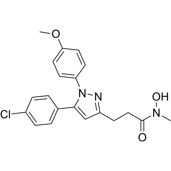
Tepoxalin is an orally active dual inhibitor of Cyclooxygenase/Lipoxygenase, with IC50 values of 4.6 μM (sheep cyclooxygenase), 2.85 μM (rat cyclooxygenase), 0.15 μM (rat 5-lipoxygenase), and 3.0 μM (h12-lipoxygenase), respectively. Tepoxalin inhibits ROS production and NF-κB activation. Tepoxalin suppresses the production of thromboxane B2, leukotriene B4, prostaglandins and cytokines, and blocks platelet aggregation. Tepoxalin exhibits potent anti-inflammatory activity in rats with adjuvant-induced arthritis. Tepoxalin possesses analgesic activity. Tepoxalin shows no ulcerogenic activity within the anti-inflammatory dose range. Tepoxalin can be used in studies related to adjuvant-induced arthritis, skin inflammation and Alzheimer's disease.
For research use only. We do not sell to patients.
- Purity : 98.46%
- CAS No.: 103475-41-8
- Formula: C20H20ClN3O3
- Molecular Weight:385.84
-
Storage:Powder -20°C, 3 years , 4°C, 2 years ; In solvent -80°C, 6 months , -20°C, 1 month

To place orders, for customer services and technical support, please contact: MedChemExpress USA
Tel: 609-228-6898 E-mail: [email protected] [email protected]


-
 Biological Activity
-
Chemical Information
-
Solvent & Solubility
- Protocols
- Purity & Documentation
- References
-
 Help & FAQs


-
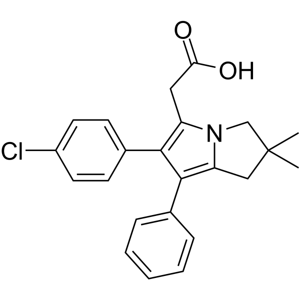
HY-B1452
LicofeloneCOX/5-LOX Inhibitor -
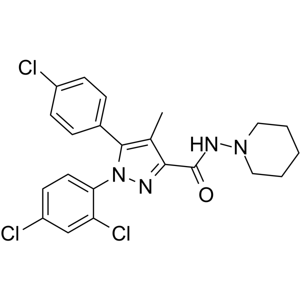
HY-14136
Rimonabant -
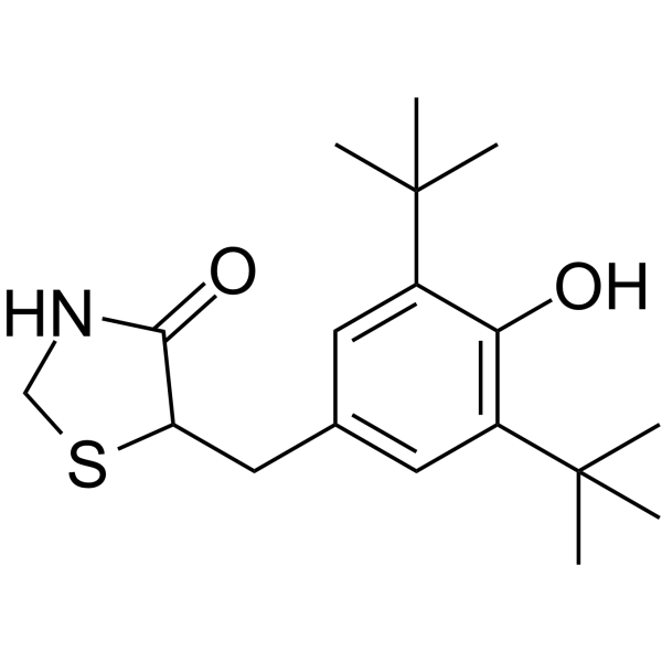
HY-137789
TazofeloneCOX-II Inhibitor -

HY-13219R
Tepoxalin (Standard)COX Inhibitor -
HY-N0172
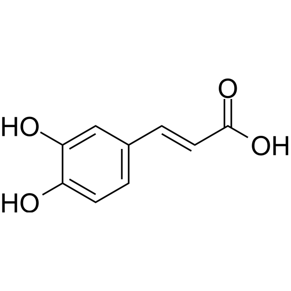
Caffeic acidEndogenous Metabolite -
HY-103157
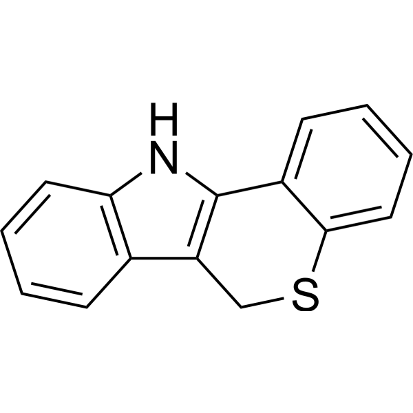
PD14617615-LO Inhibitor -
HY-138939
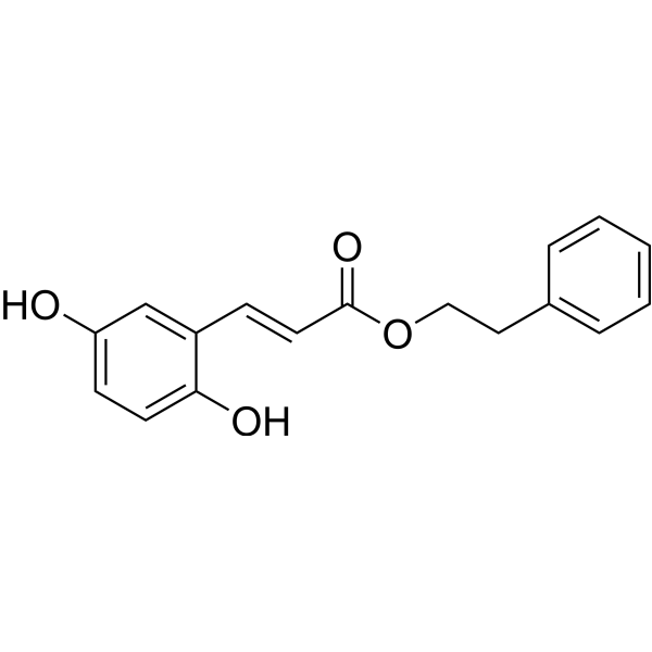
5-LOX-IN-25-LOX Inhibitor -

HY-N0172R
Caffeic acid (Standard)Endogenous Metabolite -
HY-N2075
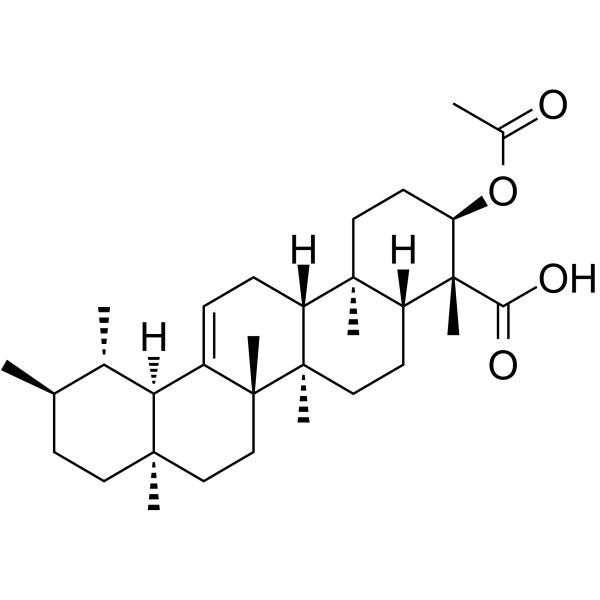
3-Acetyl-beta-boswellic acidNatural Product -
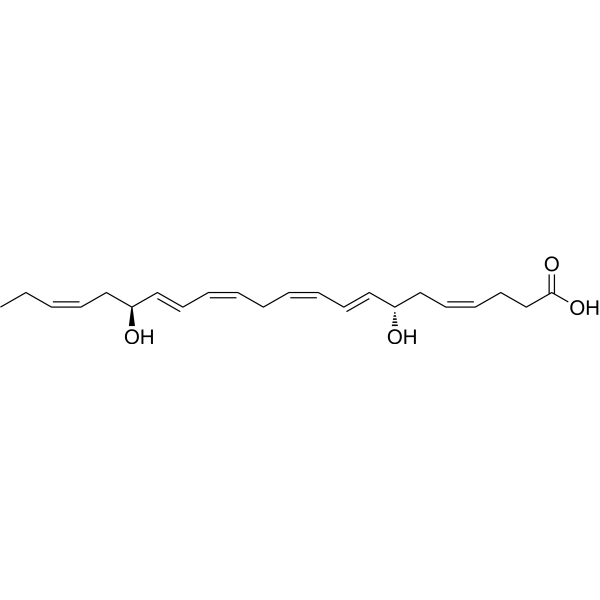
HY-145491
Resolvin D5Inflammation Inhibitor -
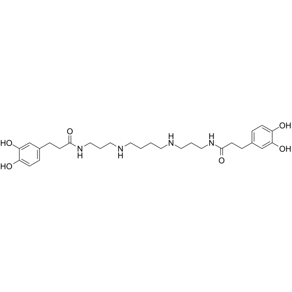
HY-N2392
Kukoamine AAlkaloid -

HY-130675A
15(S)-HEPEAsthma Marker


Biological Activity
Description
IC50 & Target
[1]|
Sheep cyclooxygenase 4.6 μM (IC50) |
Rat cyclooxygenase 2.85 μM (IC50) |
Rat 5-lipoxygenase 0.15 μM (IC50) |
h12-lipoxygenase 3.0 μM (IC50) |
Cellular Effect
|
Cell Line

|
Type | Value | Description | References |
|---|---|---|---|---|
| RBL-1 | IC50 |
0.15 μM
Compound: Tepoxalin
|
Inhibitory activity against 5-lipoxygenase (5-LO) in intact rat barophilic leukemia cells (RBL-1)
Inhibitory activity against 5-lipoxygenase (5-LO) in intact rat barophilic leukemia cells (RBL-1)
|
[PMID: 10230624] |
| RBL-1 | IC50 |
0.15 μM
Compound: Tepoxalin
|
The compound was tested for inhibitory activity against 5-Lipoxygenase in rat RBL-1
The compound was tested for inhibitory activity against 5-Lipoxygenase in rat RBL-1
|
[PMID: 1635053] |
| RBL-1 | IC50 |
1.7 μM
Compound: Tepoxalin
|
Inhibitory activity against 5-lipoxygenase (5-LO) using broken rat barophilic leukemia cells (RBL-1)
Inhibitory activity against 5-lipoxygenase (5-LO) using broken rat barophilic leukemia cells (RBL-1)
|
[PMID: 10230624] |
| RBL-1 | IC50 |
2.85 μM
Compound: Tepoxalin
|
Inhibitory activity against cyclooxygenase (COX) in intact rat barophilic leukemia cells (RBL-1)
Inhibitory activity against cyclooxygenase (COX) in intact rat barophilic leukemia cells (RBL-1)
|
[PMID: 10230624] |
| RBL-1 | IC50 |
4.2 μM
Compound: Tepoxalin
|
Inhibitory activity against Cyclooxygenase (COX) using broken rat barophilic leukemia cells (RBL-1)
Inhibitory activity against Cyclooxygenase (COX) using broken rat barophilic leukemia cells (RBL-1)
|
[PMID: 10230624] |
In Vitro
Tepoxalin inhibits ovine seminal vesicle cyclooxygenase with an IC50 of 4.6 μM[1].
Tepoxalin inhibits cyclooxygenase in RBL-1 lysates with an IC50 of 2.85 μM[1].
Tepoxalin inhibits cyclooxygenase activity in intact RBL-1 cells with an IC50 of 4.2 μM[1].
Tepoxalin inhibits the production of thromboxane B2 in peripheral blood leukocytes stimulated by calcium ionophore A-23187, with an IC50 of 0.01 μM[1].
Tepoxalin inhibits the production of thromboxane B2 in whole blood stimulated by calcium ionophore A-23187, with an IC50 of 0.08 μM[1].
Tepoxalin inhibits epinephrine-induced platelet aggregation with an IC50 of 0.045 μM[1].
Tepoxalin inhibits lipoxygenase in RBL-1 lysates with an IC50 of 0.15 μM[1].
Tepoxalin inhibits lipoxygenase activity in intact RBL-1 cells with an IC50 of 1.7 μM[1].
Tepoxalin inhibits the production of leukotriene B4 in peripheral blood leukocytes stimulated by calcium ionophore A-23187, with an IC50 of 0.07 μM[1].
Tepoxalin inhibits leukotriene B4 production in whole blood stimulated by the calcium ionophore A-23187, with an IC50 of 1.57 μM[1].
Tepoxalin inhibits platelet 12-lipoxygenase with an IC50 of 3.0 μM[1].
Tepoxalin weakly inhibits 15-lipoxygenase with an IC50 of 157 μM[1].
Tepoxalin inhibits PMA-induced Mac-1 upregulation and PMA-induced IL-8 production in HL60 granulocytes[2].
Tepoxalin (1-30 μM; 16 h-96 h) potently inhibits the proliferation of activated peripheral blood lymphocytes (PBL) and IL-2-dependent T lymphoblasts, with the strongest inhibitory activity against PBL stimulated by PMA, PMA + ionomycin, and IL-2 (IC50 values 1.36-2.75 μM), while exerting minimal effects on spontaneously proliferating or growth factor-dependent cell lines; in addition, it synergizes with suboptimal doses of cyclosporin A (CsA) to inhibit OKT3-stimulated PBL proliferation[3].
Tepoxalin (1-50 μM; 30 min pre-incubation; 24 h LPS stimulation) potently inhibits LPS-induced IL-1β and IL-6 synthesis in primary rat microglia, with the maximum inhibitory effect observed at 10 μM[4].
Tepoxalin (1-50 μM; 30 min pre-incubation; 24 h IL-1β stimulation) potently inhibits IL-1β-induced IL-6 synthesis in primary rat astrocytes, with the maximum inhibitory effect observed at 10 μM[4].
Tepoxalin (1-50 μM; pre-incubation; 60 min IL-1β stimulation) dose-dependently inhibits IL-1β-induced IκB-α degradation in human U373 MG astrocytoma cells[4].
Tepoxalin inhibits cyclooxygenase activity in purified ovine PGHS1 in a non-competitive and reversible manner, with an IC50 of 0.1 μM, and this inhibitory effect cannot be reversed by excess heme[5].
Tepoxalin inhibits the peroxidase activity of purified ovine PGHS1 with an IC50 of 4 μM. This effect is independent of cyclooxygenase activity, can be reversed by excess heme, and does not inhibit unrelated peroxidases[5].
Tepoxalin (20 μM; 1 h at 37°C) inhibits tert-butyl hydroperoxide-induced reactive oxygen species production and prostaglandin E2 synthesis in Jurkat cells at a concentration of 20 μM[5].
MedChemExpress (MCE) has not independently confirmed the accuracy of these methods. They are for reference only. Further protocols information, click here.
-
Cell Line:human peripheral blood lymphocytes (PBLs), IL-2-dependent T cell blasts, Jurkat cells, PDGF-supplemented 3T3 cells, T47D cells, MCF7 cells
-
Concentration:1-30 μM (stimulated PBLs, IL-2-dependent blasts); up to 20 μM (Jurkat, PDGF-supplemented 3T3, T47D, MCF7 cells); 10, 30 μM (timed addition assays); 2 μM (combination assay with CsA)
-
Incubation Time:72-96 h (stimulated PBLs); 16 h (IL-2-dependent blasts); 3 days (Jurkat, PDGF-supplemented 3T3, T47D, MCF7 cells, combination assay); 48-72 h total (timed addition assays); 24 h (viability assays)
-
Result:Suppressed PBL proliferation stimulated by OKT3, PMA, PMA + ionomycin, or IL-2, with mean IC50 values of 5.9 μM, 1.36 μM, 1.6 μM, and 2.75 μM, respectively.
Caused near-complete suppression of IL-2-dependent blasts at 10 μM.
Exerted minimal effect on proliferation of Jurkat, PDGF-supplemented 3T3, T47D, or MCF7 cells at concentrations up to 20 μM.
Showed equivalent potency when added at culture initiation or 48 hours post-OKT3 stimulation.
Co-administration of 2 μM tepoxalin with 0.1 nM CsA resulted in 40% suppression of OKT3-stimulated PBL proliferation, and with 0.4 nM CsA resulted in 45% suppression.
Maintained 90-95% cell viability at 20 μM after 24 hours.
-
Cell Line:human PBLs, IL-2-dependent T cell blasts
-
Concentration:30 μM (OKT3-stimulated PBLs); 1-30 μM (PMA + ionomycin-stimulated PBLs); 15, 30 μM (PMA-stimulated PBLs); 10 μM (IL-2-dependent blasts)
-
Incubation Time:2 h pre-incubation, then 2, 4, or 21 h post-OKT3 stimulation; 2 h pre-incubation, then 4 h post-PMA + ionomycin stimulation; 2 h pre-incubation, then 4 h post-PMA stimulation; 2 h (IL-2-dependent blasts)
-
Result:Caused only minimal (≤1.8-fold) reduction in OKT3-induced IL-2 mRNA at 2, 4, and 21 hours post-stimulation.
Induced dose-dependent reduction of IL-2 mRNA in PMA + ionomycin-stimulated PBLs, with 2.7-fold reduction at 1 μM, 4.2-fold at 5 μM, 8.3-fold at 10 μM, and 12.5-fold at 30 μM.
Caused 6.1-fold reduction of IL-2 mRNA at 15 μM and 27-fold reduction at 30 μM in PMA-stimulated PBLs, with no effect on G3PDH mRNA levels.
Caused more than 4.5-fold reduction of IFN-γ mRNA in IL-2-dependent blasts at 10 μM.
-
Cell Line:human U373 MG astrocytoma cells
-
Concentration:1, 10, 25, 50 μM
-
Incubation Time:pre-incubation; 60 min IL-1β stimulation
-
Result:Dose-dependently inhibited IL-1β-induced degradation of 35 kDa IkB-α, with increasing recovery of IkB-α protein observed at higher concentrations.
In Vivo
Tepoxalin (0.45 mg/kg; p.o.) exhibits potent oral analgesic activity in mice (ED50 = 0.45 mg/kg) in the acetic acid-induced abdominal constriction assay[1].
Tepoxalin (0.015-2.37 mg/kg; p.o.) exhibits oral dose-dependent inhibition of ex vivo eicosanoid production in dogs, with greater potency for prostaglandin synthesis (ED50 = 0.015 mg/kg) than leukotriene synthesis (ED50 = 2.37 mg/kg)[1].
Tepoxalin (3-50 mg/kg; p.o.; 18 h and 1 h before stimulant injection) inhibits neutrophil migration into LPS- or TNF-α-induced murine cutaneous inflammatory sites in a dose-dependent manner, with a maximum 64.7% inhibition of LPS-induced MPO activity at 50 mg/kg, by suppressing NFκB-mediated upregulation of Mac-1 and E-selectin, and at the highest dose, ICAM-1[2].
MedChemExpress (MCE) has not independently confirmed the accuracy of these methods. They are for reference only.
-
Animal Model:C57BL/6, B6D2F1 (male, 8-12 weeks of age, intradermal injection of LPS or recombinant murine TNF-α-induced acute cutaneous inflammation)[2]
-
Dosage:3 mg/kg; 6 mg/kg; 12 mg/kg; 25 mg/kg; 50 mg/kg
-
Administration:p.o.; 18 h and 1 h before stimulant injection
-
Result:Inhibited LPS-induced MPO activity by 0% at 3 mg/kg, 33.6% at 6 mg/kg, 50.0% at 12 mg/kg, 54.3% at 25 mg/kg, and 64.7% at 50 mg/kg.
Inhibited TNF-α-induced MPO activity by 12.0% at 12 mg/kg, 54.7% at 25 mg/kg, and 61.2% at 50 mg/kg.
Reduced LPS-induced neutrophil extravasation and edema in skin tissue at 12, 25, and 50 mg/kg.
Suppressed LPS-induced upregulation of Mac-1 (CD18) on neutrophils and reduced E-selectin protein expression on vascular endothelial cells at 12, 25, and 50 mg/kg.
Reduced LPS-induced E-selectin mRNA levels to 6.3 × 10⁻²² moles/μg RNA at 25 mg/kg and did not alter ICAM-1 mRNA levels.
Reduced LPS-induced E-selectin mRNA levels to 4.8 × 10⁻²² moles/μg RNA and ICAM-1 mRNA levels to 9.7 × 10⁻²² moles/μg RNA at 50 mg/kg.
Caused no change in total leukocyte numbers or lymphocyte:monocyte:neutrophil ratios at 50 mg/kg compared to vehicle controls.


Chemical Information
-
CAS No. 103475-41-8
-
Appearance Solid
-
Molecular Weight 385.84
-
Formula C20H20ClN3O3
-
Color Off-white to yellow
-
SMILES
O=C(N(O)C)CCC1=NN(C2=CC=C(OC)C=C2)C(C3=CC=C(Cl)C=C3)=C1
-
Shipping
Room temperature in continental US; may vary elsewhere.
-
Storage
Powder -20°C 3 years 4°C 2 years In solvent -80°C 6 months -20°C 1 month


Solvent & Solubility
In Vitro:
DMSO : 100 mg/mL (259.17 mM; Need ultrasonic; Hygroscopic DMSO has a significant impact on the solubility of product, please use newly opened DMSO)
Please refer to the solubility information to select the appropriate solvent. Once prepared, please aliquot and store the solution to prevent product inactivation from repeated freeze-thaw cycles.
Storage method and period of stock solution: -80°C, 6 months; -20°C, 1 month. When stored at -80°C, please use it within 6 months. When stored at -20°C, please use it within 1 month.
Please refer to the solubility information to select the appropriate solvent. Once prepared, please aliquot and store the solution to prevent product inactivation from repeated freeze-thaw cycles.
Storage method and period of stock solution: -80°C, 6 months; -20°C, 1 month. When stored at -80°C, please use it within 6 months. When stored at -20°C, please use it within 1 month.
Concentration (start) × Volume (start) = Concentration (final) × Volume (final)
In Vivo:
Select the appropriate dissolution method based on your experimental animal and administration route.
- For the following dissolution methods, please ensure to first prepare a clear stock solution using an In Vitro approach and then sequentially add co-solvents:
- To ensure reliable experimental results, the clarified stock solution can be appropriately stored based on storage conditions. As for the working solution for In Vivo experiments, it is recommended to prepare freshly and use it on the same day.
- The percentages shown for the solvents indicate their volumetric ratio in the final prepared solution. If precipitation or phase separation occurs during preparation, heat and/or sonication can be used to aid dissolution.
Add each solvent one by one: 10% DMSO 40% PEG300 5% Tween-80 45% Saline
Solubility: ≥ 2.5 mg/mL (6.48 mM); Clear solution
This protocol yields a clear solution of ≥ 2.5 mg/mL (saturation unknown).
Taking 1 mL working solution as an example, add 100 μL DMSO stock solution (25.0 mg/mL) to 400 μL PEG300, and mix evenly; then add 50 μL Tween-80 and mix evenly; then add 450 μL Saline to adjust the volume to 1 mL.
Preparation of Saline: Dissolve 0.9 g sodium chloride in ddH₂O and dilute to 100 mL to obtain a clear Saline solution.
Add each solvent one by one: 10% DMSO 90% (20% SBE-β-CD in Saline)
Solubility: ≥ 2.5 mg/mL (6.48 mM); Clear solution
This protocol yields a clear solution of ≥ 2.5 mg/mL (saturation unknown).
Taking 1 mL working solution as an example, add 100 μL DMSO stock solution (25.0 mg/mL) to 900 μL 20% SBE-β-CD in Saline, and mix evenly.
Preparation of 20% SBE-β-CD in Saline (4°C, storage for one week): 2 g SBE-β-CD powder is dissolved in 10 mL Saline, completely dissolve until clear.
In Vivo Dissolution Calculator
Please enter the basic information of animal experiments:
-
-
-
-
Recommended: Prepare an additional quantity of animals to account for potential losses during experiments.
Please enter your animal formula composition:
-
%DMSO +
Recommended: Keep the proportion of DMSO in working solution below 2% if your animal is weak.
-
%+
-
+%Tween-80 + +
-
%Saline +
The co-solvents required include: DMSO, . All of co-solvents are available by MedChemExpress (MCE). , Tween 80. All of co-solvents are available by MedChemExpress (MCE).
Working solution concentration: 0.22 mg/mL
Method for preparing stock solution: mg drug dissolved in μL DMSO. Stock solution concentration: mg/mL.
1. Take μL DMSO stock solution;
2. Add μL .
μL , mix evenly;
3. Then add μL Tween 80, mix evenly;
4. Then add μL
Please ensure that the stock solution in the first step is dissolved to a clear state, and add co-solvents in sequence. You can use ultrasonic heating (ultrasonic cleaner, recommended frequency 20-40 kHz), vortexing, etc. to assist dissolution.

Protocols
-
Collagen-Induced Arthritis
Collagen-induced arthritis (CIA) is an autoimmune murine model of rheumatoid arthritis in which immunization with type II collagen (CII) emulsified in an adjuvant induces a T cell- and autoantibody-driven inflammatory arthritis characterized by synovial hyperplasia, immune cell infiltration, and joint destruction. The model typically relies on genetically susceptible mouse strains (e. g. , DBA/1) and reproduces key features of human rheumatoid arthritis, including anti-collagen immune responses and progressive joint inflammation. Disease onset generally occurs within ~3-4 weeks after immunization, depending on antigen/adjuvant combinations and protocol variation. The immunopathology is driven by adaptive immune activation against CII, leading to systemic and local joint inflammation mediated by pro-inflammatory cytokines and effector immune cells, making CIA a standard preclinical platform for evaluating immunomodulatory and anti-arthritic interventions.
-
Cell Cytotoxicity Assay
Cytotoxicity assays are usually based on the assessment of cell membrane damage, which can also be indirectly detected by measuring cell viability. Detection methods include MTT assay, CKK-8 assay, LDH assay and ATP assay, etc.
-
Imiquimod-Induced Psoriasiform Dermatitis
Imiquimod (IMQ)-induced psoriasiform dermatitis is a widely used murine model in which topical application of IMQ, a Toll-like receptor 7 (TLR7) agonist, triggers innate immune activation in the skin and induces a psoriasis-like inflammatory cascade characterized by epidermal hyperplasia, immune cell infiltration, and cytokine production dominated by the IL-23/IL-17 axis. This inflammatory response is mediated through activation of dendritic cells and downstream induction of IL-23, IL-17A, IL-22, and related pro-inflammatory mediators, recapitulating key features of human plaque psoriasis and enabling mechanistic and therapeutic studies. The model is commonly induced using Aldara (5% IMQ cream) applied topically to murine skin, resulting in rapid onset of erythema, scaling, and thickening that can be quantified as disease severity indices and validated histologically.
-
ROS/oxidative-stress fluorescent staining
ROS/oxidative-stress fluorescent staining uses cell-permeant fluorogenic probes that become fluorescent after oxidation inside cells or tissues; commonly used examples include DCFH-DA/DCFDA for broad cellular oxidant detection, DHE for superoxide-related signal detection, MitoSOX for mitochondrial superoxide-related signal detection, and CellROX probes for oxidative-stress-associated fluorescence readouts. The assay detects probe oxidation rather than a single ROS species unless the probe and analysis method have been chemically validated for that species. DCFH-DA enters cells, is deacetylated by intracellular esterases to DCFH, and produces fluorescent DCF after oxidation, so the readout is used as an operational measure of total cellular oxidative stress rather than a species-specific ROS measurement. DHE and MitoSOX can report superoxide-related oxidation, but red fluorescence alone can include non-specific ethidium-like oxidation products; HPLC or optimized spectral approaches are
-
Cotton Pellet Granuloma
Cotton pellet granuloma is a classical in vivo chronic inflammation model used to evaluate the anti-inflammatory potential of test substances by measuring their ability to inhibit granuloma tissue formation around an implanted foreign body (cotton pellet) in rodents. The method is based on the biological response to a sterile implanted material, which induces proliferative phase inflammation characterized by fibroblast proliferation and collagen-rich granuloma formation, and the final readout reflects the extent of chronic inflammatory tissue growth surrounding the pellet. In multiple preclinical pharmacological evaluations, inhibition of cotton pellet-induced granuloma formation has been used as an indicator of anti-inflammatory activity in both synthetic and natural product screening contexts.
-
Carrageenan-Induced Paw Edema
Carrageenan-induced paw edema is an acute inflammation model in which intraplantar injection of carrageenan induces localized inflammatory swelling characterized by vascular permeability, leukocyte infiltration, and production of inflammatory mediators such as prostaglandins and cytokines, making it widely used to evaluate anti-inflammatory agents in vivo. The resulting paw volume or thickness increase is quantified over time as a direct readout of inflammatory intensity and drug efficacy, typically reflecting cyclooxygenase-mediated prostaglandin-driven edema formation and immune cell recruitment in peripheral tissue[20].
-
TPA/Croton Oil Ear Edema and Dermatitis
The TPA (12-O-tetradecanoylphorbol-13-acetate) and croton oil-induced mouse ear edema model is a well-established acute cutaneous inflammation system used to evaluate topical anti-inflammatory activity by measuring edema formation, neutrophil infiltration, vascular permeability, and cytokine-mediated skin responses in vivo. The inflammatory response is triggered by topical application of phorbol esters (TPA) or croton oil constituents, leading to rapid activation of protein kinase C signaling, leukocyte recruitment, and increased vascular permeability, which can be quantified by ear thickness, weight, dye extravasation, and biochemical markers such as myeloperoxidase (MPO) activity and pro-inflammatory mediators in ear tissue homogenates. This model is widely used for screening anti-inflammatory agents, where reductions in edema and inflammatory biomarkers reflect suppression of acute dermal inflammation and immune cell infiltration. Histological evaluation typically confirms epidermal
-
Contact Hypersensitivity Dermatitis
Contact hypersensitivity (CHS) dermatitis is a T cell-mediated delayed-type (Type IV) immune reaction in which low-molecular-weight haptens applied to the skin bind host proteins to form complete antigens, triggering sensitization followed by a secondary inflammatory response upon re-exposure (elicitation phase), which is commonly quantified by ear swelling as a readout of skin inflammation in murine models. This model is widely used to study allergic contact dermatitis because it is antigen-specific, reproducible, and reflects key immunological events including dendritic cell activation, T cell priming in draining lymph nodes, and effector T cell-driven tissue inflammation. DNFB- and oxazolone-induced CHS models are standard systems for evaluating both acute and chronic T cell-dependent skin inflammation and for testing immunomodulatory interventions.
-
Research Protocol for Inflammation-related Diseases
The NLRP3 inflammasome is a cytosolic innate immune signaling platform that integrates priming signals and danger-signal activation to promote caspase-1 activation, maturation of IL-1β and IL-18, and gasdermin D-mediated pyroptotic cell death. The core experimental logic is to determine whether inflammatory disease phenotypes are driven by increased NLRP3 expression, ASC-containing inflammasome assembly, caspase-1 cleavage, GSDMD cleavage, and extracellular release of IL-1β/IL-18 rather than by nonspecific cell injury alone. The pathway is strongly linked to inflammation-related disease phenotypes because monosodium urate crystals activate NALP3/NLRP3 inflammasome signaling in gout-like crystal inflammation, cholesterol crystals activate NLRP3 inflammasomes in atherogenesis models, and DSS-induced intestinal inflammation has been reported to involve NLRP3 inflammasome activity. However, experimental colitis studies also show context-dependent protective effects of NLRP3 inflammasome co
-
Alzheimer’s Disease Modeling
Alzheimer’s Disease (AD) is a neurodegenerative disorder characterized by a progressive decline in cognitive functions and loss of specific types of neurons and synapses. Alzheimer's symptoms can be simulated in mice by injecting drugs (such as Aβ) or genetically modified.


Purity & Documentation
-
Data Sheet (289 KB)
-
SDS (393 KB)
- English - EN (393 KB)
- Français - FR (393 KB)
- Deutsch - DE (393 KB)
- Norwegian - NO (393 KB)
- Español - ES (393 KB)
- Swedish - SV (393 KB)
- Italian - IT (393 KB)
- Korean - KR (393 KB)
- Portuguese - PT (393 KB)
-
Handling Instructions (2659 KB)
References
[1]. Argentieri DC, et al. Tepoxalin: a dual cyclooxygenase/5-lipoxygenase inhibitor of arachidonic acid metabolism with potent anti-inflammatory activity and a favorable gastrointestinal profile. J Pharmacol Exp Ther. 1994 Dec;271(3):1399-408. [Content Brief]
[2]. Zhou L, et al. Tepoxalin blocks neutrophil migration into cutaneous inflammatory sites by inhibiting Mac-1 and E-selectin expression. Eur J Immunol. 1996 Jan;26(1):120-9. [Content Brief]
[3]. Zhou L, et al. Tepoxalin, a novel immunosuppressive agent with a different mechanism of action from cyclosporin A. J Immunol. 1994 Dec 1;153(11):5026-37. PMID: 7963563. [Content Brief]
[4]. Fiebich BL, et al. The non-steroidal anti-inflammatory drug tepoxalin inhibits interleukin-6 and alpha1-anti-chymotrypsin synthesis in astrocytes by preventing degradation of IkappaB-alpha. Neuropharmacology. 1999;38(9):1325-1333. [Content Brief]
[5]. Tam SS, et al. Tepoxalin, a novel dual inhibitor of the prostaglandin-H synthase cyclooxygenase and peroxidase activities. J Biol Chem. 1995;270(23):13948-13955. [Content Brief]


Complete Stock Solution Preparation Table
Please refer to the solubility information to select the appropriate solvent. Once prepared, please aliquot and store the solution to prevent product inactivation from repeated freeze-thaw cycles.
Storage method and period of stock solution: -80°C, 6 months; -20°C, 1 month. When stored at -80°C, please use it within 6 months. When stored at -20°C, please use it within 1 month.
| Optional Solvent | Concentration Solvent Mass | 1 mg | 5 mg | 10 mg | 25 mg |
|---|---|---|---|---|---|
| DMSO | 1 mM | 2.5917 mL | 12.9587 mL | 25.9175 mL | 64.7937 mL |
| 5 mM | 0.5183 mL | 2.5917 mL | 5.1835 mL | 12.9587 mL | |
| 10 mM | 0.2592 mL | 1.2959 mL | 2.5917 mL | 6.4794 mL | |
| 15 mM | 0.1728 mL | 0.8639 mL | 1.7278 mL | 4.3196 mL | |
| 20 mM | 0.1296 mL | 0.6479 mL | 1.2959 mL | 3.2397 mL | |
| 25 mM | 0.1037 mL | 0.5183 mL | 1.0367 mL | 2.5917 mL | |
| 30 mM | 0.0864 mL | 0.4320 mL | 0.8639 mL | 2.1598 mL | |
| 40 mM | 0.0648 mL | 0.3240 mL | 0.6479 mL | 1.6198 mL | |
| 50 mM | 0.0518 mL | 0.2592 mL | 0.5183 mL | 1.2959 mL | |
| 60 mM | 0.0432 mL | 0.2160 mL | 0.4320 mL | 1.0799 mL | |
| 80 mM | 0.0324 mL | 0.1620 mL | 0.3240 mL | 0.8099 mL | |
| 100 mM | 0.0259 mL | 0.1296 mL | 0.2592 mL | 0.6479 mL |


Tepoxalin Related Classifications
HY-13219 Related Classifications
 Powered by Bioz