



Tralokinumab
Based on 2 publication(s) in Google Scholar
Tralokinumab (CAT354) is a humanized IgG4 monoclonal antibody that specifically binds to and neutralizes IL-13. Tralokinumab can be used in the research of diseases such as asthma, atopic dermatitis, and pulmonary fibrosis.
For research use only. We do not sell to patients.
- Purity : 99.12%
- CAS No.: 1044515-88-9
- Molecular Weight:144.14 kDa
-
Storage:
Please store the product under the recommended conditions in the Certificate of Analysis.


To place orders, for customer services and technical support, please contact: MedChemExpress USA
Tel: 609-228-6898 E-mail: [email protected] [email protected]


-
 Biological Activity
-
Chemical Information
- Protocols
- Purity & Documentation
- References
-
 Help & FAQs


-

HY-P99025
LebrikizumabIL-13 Inhibitor -

HY-P99248
RisankizumabIL-23 IgG Monoclonal Antibody -

HY-P99496
CendakimabIL-13 Antibody -

HY-P9926
DupilumabIL-4Rα Inhibitor -

HY-P99244
FezakinumabIL-22 Antibody -

HY-P99737
MirikizumabIL-23 Antibody -

HY-P9909
Ustekinumab -

HY-108810
CanakinumabAnti-IL-1β Monoclonal Antibody -

HY-P9931
GuselkumabIL-23 Neutralizing Antibody -
HY-P99396
Spesolimab (WT IgG1)Anti-IL-36R Antibody -

HY-P99397
SonelokimabAnti-IL-17A/F Antibody -

HY-P9945
MepolizumabIL-5 Inhibitor
Publications Citing Use of MedChemExpress (MCE) Tralokinumab
More Customer Validation & Images

-
Cell Imaging/Staining
-
IF
All Caspase Isoforms
More

Biological Activity
Description
Isotype
Human IgG4 lambda
Recommend Isotype Controls
Species Reactivity
Human
IC50 & Target
[1]|
IL-13 |
In Vivo
MedChemExpress (MCE) has not independently confirmed the accuracy of these methods. They are for reference only.
-
Animal Model:Female C.B-17-scid-beige (C.B-17SCID/bg) mice received single-cell preparations of IPF and normal fibroblasts via tail vein injection[2]
-
Dosage:3 mg/kg
-
Administration:Intraperitoneal injection; once every other day; from day 35 to day 63
-
Result:Blocked aberrant lung remodeling, promoted lung repair and restored epithelial integrity.
Significantly decreased fibrotic alterations.
Reduced serum CC16 (a marker of epithelial injury) and reduced BAL caspase 3 activity.
Enhanced the expression of epithelial-associated genes, including E-cadherin and all of the surfactants.
Clinical Trial
| NCT Number | Sponsor | Condition | Start Date |
Phase

|
|---|---|---|---|---|
| NCT01329991 | 2011-05 | PHASE1 |
Gene ID
Accession
Conjugated
Unconjugated
Reconsititution
The product can be reconstituted/diluted with sterile PBS or saline.
Format
-
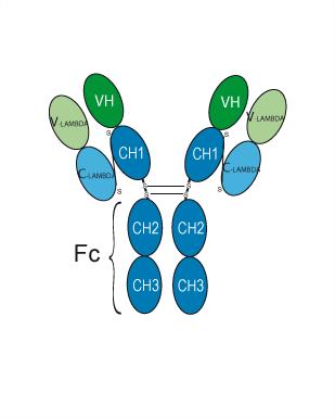
Human IgG4 lambda
Application
ELISA, FACS, Functional assay
Verified Bioactivity
-
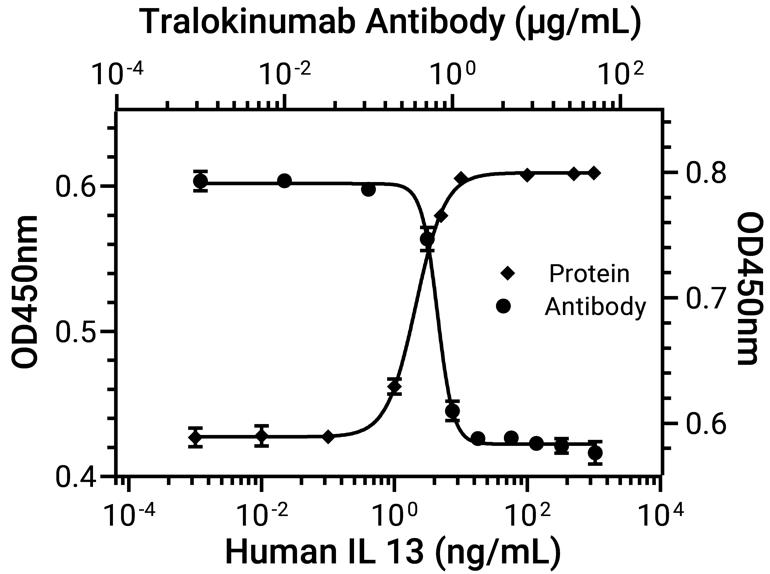 Tralokinumab (HY-P99053) neutralizes Recombinant Human IL-13 (HY-P70568) induced proliferation in the TF-1 human erythroleukemic cell line. The Neutralization Dose (ND50) for this effect is 0.6630 μg/mL in the presence of 10 ng/mL Recombinant Human IL-13.


Chemical Information
-
CAS No. 1044515-88-9
-
Appearance Liquid
-
Molecular Weight 144.14 kDa
-
Color Colorless to light yellow
-
SMILES
[Tralokinumab]
-
Synonyms
CAT354
-
Shipping
Shipping with dry ice.
-
Formulation
Please refer to the lot-specific COA for specific buffer information.
-
Storage
Please store the product under the recommended conditions in the Certificate of Analysis.


Publications (2)
-
Journal Impact Factor
-
Most Recent
-
ACS Nano
Combining Microfluidic Assessment with Encoded Hydrogel Microparticle-Based Immunoassay for Anti-Interleukin Drug Repurposing of Pulmonary Fibrosis Therapy. [Abstract]2025 Sep 16;19(36):32361-32373. PMID: 40903905
Tralokinumab purchased from MedChemExpress. Usage Cited in: ACS Nano. 2025 Sep 16;19(36):32361-32373. [Abstract]
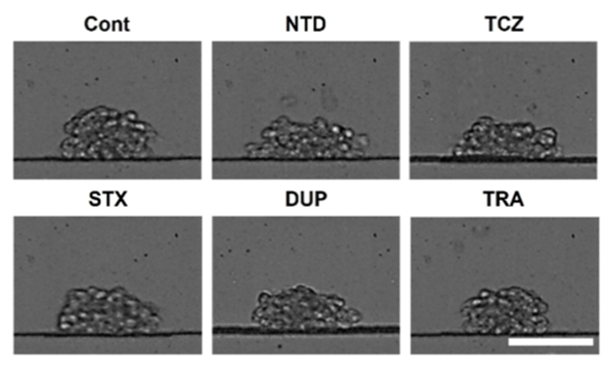
Representative images of fibrosis-induced spheroids subjected to microfluidic deformation analysis, comparing untreated control, antifibrotic drug NTD, and anti-interleukin drugs TCZ, STX, DUP, and TRA. Mab (5 μg/mL, 1 d) were added, Scale bars indicate 100 μm.
Tralokinumab purchased from MedChemExpress. Usage Cited in: ACS Nano. 2025 Sep 16;19(36):32361-32373. [Abstract]
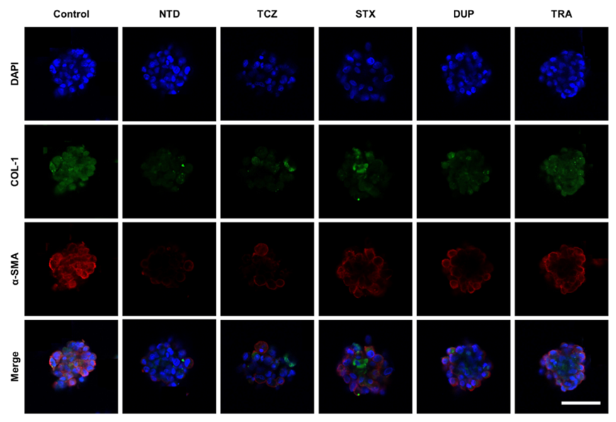
Confocal fluorescence images showing the expression of collagen-1 and α-SMA in spheroids treated with NTD, TCZ, STX, DUP, and TRA. Mab (5 μg/mL, 1 d) were added, Scale bars indicate 100 μm.
-
bioRxiv
Genome-scale spatial mapping of the Hodgkin lymphoma microenvironment identifies tumor cell survival factors. [Abstract]2025 Jan 25:2025.01.24.631210. PMID: 39896575


Purity & Documentation
-
Data Sheet (261 KB)
-
SDS (251 KB)
- English - EN (251 KB)
- Français - FR (251 KB)
- Deutsch - DE (251 KB)
- Norwegian - NO (251 KB)
- Español - ES (251 KB)
- Swedish - SV (251 KB)
- Italian - IT (251 KB)
- Korean - KR (251 KB)
- Portuguese - PT (251 KB)
-
Inhibitory Antibodies User Guide (603 KB)
References
[1]. A Wollenberg, et al. Tralokinumab for moderate-to-severe atopic dermatitis: results from two 52-week, randomized, double-blind, multicentre, placebo-controlled phase III trials (ECZTRA 1 and ECZTRA 2). Br J Dermatol. 2021 Mar;184(3):437-449. [Content Brief]
[2]. Murray LA, et al. Targeting interleukin-13 with tralokinumab attenuates lung fibrosis and epithelial damage in a humanized SCID idiopathic pulmonary fibrosis model. Am J Respir Cell Mol Biol. 2014 May;50(5):985-94. [Content Brief]

Calculators
Concentration (start) × Volume (start) = Concentration (final) × Volume (final)


Tralokinumab Related Classifications
HY-P99053 Related Classifications
 Powered by Bioz