



Tylvalosin
Based on 2 publication(s) in Google Scholar
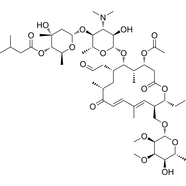
Tylvalosin (Acetylisovaleryltylo?sin) is an orally active, broad-spectrum macrolide antibiotic with antimicrobial activity. Tylvalosin is an antiviral agent used to study PRRSV infection. Tylvalosin induces apoptosis. Tylvalosin also has anti-inflammatory activity, alleviates oxidative stress, and alleviates acute lung injury by inhibiting NF-κB activation.
For research use only. We do not sell to patients.
- Purity : 99.00%
- CAS No.: 63409-12-1
- Formula: C53H87NO19
- Molecular Weight:1042.25
-
Storage:Powder -20°C, 3 years ; In solvent -80°C, 6 months , -20°C, 1 month

To place orders, for customer services and technical support, please contact: MedChemExpress USA
Tel: 609-228-6898 E-mail: [email protected] [email protected]


-
 Biological Activity
-
Chemical Information
-
Solvent & Solubility
- Protocols
- Purity & Documentation
- References
-
 Help & FAQs


Publications Citing Use of MedChemExpress (MCE) Tylvalosin
MoreAll Antibiotic Isoforms
More

Biological Activity
Description
In Vitro
Tylvalosin (10 μg/mL; 0.5, 1 h) increases caspase-3 cleavage in porcine neutrophils, leading to DNA fragmentation during apoptosis[4].
Tylvalosin (0.1-10 μg/mL; 0.5 h) promotes endocytosis of porcine neutrophils by macrophages without changing phagocytosis[4].
MedChemExpress (MCE) has not independently confirmed the accuracy of these methods. They are for reference only. Further protocols information, click here.
-
Cell Line:Porcine neutrophils
-
Concentration:0.1, 1.0, or 10 µg/mL
-
Incubation Time:0.5, 1 h
-
Result:Resulted induction of concentration- and time-dependent apoptosis in porcine monocyte-derived macrophages.
In Vivo
MedChemExpress (MCE) has not independently confirmed the accuracy of these methods. They are for reference only.
-
Animal Model:PRRSV-free commercial breed piglets[5]
-
Dosage:800 mg/kg Aivlosin (20% Tylvalosin Tartrate Premix)
-
Administration:po; 7-14 days
-
Result:Attenuated the increase in total white blood cells induced by immunization at day one post-immunization (DPI) and induced an increase in monocyte counts after seven DPI.
Attenuate the reduction in the percentage of CD8+ T cells induced by PRRSV-inactivated vaccine immunization at seven DPI.


Chemical Information
-
CAS No. 63409-12-1
-
Appearance Solid
-
Molecular Weight 1042.25
-
Formula C53H87NO19
-
Color White to off-white
-
SMILES
O[C@@H]1[C@@H](C)O[C@@H](OC[C@H]([C@@H](CC)OC(C[C@@H](OC(C)=O)[C@H](C)[C@@H](O[C@@H]2O[C@H](C)[C@@H](O[C@@H]3O[C@@H](C)[C@H](OC(CC(C)C)=O)[C@@](O)(C)C3)[C@H](N(C)C)[C@H]2O)[C@@H](CC=O)C[C@H]4C)=O)/C=C(C)/C=C/C4=O)[C@H](OC)[C@@H]1OC
-
Synonyms
Acetylisovaleryltylosin
-
Shipping
Room temperature in continental US; may vary elsewhere.
-
Storage
Powder -20°C 3 years In solvent -80°C 6 months -20°C 1 month


Publications (2)
-
Journal Impact Factor
-
Most Recent
-
Int J Biol Macromol
De novo design mini-binder proteins targeting the capsid-forming domain of type 2 PRRSV nucleocapsid protein to inhibit PRRSV replication in vitro. [Abstract]2025 Aug 26;323(Pt 2):147118. PMID: 40865823 -
Rapid Commun Mass Spectrom
2025;39(20):e10103. PMID: 40589310


Solvent & Solubility
In Vitro:
DMSO : ≥ 100 mg/mL (95.95 mM; Hygroscopic DMSO has a significant impact on the solubility of product, please use newly opened DMSO)
* "≥" means soluble, but saturation unknown.
Please refer to the solubility information to select the appropriate solvent. Once prepared, please aliquot and store the solution to prevent product inactivation from repeated freeze-thaw cycles.
Storage method and period of stock solution: -80°C, 6 months; -20°C, 1 month. When stored at -80°C, please use it within 6 months. When stored at -20°C, please use it within 1 month.
Please refer to the solubility information to select the appropriate solvent. Once prepared, please aliquot and store the solution to prevent product inactivation from repeated freeze-thaw cycles.
Storage method and period of stock solution: -80°C, 6 months; -20°C, 1 month. When stored at -80°C, please use it within 6 months. When stored at -20°C, please use it within 1 month.
Concentration (start) × Volume (start) = Concentration (final) × Volume (final)

Protocols
-
Research Protocol for Infectious Diseases
Infectious-disease experiments test how pathogens interact with host barriers, innate immune receptors, inflammatory signaling, pathogen replication, and tissue injury; pattern-recognition receptors such as TLRs, RIG-I-like receptors, NOD-like receptors, and inflammasomes detect microbial molecules and activate NF-κB, interferon, and cytokine responses. The central hypothesis is that infection severity reflects the balance between pathogen burden and host response: protective inflammation restricts pathogen growth, whereas excessive or mislocalized inflammation contributes to tissue damage and disease phenotype. Unresolved questions include which host pathways are protective versus pathogenic, why some infection models fail to translate to human disease, and which combined readouts best predict clinically relevant infection outcomes.
-
Detection of Apoptosis
Apoptosis, also called programmed cell death, is generally characterized by distinct morphological characteristics.
-
TUNEL staining for apoptotic DNA fragmentation
TUNEL staining detects DNA strand breaks by using terminal deoxynucleotidyl transferase to add labeled nucleotides to exposed 3′-OH DNA termini, generating either microscopic staining in fixed cells or tissue sections, or fluorescence/cytometric signal in cell suspensions. TUNEL positivity reflects DNA fragmentation but should not be interpreted alone as definitive apoptosis, because TUNEL can also label necrotic, autolytic, mechanically damaged, or DNA-repair-associated DNA breaks.
-
Annexin V plus membrane-impermeant dye apoptosis staining
Annexin V-based apoptosis assays rely on the detection of phosphatidylserine (PS) externalization from the inner leaflet of the plasma membrane to the outer leaflet, an early biochemical hallmark of apoptosis. Fluorescently labeled Annexin V binds PS in a calcium-dependent manner, enabling identification of early apoptotic cells by flow cytometry or fluorescence microscopy. When combined with a membrane-impermeant DNA-binding dye (e. g. , propidium iodide), this approach allows discrimination between viable (Annexin V−/dye−), early apoptotic (Annexin V+/dye−), and late apoptotic or necrotic (Annexin V+/dye+) cell populations by assessing membrane integrity and PS exposure.
-
Cotton Pellet Granuloma
Cotton pellet granuloma is a classical in vivo chronic inflammation model used to evaluate the anti-inflammatory potential of test substances by measuring their ability to inhibit granuloma tissue formation around an implanted foreign body (cotton pellet) in rodents. The method is based on the biological response to a sterile implanted material, which induces proliferative phase inflammation characterized by fibroblast proliferation and collagen-rich granuloma formation, and the final readout reflects the extent of chronic inflammatory tissue growth surrounding the pellet. In multiple preclinical pharmacological evaluations, inhibition of cotton pellet-induced granuloma formation has been used as an indicator of anti-inflammatory activity in both synthetic and natural product screening contexts.
-
ROS/oxidative-stress fluorescent staining
ROS/oxidative-stress fluorescent staining uses cell-permeant fluorogenic probes that become fluorescent after oxidation inside cells or tissues; commonly used examples include DCFH-DA/DCFDA for broad cellular oxidant detection, DHE for superoxide-related signal detection, MitoSOX for mitochondrial superoxide-related signal detection, and CellROX probes for oxidative-stress-associated fluorescence readouts. The assay detects probe oxidation rather than a single ROS species unless the probe and analysis method have been chemically validated for that species. DCFH-DA enters cells, is deacetylated by intracellular esterases to DCFH, and produces fluorescent DCF after oxidation, so the readout is used as an operational measure of total cellular oxidative stress rather than a species-specific ROS measurement. DHE and MitoSOX can report superoxide-related oxidation, but red fluorescence alone can include non-specific ethidium-like oxidation products; HPLC or optimized spectral approaches are
-
Carrageenan-Induced Paw Edema
Carrageenan-induced paw edema is an acute inflammation model in which intraplantar injection of carrageenan induces localized inflammatory swelling characterized by vascular permeability, leukocyte infiltration, and production of inflammatory mediators such as prostaglandins and cytokines, making it widely used to evaluate anti-inflammatory agents in vivo. The resulting paw volume or thickness increase is quantified over time as a direct readout of inflammatory intensity and drug efficacy, typically reflecting cyclooxygenase-mediated prostaglandin-driven edema formation and immune cell recruitment in peripheral tissue[20].
-
Apoptosis Solutions
Apoptosis is a regulated, generally non-lytic cell-death pathway that removes unwanted, damaged, infected, or abnormal cells through coordinated morphological changes, caspase activation, DNA fragmentation, and membrane remodeling. The intrinsic apoptosis pathway is controlled mainly by mitochondrial outer membrane permeabilization, BCL-2 family proteins, cytochrome c release, apoptosome formation, caspase-9 activation, and downstream executioner caspase-3/7 activation. The extrinsic apoptosis pathway is initiated by death receptors such as Fas, TNFR, and TRAIL receptors, which recruit adaptor proteins and activate caspase-8 before engaging executioner caspases or mitochondrial amplification through BID cleavage. Apoptosis is linked to many phenotypes, including cancer cell killing, tissue homeostasis, immune regulation, neurodegeneration, infection response, and treatment-induced cytotoxicity; unresolved questions include how apoptosis interacts with necroptosis, pyroptosis, ferroptos
-
Research Protocol for Inflammation-related Diseases
The NLRP3 inflammasome is a cytosolic innate immune signaling platform that integrates priming signals and danger-signal activation to promote caspase-1 activation, maturation of IL-1β and IL-18, and gasdermin D-mediated pyroptotic cell death. The core experimental logic is to determine whether inflammatory disease phenotypes are driven by increased NLRP3 expression, ASC-containing inflammasome assembly, caspase-1 cleavage, GSDMD cleavage, and extracellular release of IL-1β/IL-18 rather than by nonspecific cell injury alone. The pathway is strongly linked to inflammation-related disease phenotypes because monosodium urate crystals activate NALP3/NLRP3 inflammasome signaling in gout-like crystal inflammation, cholesterol crystals activate NLRP3 inflammasomes in atherogenesis models, and DSS-induced intestinal inflammation has been reported to involve NLRP3 inflammasome activity. However, experimental colitis studies also show context-dependent protective effects of NLRP3 inflammasome co
-
Inhalation Toxicity Study
Inhalation toxicity studies expose rodents to a controlled aerosol, vapor, gas, or smoke atmosphere and assess respiratory and systemic toxicity using exposure-atmosphere characterization, clinical observations, body and organ weights, bronchoalveolar lavage fluid, histopathology, blood chemistry, hematology, and, when included, molecular endpoints such as transcriptomics, proteomics, lipidomics, or tissue burden analysis. The primary biological readouts are airway irritation, pulmonary inflammation, cytotoxicity, altered surfactant or lipid homeostasis, impaired particle clearance, and tissue remodeling, reflected by BALF cell differentials, BALF protein, LDH, phosphatase activities, cytokines, lung weight, microscopic respiratory-tract lesions, and retained lung burden.


Purity & Documentation
-
Data Sheet (281 KB)
-
SDS (251 KB)
- English - EN (251 KB)
- Français - FR (251 KB)
- Deutsch - DE (251 KB)
- Norwegian - NO (251 KB)
- Español - ES (251 KB)
- Swedish - SV (251 KB)
- Italian - IT (251 KB)
- Korean - KR (251 KB)
- Portuguese - PT (251 KB)
-
Handling Instructions (2659 KB)
References
[1]. Albert Philip Adrian Mockett, et al. Use of tylvalosin as antiviral agent. Patent. WO2008007104.
[2]. Zhao Z, et al. Tylvalosin exhibits anti-inflammatory property and attenuates acute lung injury in different models possibly through suppression of NF-κB activation. Biochem Pharmacol. 2014 Jul 1;90(1):73-87. [Content Brief]
[3]. Moges R, et al. Anti-Inflammatory Benefits of Antibiotics: Tylvalosin Induces Apoptosis of Porcine Neutrophils and Macrophages, Promotes Efferocytosis, and Inhibits Pro-Inflammatory CXCL-8, IL1α, and LTB4 Production, While Inducing the Release of Pro-Resolving Lipoxin A4 and Resolvin D1. Front Vet Sci. 2018 Apr 11;5:57. [Content Brief]


Complete Stock Solution Preparation Table
Please refer to the solubility information to select the appropriate solvent. Once prepared, please aliquot and store the solution to prevent product inactivation from repeated freeze-thaw cycles.
Storage method and period of stock solution: -80°C, 6 months; -20°C, 1 month. When stored at -80°C, please use it within 6 months. When stored at -20°C, please use it within 1 month.
| Optional Solvent | Concentration Solvent Mass | 1 mg | 5 mg | 10 mg | 25 mg |
|---|---|---|---|---|---|
| DMSO | 1 mM | 0.9595 mL | 4.7973 mL | 9.5946 mL | 23.9866 mL |
| 5 mM | 0.1919 mL | 0.9595 mL | 1.9189 mL | 4.7973 mL | |
| 10 mM | 0.0959 mL | 0.4797 mL | 0.9595 mL | 2.3987 mL | |
| 15 mM | 0.0640 mL | 0.3198 mL | 0.6396 mL | 1.5991 mL | |
| 20 mM | 0.0480 mL | 0.2399 mL | 0.4797 mL | 1.1993 mL | |
| 25 mM | 0.0384 mL | 0.1919 mL | 0.3838 mL | 0.9595 mL | |
| 30 mM | 0.0320 mL | 0.1599 mL | 0.3198 mL | 0.7996 mL | |
| 40 mM | 0.0240 mL | 0.1199 mL | 0.2399 mL | 0.5997 mL | |
| 50 mM | 0.0192 mL | 0.0959 mL | 0.1919 mL | 0.4797 mL | |
| 60 mM | 0.0160 mL | 0.0800 mL | 0.1599 mL | 0.3998 mL | |
| 80 mM | 0.0120 mL | 0.0600 mL | 0.1199 mL | 0.2998 mL |


Tylvalosin Related Classifications
HY-128423A Related Classifications
 Powered by Bioz