



Vicriviroc malate
Vicriviroc (SCH 417690) malate is an orally active CCR5 antagonist with the IC50 of 10 nM, and also inhibts MIP-1α and intracellular calcium release induced by the ligand RANTES (10 nM) with the IC50 values of 0.91 nM and 16 nM,,respectively. Vicriviroc malate can inhibits human immunodeficiency virus type 1 (HIV-1) infection, and can also used for study of cancer.
For research use only. We do not sell to patients.
- CAS No.: 541503-81-5
- Formula: C32H44F3N5O7
- Molecular Weight:667.72
-
Storage:
Please store the product under the recommended conditions in the Certificate of Analysis.


To place orders, for customer services and technical support, please contact: MedChemExpress USA
Tel: 609-228-6898 E-mail: [email protected] [email protected]


-
 Biological Activity
-
Chemical Information
- Protocols
- Purity & Documentation
- References
-
 Help & FAQs


-
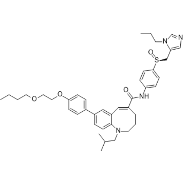
HY-14882
CenicrivirocCCR2/5 Antagonist -
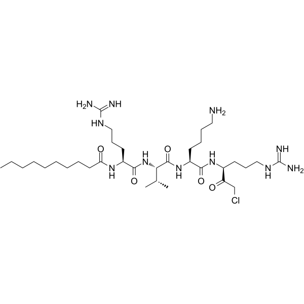
HY-107760
Decanoyl-RVKR-CMKHIV Inhibitor -
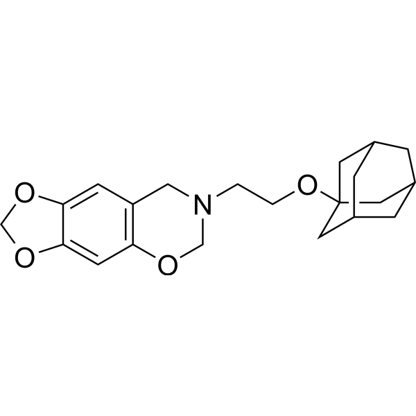
HY-122219
R243CCR8 Antagonist -
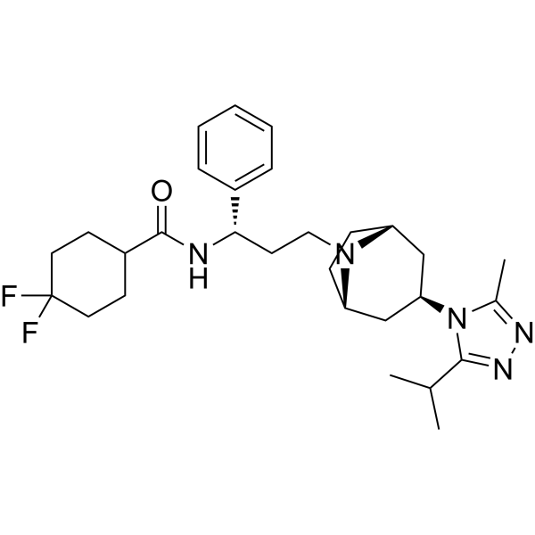
HY-13004
MaravirocCCR5 Antagonist -
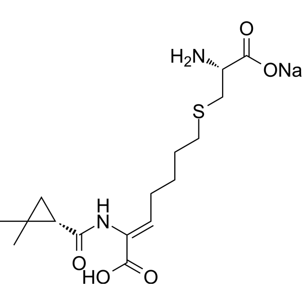
HY-A0166A
Cilastatin sodiumRenal Dehydropeptidase I Inhibitor -
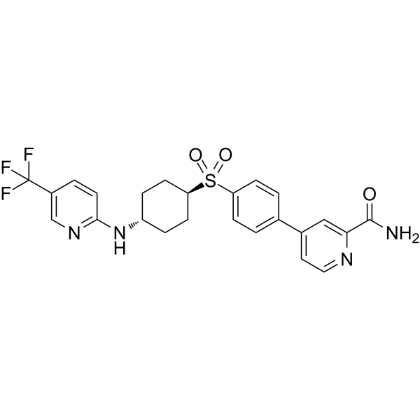
HY-112701
CCR6 inhibitor 1CCR Inhibitor -
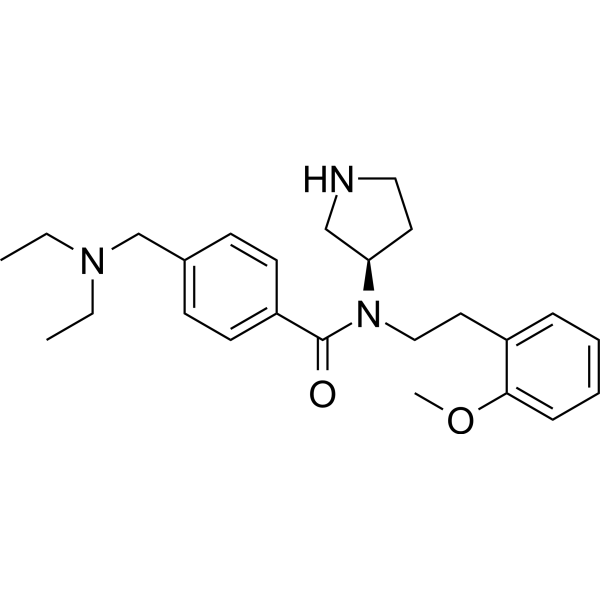
HY-13447
PF-429242S1P Inhibitor -
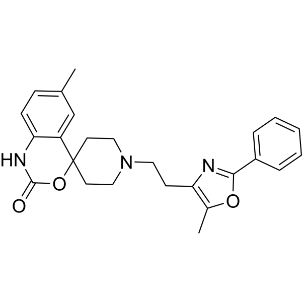
HY-15418
RS 504393CCR2 Antagonist -
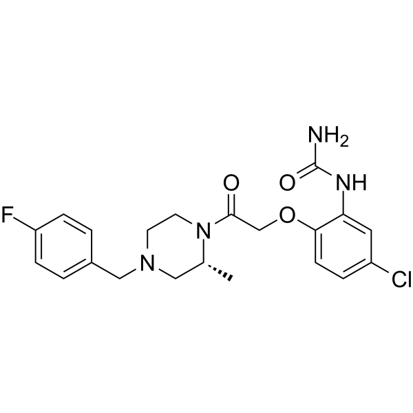
HY-12080
BX471CCR1 Antagonist -
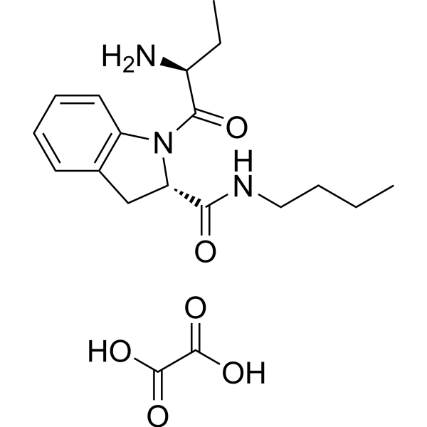
HY-107759
Butabindide oxalateTPP II Inhibitor -
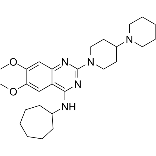
HY-103364
C-021CCR4 Antagonist -
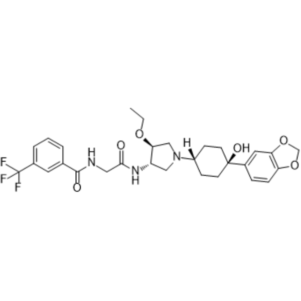
HY-50674
INCB3344CCR2 Antagonis


Biological Activity
Description


Chemical Information
-
CAS No. 541503-81-5
-
Molecular Weight 667.72
-
Formula C32H44F3N5O7
-
SMILES
COC[C@@H](C1=CC=C(C=C1)C(F)(F)F)N2[C@H](CN(C3(CCN(CC3)C(C4=C(C)N=CN=C4C)=O)C)CC2)C.O=C(O)C(O)CC(O)=O
-
Synonyms
SCH 417690 malate; SCH-D malate; MK-7690
-
Shipping
Room temperature in continental US; may vary elsewhere.
-
Storage
Please store the product under the recommended conditions in the Certificate of Analysis.


Purity & Documentation
References
[1]. Strizki JM, et al. Discovery and characterization of vicriviroc (SCH 417690), a CCR5 antagonist with potent activity against human immunodeficiency virus type 1. Antimicrob Agents Chemother. 2005;49(12):4911-4919. [Content Brief]
[2]. Bessudo A, et al. Safety and Efficacy of Vicriviroc (MK-7690) in Combination With Pembrolizumab in Patients With Advanced or Metastatic Microsatellite Stable Colorectal Cancer. Clin Colorectal Cancer. 2024;23(3):285-294. [Content Brief]

Calculators
Concentration (start) × Volume (start) = Concentration (final) × Volume (final)


Vicriviroc malate Related Classifications
HY-B0155B Related Classifications
 Powered by Bioz