



PROTAC KDM3 degrader-1

Based on 1 Customer Validation
PROTAC KDM3 degrader-1 is a KDM3A/KDM3B PROTAC degrader with DC50 values of 13.73 nM and 172.6 nM in SW480 cells, respectively. PROTAC KDM3 degrader-1 induces cereblon-dependent proteasomal degradation of KDM3A and KDM3B. PROTAC KDM3 degrader-1 inhibits the oncogenic Wnt/β-catenin signaling pathway. PROTAC KDM3 degrader-1 eliminates colorectal cancer stem cells, suppresses their self-renewal, and blocks colony formation of colorectal cancer cells. PROTAC KDM3 degrader-1 inhibits tumor growth in a colorectal cancer xenograft mouse model. PROTAC KDM3 degrader-1 can be used for the research of colorectal cancer.
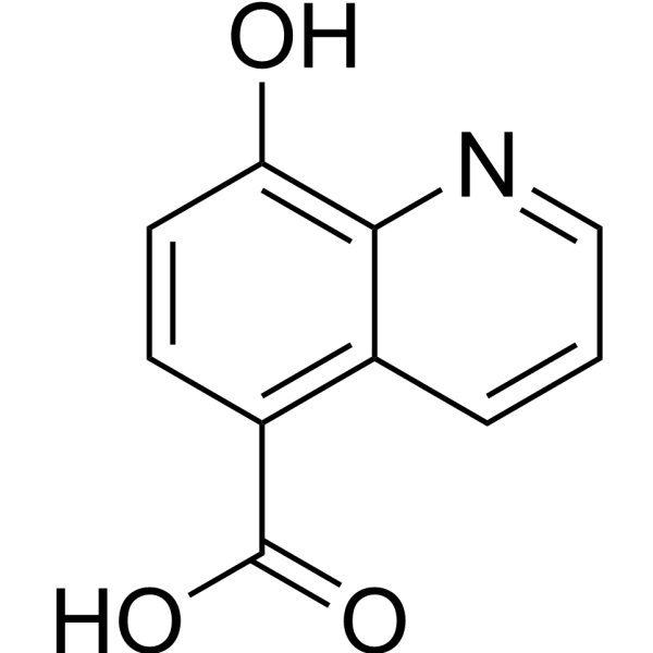
(Pink: KDM3A and KDM3B ligand (HY-12304); Blue: Cereblon ligand (HY-41547); Black: linker (HY-22335)).
For research use only. We do not sell to patients.
- Purity : 99.07%
- CAS No.: 3119090-33-1
- Formula: C33H37N5O10
- Molecular Weight:663.67
-
Storage:Powder -20°C, 3 years , 4°C, 2 years ; In solvent -80°C, 6 months , -20°C, 1 month


To place orders, for customer services and technical support, please contact: MedChemExpress USA
Tel: 609-228-6898 E-mail: [email protected] [email protected]


-
 Biological Activity
-
Chemical Information
-
Solvent & Solubility
- Protocols
- Purity & Documentation
- References
-
 Help & FAQs


All PROTACs Isoforms
More

Biological Activity
Description
IC50 & Target
[1]|
KDM3A |
KDM3B |
In Vitro
PROTAC KDM3 degrader-1 (Compound 4) (16 h) potently inhibits the Wnt/β-catenin signaling pathway in 293T-TCF-Luc cells, with an IC50 of 8.9 μM[1].
PROTAC KDM3 degrader-1 (48 h) exhibits low basal toxicity in normal human colonic epithelial CRL-1790 cells, with a TD50 of 285.4 μM[1].
PROTAC KDM3 degrader-1 (0-30 μM) exhibits low hERG inhibitory activity, with an IC50 > 30 μM[1].
PROTAC KDM3 degrader-1 (0.001-100 μM; 16 h) selectively degrades KDM3A and KDM3B in SW480 and HCP-1 colon cancer cells via a CRBN-dependent proteasomal pathway, with no effect on KDM4 family proteins or the novel substrate GSPT1, and its DC50 values are 13.73 nM and 172.6 nM, respectively[1].
PROTAC KDM3 degrader-1 (50 μM; 16 h) potently inhibits the expression of Wnt target genes and colorectal cancer CSC signature genes in SW480 and HCP-1 cells[1].
PROTAC KDM3 degrader-1 (2 weeks) potently inhibits the self-renewal of ALDHHigh colorectal cancer stem cells, with an ED50 value of 0.95 μM in SW480 cells and 0.28 μM in HCP-1 cells[1].
MedChemExpress (MCE) has not independently confirmed the accuracy of these methods. They are for reference only. Further protocols information, click here.
-
Cell Line:SW480 cells, HCP-1 cells
-
Concentration:0.001, 0.01, 0.1, 1, 10, 100 μM
-
Incubation Time:16 h
-
Result:Degraded KDM3A and KDM3B in a dose-dependent manner, with DC50 values of 13.73 nM for KDM3A and 172.6 nM for KDM3B; Dₘₐₓ values were 65.34% for KDM3A and 88.55% for KDM3B.
Observed a "hook effect" for KDM3A degradation at higher doses.
Did not dramatically affect KDM4 family proteins (KDM4A, KDM4B, KDM4C).
Reversed degradation of KDM3A and KDM3B by pre-treatment with 1 μM MLN4924, 100 μM pomalidomide, or IOX1, confirming proteasomal degradation dependent on CRBN binding and target protein interaction.
Degraded KDM3A and KDM3B in freshly isolated human CRC HCP-1 cells.
Did not induce degradation of the known neo-substrate GSPT1 in SW480 cells.
-
Cell Line:human colorectal cancer SW480 and HCP-1 cells
-
Concentration:50 μM
-
Incubation Time:16 h
-
Result:Profoundly suppressed expression of Wnt target genes (AXIN2, DKK1, CCND1) and colorectal CSC signature genes (ASCL2, RNF43, ZNRF3, LGR5) by 50% or more in both SW480 and HCP-1 cells.
Showed a more dramatic inhibitory effect than the parent compound IOX1.
In Vivo
MedChemExpress (MCE) has not independently confirmed the accuracy of these methods. They are for reference only.
-
Animal Model:NSG mice[1]
-
Dosage:3 mg/kg; 10 mg/kg; 35 mg/kg
-
Administration:i.p.; daily; 16 days
-
Result:Significantly inhibited tumor growth compared to the control group.
Reduced tumor volume and weight at 10 mg/kg and 35 mg/kg doses.
Confirmed degradation of KDM3A and KDM3B in tumor tissues, along with downregulated expression of the Wnt target gene CCND1.
Slightly upregulated KDM3A (but not KDM3B) in tumors from mice treated with the 35 mg/kg dose, consistent with an in vitro "hook effect," though tumor growth remained drastically reduced.
Had no significant impact on mouse body weight, indicating good tolerability.


Chemical Information
-
CAS No. 3119090-33-1
-
Appearance Solid
-
Molecular Weight 663.67
-
Formula C33H37N5O10
-
Color Light yellow to green yellow
-
SMILES
O=C(C1=C2C=CC=NC2=C(O)C=C1)NCCOCCOCCOCCOCCNC3=CC=CC(C(N4C(CC5)C(NC5=O)=O)=O)=C3C4=O
-
Shipping
Room temperature in continental US; may vary elsewhere.
-
Storage
Powder -20°C 3 years 4°C 2 years In solvent -80°C 6 months -20°C 1 month


Solvent & Solubility
In Vitro:
DMSO : 100 mg/mL (150.68 mM; Need ultrasonic; Hygroscopic DMSO has a significant impact on the solubility of product, please use newly opened DMSO)
Please refer to the solubility information to select the appropriate solvent. Once prepared, please aliquot and store the solution to prevent product inactivation from repeated freeze-thaw cycles.
Storage method and period of stock solution: -80°C, 6 months; -20°C, 1 month. When stored at -80°C, please use it within 6 months. When stored at -20°C, please use it within 1 month.
Please refer to the solubility information to select the appropriate solvent. Once prepared, please aliquot and store the solution to prevent product inactivation from repeated freeze-thaw cycles.
Storage method and period of stock solution: -80°C, 6 months; -20°C, 1 month. When stored at -80°C, please use it within 6 months. When stored at -20°C, please use it within 1 month.
Concentration (start) × Volume (start) = Concentration (final) × Volume (final)

Protocols
-
Patient-Derived Xenograft (PDX)
Patient-derived xenograft (PDX) models are generated by engrafting primary human tumor tissue directly into immunodeficient mice, allowing in vivo propagation of patient tumor biology without initial in vitro adaptation. These models are used to preserve key histopathological and molecular characteristics of the original tumor and enable assessment of tumor growth dynamics and therapeutic response in a living organism. The biological readout is tumor engraftment and subsequent growth in the murine host, which reflects the ability of human tumor cells to survive, vascularize, and expand in an immunocompromised microenvironment.
-
Patient-Derived Orthotopic Xenograft (PDOX)
Patient-derived orthotopic xenograft (PDOX) modeling implants fresh patient tumor tissue or patient-derived tumor cells into the anatomically corresponding organ or tissue site of immunodeficient mice, usually by surgical orthotopic implantation, to preserve patient tumor histology, local microenvironmental context, invasion, metastatic behavior, and treatment-response features better than subcutaneous implantation. PDOX readouts include tumor engraftment, orthotopic tumor growth, local invasion, metastasis, recurrence after resection, histologic similarity to the donor tumor, biomarker retention, molecular concordance, survival, and response or resistance to therapy. PDOX models are used for preclinical drug testing and individualized therapy evaluation, but engraftment success varies by tumor type and specimen quality.
-
Splenic/Portal-Vein Liver Metastasis Xenograft
Splenic and portal-vein liver metastasis xenograft models deliver tumor cells into the portal circulation so that cells reach the liver first and form hepatic metastatic lesions; splenic injection uses the spleen as an access route to the portal system, while direct portal-vein injection introduces cells into the portal vein without requiring splenectomy. The assay detects liver colonization, intrahepatic tumor growth, tumor distribution, treatment response, survival, and liver-metastasis microenvironment changes; readouts include bioluminescence or fluorescence imaging, gross liver nodule counts, liver weight or tumor burden, histology, and survival.
-
Soft Agar Colony Formation Assay
Soft agar colony formation assay measures anchorage-independent growth, in which transformed or tumorigenic cells proliferate as colonies in a semisolid agar matrix while many non-transformed adherent cells fail to proliferate without attachment; classic studies showed that growth in semisolid medium correlates with tumorigenicity in nude mice, and later protocol papers describe the method as a stringent in vitro assay for malignant transformation. The readout is the number, size, morphology, or signal intensity of colonies formed within agar after incubation; published formats include manual colony counting after staining, 96-well or 384-well quantitative formats, DNA-binding dye detection, MTT/tetrazolium-based detection, digital image analysis, and PCR-based marker detection from soft agar cultures.
-
Colony Formation (Clonogenic) Assay
The clonogenic (colony formation) assay measures the ability of a single cell to retain reproductive viability and form a macroscopic colony, typically defined as a cluster derived from one progenitor cell after a defined growth period. This assay is widely used to evaluate cell survival after exposure to ionizing radiation or cytotoxic treatments and is considered a standard method in radiation biology for generating dose-response relationships of reproductive cell death. Colony formation reflects long-term proliferative capacity rather than short-term metabolic activity, and survival is quantified by comparing treated versus untreated conditions based on colony number and derived survival fractions.
-
Subcutaneous Cell-Line-Derived Xenograft
Subcutaneous cell-line-derived xenograft (CDX) models are established by implanting cultured human cancer cell lines into immunodeficient mice, where the injected cells form localized tumors that can be monitored in vivo as a measure of tumorigenic potential, growth kinetics, and treatment response. These models are widely used in oncology research because they allow reproducible tumor formation and enable comparative assessment of tumor growth between different cell lines or genetic manipulations in a controlled in vivo microenvironment. Subcutaneous implantation of cancer cells in immunodeficient mice is a standard approach for evaluating tumor growth behavior and therapeutic response across multiple cancer types, including prostate, esophageal, pancreatic, and colon cancer models.
-
hPSC maintenance and expansion
This protocol maintains and expands human pluripotent stem cells under feeder-free, chemically defined conditions using E8 medium and vitronectin-coated culture surfaces; the readout is sustained adherent colony growth with undifferentiated morphology and retained pluripotency-marker expression during serial passaging. E8-based hPSC culture relies on defined soluble factors and matrix-dependent adhesion rather than feeder cells; vitronectin supports hPSC attachment through integrin-mediated interactions, and EDTA passaging dissociates colonies as small aggregates without enzymatic digestion, centrifugation, or routine ROCK-inhibitor treatment.
-
Orthotopic Cell-Line Xenograft
Orthotopic cell-line xenograft models involve implantation of human cancer cell lines into the anatomically corresponding organ of immunodeficient mice to reproduce tumor growth within a native microenvironment, enabling more clinically relevant tumor behavior compared with subcutaneous models. These models are widely used because orthotopic placement better recapitulates tumor progression, including invasion and metastatic spread, which are often underrepresented in heterotopic implantation systems. Compared with conventional xenografts, orthotopic implantation is described as more technically complex but provides improved simulation of tumor-microenvironment interactions and metastatic behavior, making it particularly valuable for translational oncology research. Surgical orthotopic implantation approaches have been emphasized as enabling faithful reproduction of clinical cancer features, including metastasis and disease progression patterns that align with the tumor’s organ of origi
-
Intraperitoneal/Peritoneal Dissemination Xenograft
Intraperitoneal (IP) or peritoneal dissemination xenograft models are based on the introduction of human cancer cells into the peritoneal cavity of immunodeficient mice, where they attach to peritoneal surfaces, form multicellular aggregates or spheroids, and progressively generate disseminated tumor nodules that mimic advanced peritoneal metastatic disease. These models are widely used to study ovarian cancer progression, tumor-microenvironment interactions, and intraperitoneal therapeutic responses, often incorporating bioluminescence or fluorescence imaging to longitudinally monitor tumor burden in vivo. The biological principle relies on the capacity of tumor cells such as SKOV3 or related ovarian carcinoma lines to survive in suspension, aggregate within ascites-like fluid, adhere to mesothelial surfaces, and invade peritoneal organs, thereby recapitulating human peritoneal carcinomatosis patterns observed in advanced disease.
-
How to Choose the Right Model Animal
Choosing the right model animal is a validity-driven decision in which the species, strain, sex, age, genetic background, disease-induction method, outcome measures, and welfare burden must match the scientific question rather than laboratory tradition or convenience. A model should be selected by judging face validity, construct validity, and predictive validity: whether it resembles the human phenotype, whether it reproduces relevant mechanisms, and whether results are likely to predict human biology or treatment response. Animal studies often fail to translate because of species differences, weak disease resemblance, poor experimental design, inadequate reporting, publication bias, and underuse of randomization, blinding, and sample-size justification. Unresolved questions include how to rank competing models objectively, how much human-disease complexity must be reproduced for a given objective, and when non-animal systems such as organoids, ex vivo tissue, or computational models


Purity & Documentation
-
Data Sheet (276 KB)
-
SDS (254 KB)
- English - EN (254 KB)
- Français - FR (254 KB)
- Deutsch - DE (254 KB)
- Norwegian - NO (254 KB)
- Español - ES (254 KB)
- Swedish - SV (254 KB)
- Italian - IT (254 KB)
- Korean - KR (254 KB)
- Portuguese - PT (254 KB)
-
Handling Instructions (2659 KB)
References


Complete Stock Solution Preparation Table
Please refer to the solubility information to select the appropriate solvent. Once prepared, please aliquot and store the solution to prevent product inactivation from repeated freeze-thaw cycles.
Storage method and period of stock solution: -80°C, 6 months; -20°C, 1 month. When stored at -80°C, please use it within 6 months. When stored at -20°C, please use it within 1 month.
| Optional Solvent | Concentration Solvent Mass | 1 mg | 5 mg | 10 mg | 25 mg |
|---|---|---|---|---|---|
| DMSO | 1 mM | 1.5068 mL | 7.5339 mL | 15.0677 mL | 37.6693 mL |
| 5 mM | 0.3014 mL | 1.5068 mL | 3.0135 mL | 7.5339 mL | |
| 10 mM | 0.1507 mL | 0.7534 mL | 1.5068 mL | 3.7669 mL | |
| 15 mM | 0.1005 mL | 0.5023 mL | 1.0045 mL | 2.5113 mL | |
| 20 mM | 0.0753 mL | 0.3767 mL | 0.7534 mL | 1.8835 mL | |
| 25 mM | 0.0603 mL | 0.3014 mL | 0.6027 mL | 1.5068 mL | |
| 30 mM | 0.0502 mL | 0.2511 mL | 0.5023 mL | 1.2556 mL | |
| 40 mM | 0.0377 mL | 0.1883 mL | 0.3767 mL | 0.9417 mL | |
| 50 mM | 0.0301 mL | 0.1507 mL | 0.3014 mL | 0.7534 mL | |
| 60 mM | 0.0251 mL | 0.1256 mL | 0.2511 mL | 0.6278 mL | |
| 80 mM | 0.0188 mL | 0.0942 mL | 0.1883 mL | 0.4709 mL | |
| 100 mM | 0.0151 mL | 0.0753 mL | 0.1507 mL | 0.3767 mL |


PROTAC KDM3 degrader-1 Related Classifications
HY-168593 Related Classifications
 Powered by Bioz