
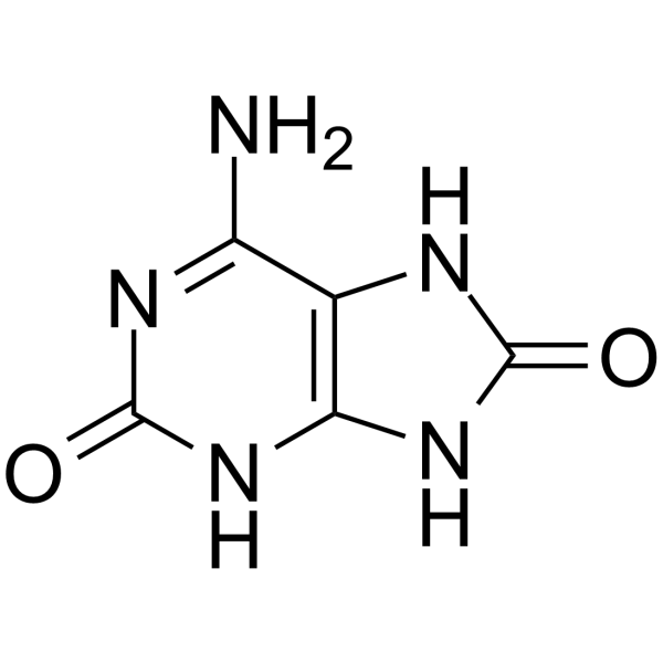
2,8-Dihydroxyadenine
2,8-Dihydroxyadenine is an endogenous metabolite that forms crystals in urine, leading to kidney stone formation and crystal deposition in the kidney. 2,8-Dihydroxyadenine induces crystal-induced tubular injury, inflammation, and fibrosis through crystal deposition in renal tubules, where crystals are excreted in urine, internalized by tubular epithelial cells, and transported to the interstitium. 2,8-Dihydroxyadenine upregulates CD44 expression near crystals, TNF-α signaling through NF-κB, and mTORC1 signaling, while inducing actin stress fiber formation and cytoskeletal remodeling. 2,8-Dihydroxyadenine downregulates epithelial-mesenchymal transition pathways and oxidative phosphorylation, and induces changes affecting inflammation, metabolism, and cell cycle regulation. 2,8-Dihydroxyadenine can be used in research on kidney disease, adenine phosphoribosyltransferase deficiency, and kidney stone disease.
For research use only. We do not sell to patients.
- CAS No.: 30377-37-8
- Formula: C5H5N5O2
- Molecular Weight:167.13
-
Storage:Powder -20°C, 3 years , 4°C, 2 years ; In solvent -80°C, 6 months , -20°C, 1 month
All Endogenous Metabolite Isoforms
More
Biological Activity
Description
|
CD44 |
NF-κB |
mTORC1 |
TNF-α |
In Vitro
2,8-Dihydroxyadenine (50 μM) undergoes quasi-reversible oxidation at a glassy carbon electrode in pH 4.5 acetate buffer, forming an unstable oxidation product and, at high scan rates, detectable dimers[2].
2,8-Dihydroxyadenine (50 μM) oxidation at a GCE is pH-dependent, occurring with a 2-electron transfer and forming a strongly adsorbing electroactive product P2,8-DHA, with maximum oxidation current at pH 4.5[2].
2,8-Dihydroxyadenine reduction at a GCE is reversible in the absence of oxygen, but molecular oxygen interferes with the reductive electron transfer, shifting the reduction potential and increasing the reduction current[2].
2,8-Dihydroxyadenine (DHA) (60-480 µg/mL; 72 h) reduces viability of HEK293, MDCK, and HK-2 cells in a dose-dependent manner, with HEK293 cells showing the greatest decrease to approximately 40% at 480 µg/mL[3].
2,8-Dihydroxyadenine (120-480 µg/mL; 24 h) impairs wound closure in HEK293, MDCK, and HK-2 cells, with HEK293 cells treated with 480 µg/mL DHA achieving only ~50% closure at 72 h compared to full closure in controls, and HK-2 cells reaching ~70-80% closure at 72 h[3].
2,8-Dihydroxyadenine (120-480 µg/mL; 72 h) upregulates CD44 gene expression in HK-2 cells, while HEK293 cells show increased CD44 gene expression at higher DHA concentrations, but MDCK cells do not show increased CD44 gene expression[3].
2,8-Dihydroxyadenine (120-480 µg/mL; 72 h) increases CD44 protein expression in HEK293, MDCK, and HK-2 cells, with CD44 localized adjacent to DHA crystals[3].
2,8-Dihydroxyadenine (60-240 µg/mL; 72 h) induces dose-dependent transcriptional changes in HK-2 cells, with upregulation of TNF-α, mTORC1, E2F targets, and G2M checkpoint pathways, and downregulation of EMT and oxidative phosphorylation, highlighting inflammation, metabolic stress, and cell cycle dysregulation[3].
MedChemExpress (MCE) has not independently confirmed the accuracy of these methods. They are for reference only. Further protocols information, click here.
-
Cell Line:HEK293, MDCK, and HK-2
-
Concentration:60 µg/mL, 120 µg/mL, 240 µg/mL, 480 µg/mL
-
Incubation Time:72 h
-
Result:Decreased viability in a dose-dependent manner.
Reduced viability to approximately 40% in HEK293 cells at 480 µg/mL.
Reduced viability to 80% in MDCK cells at 480 µg/mL.
Reduced viability to 70% in HK-2 cells at 480 µg/mL.
-
Cell Line:HEK293, MDCK, and HK-2
-
Concentration:120 µg/mL, 480 µg/mL
-
Incubation Time:24 h
-
Result:Impaired wound closure.
HEK293 cells treated with 480 µg/mL DHA closed ~50% of the wound at 72 h.
HK-2 cells reached ~80% closure at 120 µg/mL and ~70% closure at 480 µg/mL at 72 h.
-
Cell Line:HEK293, HK-2, and MDCK
-
Concentration:120 µg/mL, 480 µg/mL
-
Incubation Time:72 h
-
Result:Induced a statistically significant upregulation of CD44 expression in HK-2 cells at 480 µg/mL.
Increased CD44 gene expression in HEK293 cells at 480 µg/mL.
Did not increase CD44 gene expression in MDCK cells.
-
Cell Line:HEK293, MDCK, and HK-2
-
Concentration:120, 480 µg/mL
-
Incubation Time:72 h
-
Result:Increased CD44 protein expression in HEK293, MDCK, and HK-2 cells.
CD44 expression was localized adjacent to DHA crystals.
Chemical Information
-
CAS No. 30377-37-8
-
Appearance Solid
-
Molecular Weight 167.13
-
Formula C5H5N5O2
-
Color White to light yellow
-
SMILES
O=C1NC2=C(NC(N2)=O)C(N)=N1
-
Structure Classification
-
Initial Source
-
Shipping
Room temperature in continental US; may vary elsewhere.
-
Storage
Powder -20°C 3 years 4°C 2 years In solvent -80°C 6 months -20°C 1 month
Solvent & Solubility
In Vitro:
H2O : 2.2 mg/mL (13.16 mM; Need ultrasonic and warming)
Please refer to the solubility information to select the appropriate solvent. Once prepared, please aliquot and store the solution to prevent product inactivation from repeated freeze-thaw cycles.
Storage method and period of stock solution: -80°C, 6 months; -20°C, 1 month. When stored at -80°C, please use it within 6 months. When stored at -20°C, please use it within 1 month.
* Note: If you choose water as the stock solution, please dilute it to the working solution, then filter and sterilize it with a 0.22 μm filter before use.
Please refer to the solubility information to select the appropriate solvent. Once prepared, please aliquot and store the solution to prevent product inactivation from repeated freeze-thaw cycles.
Storage method and period of stock solution: -80°C, 6 months; -20°C, 1 month. When stored at -80°C, please use it within 6 months. When stored at -20°C, please use it within 1 month.
* Note: If you choose water as the stock solution, please dilute it to the working solution, then filter and sterilize it with a 0.22 μm filter before use.
Concentration (start) × Volume (start) = Concentration (final) × Volume (final)
Protocols
-
Fibrosis/Collagen Morphometry
Fibrosis and collagen morphometry is based on the quantitative visualization of fibrillar collagen deposition in tissue sections using histochemical stains such as Sirius Red (Picrosirius Red) or Masson's trichrome, followed by image-based or polarization-enhanced analysis to estimate collagen proportional area as a surrogate of extracellular matrix accumulation during fibrotic remodeling. Sirius Red combined with polarized light microscopy enhances detection of collagen fibers due to birefringence properties, enabling more specific visualization of collagen type I and III fibrils compared to conventional bright-field histology, while whole-section or region-restricted digital morphometry reduces field-selection bias in fibrosis assessment. Alternative quantitative approaches include second harmonic generation (SHG) and two-photon excited fluorescence microscopy, which enable label-free detection of fibrillar collagen and have been validated against histological staining and biochemica
-
Kinase activity and phosphorylation assays
Kinase activity assays measure the ability of kinases to transfer phosphate groups from ATP to specific substrates, while phosphorylation assays detect the presence and levels of phosphorylated proteins. Common methods include radiolabeled ATP incorporation (e. g. ,), ADP release detection via bioluminescence (e. g. ,[3]), enzyme-linked immunosorbent assays (ELISA) for phospho-specific epitopes (e. g. ,[6]), and microtiter-based formats for high-throughput screening (e. g. ,[8]). The ADP-Glo assay quantifies kinase activity by measuring ADP produced during phosphorylation using a luciferase-based system. Radiometric assays involve autoradiography or scintillation counting after incorporation of 32P-labeled ATP into substrate proteins. ELISA-based approaches rely on phospho-specific antibodies to detect activated kinases in cell lysates or purified samples.
-
Collagen: Sirius Red Staining
Sirius Red or picrosirius red staining is a histochemical method for visualizing collagen-rich extracellular matrix in tissue sections, and collagen fibers are detected as red-stained structures under bright-field microscopy with enhanced birefringence under polarized light. Picrosirius red is useful for assessing total collagen organization, distribution, and fibrosis burden, but polarized color should not be interpreted as a definitive collagen type I versus type III readout because color is affected by fiber orientation, thickness, and packing.
-
RNA extraction experimental
By lysing cells, releasing RNA, and removing impurities such as proteins and DNA, high-purity RNA products are finally obtained. The commonly used traditional method is the guanidine isothiocyanate/phenol/chloroform method (Trizol), which is suitable for a variety of animal materials including animal tissues, microorganisms, cultured cells, etc., and most plant materials.
-
Western Blot
Western blotting (WB) is a commonly used experimental method in molecular biology, biochemistry, and immunogenetics for identifying and quantifying target proteins. It combines gel electrophoresis with immunoassay, enabling researchers to analyze protein expression, post-translational modifications, and molecular weight.
-
Flow cytometric DNA-content cell-cycle staining
Flow cytometric DNA-content cell-cycle staining measures the fluorescence intensity of DNA-bound fluorochromes in single cells or nuclei to estimate DNA content distributions, allowing assignment of populations to G0/G1, S, and G2/M phases by DNA histogram deconvolution. Propidium iodide (PI) intercalates into DNA, and PI fluorescence is proportional to cellular DNA content when staining is performed under conditions that make DNA accessible and minimize non-DNA signal. Cells with G2/M DNA content are expected to show approximately twice the fluorescence intensity of G0/G1 cells, while S-phase cells occupy intermediate fluorescence values. PI-based DNA-content analysis can also detect cells with fractional DNA content, often reported as sub-G1, when DNA fragmentation and extraction during staining reduce retained DNA signal in apoptotic cells. DAPI is an alternative DNA fluorochrome for univariate DNA-content analysis, while bivariate approaches combining DNA content with proliferation
-
BrdU Incorporation Assay
Bromodeoxyuridine (BrdU) incorporation assay is based on the principle that BrdU, a thymidine analog, is incorporated into newly synthesized DNA during the S phase of the cell cycle, thereby serving as a marker of DNA replication and cellular proliferation. Incorporated BrdU can be detected using anti-BrdU antibodies following DNA denaturation, enabling visualization or quantification of proliferating cells through immunochemical detection methods such as immunofluorescence or immunohistochemistry.
-
Connective Tissue: Masson's Trichrome/Collagen Trichrome Staining
Masson’s Trichrome (collagen/trichrome staining) is a histological technique that differentially stains tissue compartments using sequential acidic dyes to distinguish collagen from muscle and cytoplasmic components based on dye affinity and tissue permeability differences, enabling visualization of fibrosis and connective tissue architecture in histological sections. The classical formulation typically uses Weigert's iron hematoxylin for nuclear staining, Biebrich scarlet-acid fuchsin for cytoplasm and muscle, and aniline blue (or light green variants) for collagen, producing a characteristic blue/green collagen signal contrasted against red cytoplasm and dark nuclei. The staining principle relies on selective displacement of smaller dye molecules by larger anionic dyes in collagen-rich regions under controlled acidified conditions, which enhances collagen-specific dye retention. This property makes the method widely used for fibrosis assessment in organs such as heart, liver, lung, a
-
Protocol for Cell Cycle
Cell-cycle analysis by flow cytometry measures DNA content in single cells to estimate the fraction of cells in G0/G1, S, and G2/M phases. Propidium iodide intercalates into DNA, and after RNA removal with RNase, fluorescence intensity reflects cellular DNA content: 2N cells are assigned to G0/G1, cells between 2N and 4N to S phase, and 4N cells to G2/M. DNA-content analysis alone cannot reliably separate G0 from G1 or G2 from M. Ki-67 can distinguish quiescent G0 cells from cycling cells, EdU or BrdU incorporation marks active DNA synthesis in S phase, and phospho-histone H3 staining identifies mitotic cells within the 4N population.
-
Research Protocol for Inflammation-related Diseases
The NLRP3 inflammasome is a cytosolic innate immune signaling platform that integrates priming signals and danger-signal activation to promote caspase-1 activation, maturation of IL-1β and IL-18, and gasdermin D-mediated pyroptotic cell death. The core experimental logic is to determine whether inflammatory disease phenotypes are driven by increased NLRP3 expression, ASC-containing inflammasome assembly, caspase-1 cleavage, GSDMD cleavage, and extracellular release of IL-1β/IL-18 rather than by nonspecific cell injury alone. The pathway is strongly linked to inflammation-related disease phenotypes because monosodium urate crystals activate NALP3/NLRP3 inflammasome signaling in gout-like crystal inflammation, cholesterol crystals activate NLRP3 inflammasomes in atherogenesis models, and DSS-induced intestinal inflammation has been reported to involve NLRP3 inflammasome activity. However, experimental colitis studies also show context-dependent protective effects of NLRP3 inflammasome co
-
Protocol for Kinase activity and phosphorylation assays
Kinase activity assays measure transfer of phosphate from ATP to a protein or peptide substrate, generating phosphorylated substrate, ADP, or incorporated radiolabeled phosphate as the readout; phosphorylation assays measure site-specific phosphorylation in cells or tissues as a proxy for kinase-pathway activation, inhibition, or substrate regulation. Phosphorylation can be detected by phospho-specific Western blot, immunoprecipitation kinase assay, phospho-immunofluorescence, phospho-flow cytometry, luminescent ADP detection, radiolabeled ATP incorporation, or reporter-based pathway assays, and these readouts can be applied to cancer cells, primary neurons, mouse tumors, organoids, inflammatory macrophages, ferroptosis studies, and mitophagy studies when the kinase target is biologically relevant.
-
Phalloidin F-actin cytoskeleton staining
Phalloidin F-actin staining detects polymerized filamentous actin in fixed and permeabilized specimens by using fluorescent phalloidin or phalloidin-derived phallotoxins that bind actin filaments and generate a fluorescence microscopy readout corresponding to F-actin organization, including stress fibers, cortical actin, filament bundles, and tissue-specific actin networks. Phalloidin stabilizes F-actin by reducing actin subunit dissociation from filament ends, and fluorescent phallotoxins were established as tools for visualizing actin-containing structures in eukaryotic cells.
Purity & Documentation
-
Data Sheet (291 KB)
-
SDS (393 KB)
- English - EN (393 KB)
- Français - FR (393 KB)
- Deutsch - DE (393 KB)
- Norwegian - NO (393 KB)
- Español - ES (393 KB)
- Swedish - SV (393 KB)
- Italian - IT (393 KB)
- Korean - KR (393 KB)
- Portuguese - PT (393 KB)
-
Handling Instructions (2659 KB)
References
Complete Stock Solution Preparation Table
Please refer to the solubility information to select the appropriate solvent. Once prepared, please aliquot and store the solution to prevent product inactivation from repeated freeze-thaw cycles.
Storage method and period of stock solution: -80°C, 6 months; -20°C, 1 month. When stored at -80°C, please use it within 6 months. When stored at -20°C, please use it within 1 month.
| Optional Solvent | Concentration Solvent Mass | 1 mg | 5 mg | 10 mg | 25 mg |
|---|---|---|---|---|---|
| H2O | 1 mM | 5.9834 mL | 29.9168 mL | 59.8337 mL | 149.5842 mL |
| 5 mM | 1.1967 mL | 5.9834 mL | 11.9667 mL | 29.9168 mL | |
| 10 mM | 0.5983 mL | 2.9917 mL | 5.9834 mL | 14.9584 mL |
* Note: If you choose water as the stock solution, please dilute it to the working solution, then filter and sterilize it with a 0.22 μm filter before use.

