



7-Aminoactinomycin D (solution)

Based on 1 Customer Validation
7-Aminoactinomycin D (solution) (7-AAD (solution)) a cell-impermeant fluorescent DNA stain, is a potent RNA polymerase inhibitor. 7-Aminoactinomycin D selectively binds to GC regions of the DNA. 7-Aminoactinomycin D also has antibacterial effects.
Solvent and concentration: DMSO: 1 mg/mL
For research use only. We do not sell to patients.
- CAS No.: 7240-37-1
- Formula: C62H87N13O16
- Molecular Weight:1270.43
-
Storage:
Please store the product under the recommended conditions in the Certificate of Analysis.


To place orders, for customer services and technical support, please contact: MedChemExpress USA
Tel: 609-228-6898 E-mail: [email protected] [email protected]


-
 Biological Activity
-
Chemical Information
- Protocols
- Purity & Documentation
- References
-
 Help & FAQs


-
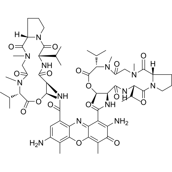
HY-D1020
7-Aminoactinomycin DDNA Stain -
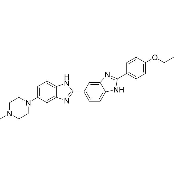
HY-15559
Hoechst 33342DNA Fluorescent Stain -
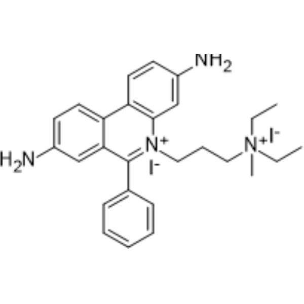
HY-D0815
Propidium IodideFluorochrome -
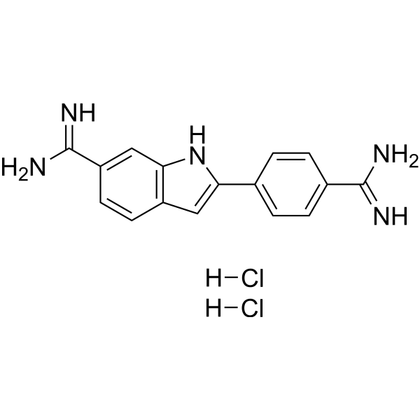
HY-D0814
DAPI dihydrochlorideFluorescent DNA Stain -

HY-DY1006
Propidium Iodide (solution)Fluorochrome -

HY-DY1081
DAPI dihydrochloride (solution)Fluorescent DNA Stain -
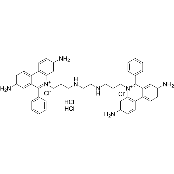
HY-D0093
Ethidium homodimerDNA Fluorochrome -
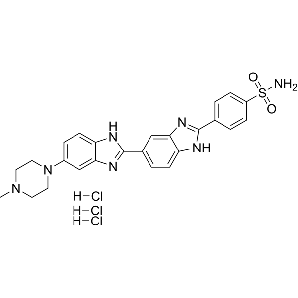
HY-15619
Hoechst S 769121DNA Dye -
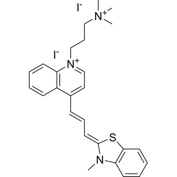
HY-117070
TO-PRO-3 iodideFluorochrome -
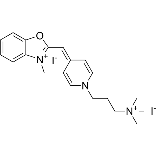
HY-D1245
Oxazole blueDye -

HY-D3374
PODi-1Fluorescent Dye


Biological Activity
Description
Solvent and concentration: DMSO: 1 mg/mL
IC50 & Target
|
RNA Polymerase |
In Vitro
Guide (The following is our recommended protocol. This protocol is only a guide and should be modified according to your specific needs).
1. Solution preparation
1.1 Preparation of working solution
Dilute to 0.5-5 μM with PBS or serum-free medium (optimized according to the experiment).
Note: The working solution should be prepared and used immediately. Keep it away from light.
2. Cell staining
2.1 Fix cells.
2.2 Pellet and resuspend cells.
2.3 Add 7-Aminoactinomycin D (7-AAD) staining solution (0.5 to 5 μM).
2.4 Incubate for 15 to 60 minutes.
2.5 Measure fluorescence intensity (Ex/Em = 549/648 nm).
7-Aminoactinomycin D (7-AAD) is a DNA dye that distinguishes viable, apoptotic, and late apoptotic/dead cells in flow cytometry[1].
7-Aminoactinomycin D is frequently used to stain and exclude dead cells in flow cytometry at low concentrations (0.5-5 μg/mL). At higher concentrations (10-20 μg/mL), 7-Aminoactinomycin D has also been used to distinguish between viable cells (7-AADnegative) and apoptotic cells (7-AADdim) or dead cells (7-AADbright) using the fact that permeability of the cell membrane, and hence fluorescence intensity, is low in early apoptotic cells and high in late apoptotic and dead cells[1].
MedChemExpress (MCE) has not independently confirmed the accuracy of these methods. They are for reference only. Further protocols information, click here.
Emission (Em)
648
Excitation (Ex)
549


Chemical Information
-
CAS No. 7240-37-1
-
Appearance Liquid
-
Molecular Weight 1270.43
-
Formula C62H87N13O16
-
Color Pink to red
-
SMILES
O=C(N[C@H](C(N1[C@@](C(N(C)CC(N(C)[C@@H](C(C)C)C(O[C@@H]2C)=O)=O)=O)([H])CCC1)=O)C(C)C)[C@H]2NC(C(C(C(OC3=C(C)C(N)=C4)=C5C)=NC3=C4C(N[C@@H]6C(N[C@H](C(N7[C@@](C(N(C)CC(N(C)[C@@H](C(C)C)C(O[C@@H]6C)=O)=O)=O)([H])CCC7)=O)C(C)C)=O)=O)=C(N)C5=O)=O
-
Synonyms
7-AAD (solution)
-
Shipping
Room temperature in continental US; may vary elsewhere.
-
Storage
Please store the product under the recommended conditions in the Certificate of Analysis.

Protocols
-
Research Protocol for Infectious Diseases
Infectious-disease experiments test how pathogens interact with host barriers, innate immune receptors, inflammatory signaling, pathogen replication, and tissue injury; pattern-recognition receptors such as TLRs, RIG-I-like receptors, NOD-like receptors, and inflammasomes detect microbial molecules and activate NF-κB, interferon, and cytokine responses. The central hypothesis is that infection severity reflects the balance between pathogen burden and host response: protective inflammation restricts pathogen growth, whereas excessive or mislocalized inflammation contributes to tissue damage and disease phenotype. Unresolved questions include which host pathways are protective versus pathogenic, why some infection models fail to translate to human disease, and which combined readouts best predict clinically relevant infection outcomes.
-
Annexin V plus membrane-impermeant dye apoptosis staining
Annexin V-based apoptosis assays rely on the detection of phosphatidylserine (PS) externalization from the inner leaflet of the plasma membrane to the outer leaflet, an early biochemical hallmark of apoptosis. Fluorescently labeled Annexin V binds PS in a calcium-dependent manner, enabling identification of early apoptotic cells by flow cytometry or fluorescence microscopy. When combined with a membrane-impermeant DNA-binding dye (e. g. , propidium iodide), this approach allows discrimination between viable (Annexin V−/dye−), early apoptotic (Annexin V+/dye−), and late apoptotic or necrotic (Annexin V+/dye+) cell populations by assessing membrane integrity and PS exposure.


Purity & Documentation
-
Data Sheet (283 KB)
-
SDS (480 KB)
- English - EN (480 KB)
- Français - FR (480 KB)
- Deutsch - DE (480 KB)
- Norwegian - NO (480 KB)
- Español - ES (480 KB)
- Swedish - SV (480 KB)
- Italian - IT (480 KB)
- Korean - KR (480 KB)
- Portuguese - PT (480 KB)
-
Handling Instructions (2659 KB)
References
[1]. Nadine C L Zembruski, et al. 7-Aminoactinomycin D for apoptosis staining in flow cytometry. Anal Biochem. 2012 Oct 1;429(1):79-81. [Content Brief]
[2]. Hanen Sellami, et al. Molecular detection of Chlamydia trachomatis and other sexually transmitted bacteria in semen of male partners of infertile couples in Tunisia: the effect on semen parameters and spermatozoa apoptosis markers. PLoS One. 2014 Jul 14;9(7):e98903. [Content Brief]
[3]. Wadkins RM, et al. Actinomycin D and 7-aminoactinomycin D binding to single-stranded DNA. Biochemistry. 1991 Oct 1;30(39):9469-78. [Content Brief]

Calculators
Concentration (start) × Volume (start) = Concentration (final) × Volume (final)


7-Aminoactinomycin D (solution) Related Classifications
HY-DY1099 Related Classifications
 Powered by Bioz