



AMG-47a
Based on 3 publication(s) in Google Scholar
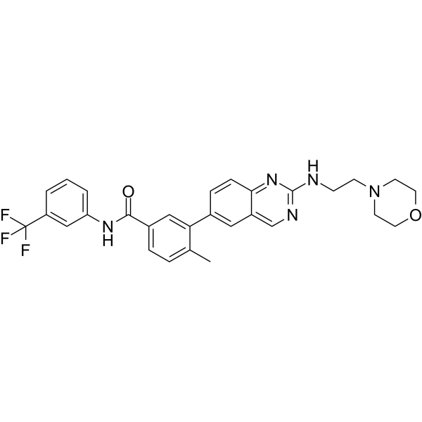
AMG-47a is an orally active, ATP-competitive Lck inhibitor (IC50=0.2 nM). AMG-47a inhibits VEGF2, p38α, p38α, Jak3, MLR, and IL-2 with IC50 of 1 nM, 3 nM, 72 nM, 30 nM, and 21 nM, respectively. AMG-47a reduces T cell activation and the production of cytokines such as TGF-β, exerting anti-inflammatory and anti-fibrotic activities. AMG-47a can be used in the research of autoimmune diseases, pulmonary fibrosis, and KRAS mutation-associated cancers[1][2][3].
For research use only. We do not sell to patients.
- Purity : 99.19%
- CAS No.: 882663-88-9
- Formula: C29H28F3N5O2
- Molecular Weight:535.56
-
Storage:Powder -20°C, 3 years , 4°C, 2 years ; In solvent -80°C, 6 months , -20°C, 1 month

To place orders, for customer services and technical support, please contact: MedChemExpress USA
Tel: 609-228-6898 E-mail: [email protected] [email protected]


-
 Biological Activity
-
Chemical Information
-
Solvent & Solubility
- Protocols
- Purity & Documentation
- References
-
 Help & FAQs


-
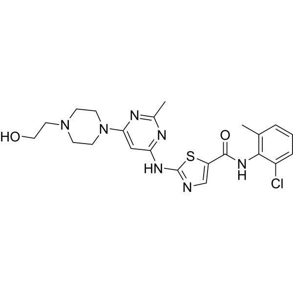
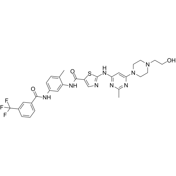
HY-10181
DasatinibSrc/Bcr-Abl Inhibitor -
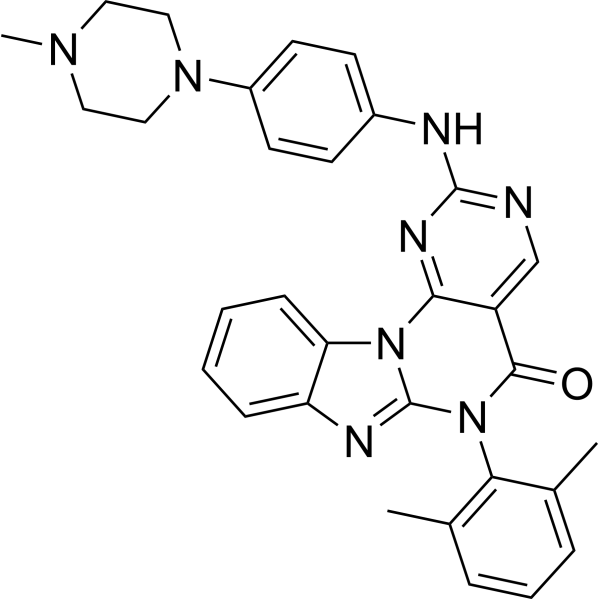
HY-12072
Lck InhibitorLck Inhibitor -
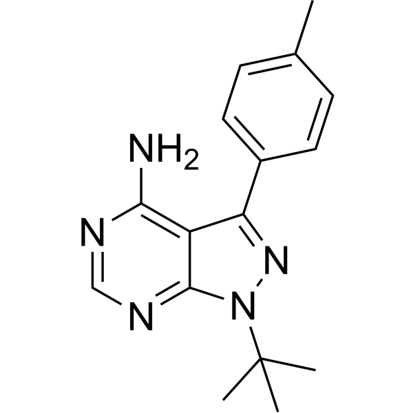
HY-13804
PP1Src Inhibitor -
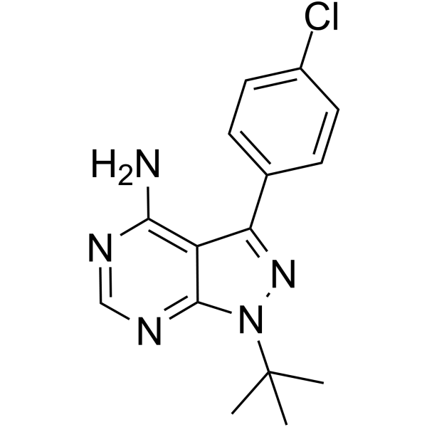
HY-13805
PP2Src Inhibitor -
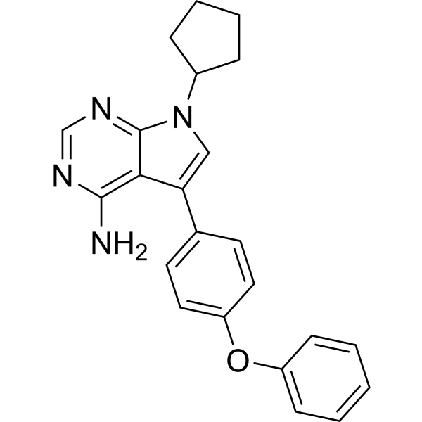
HY-108318
RK-24466Lck Inhibitor -
HY-11011
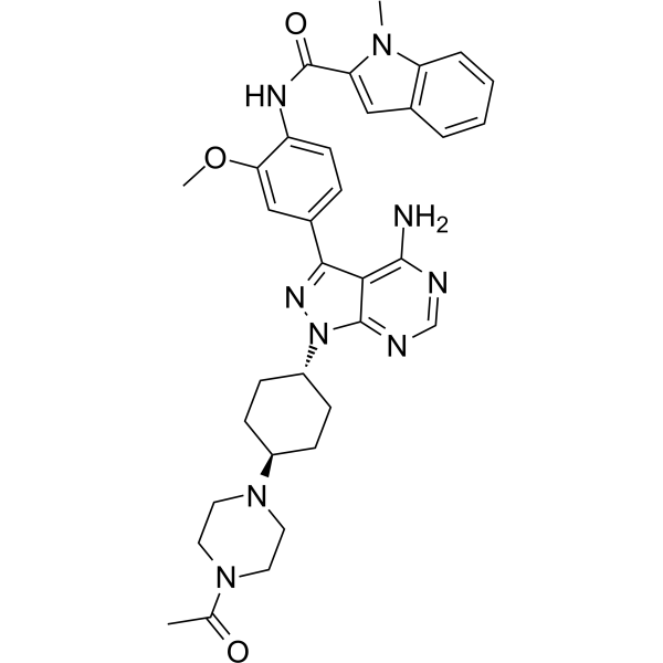
A-770041Lck Inhibitor -
HY-112182
UM-164c-Src Inhibitor -
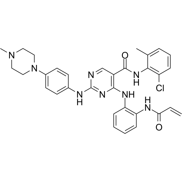
HY-136605
DGY-06-116Src Inhibitor -
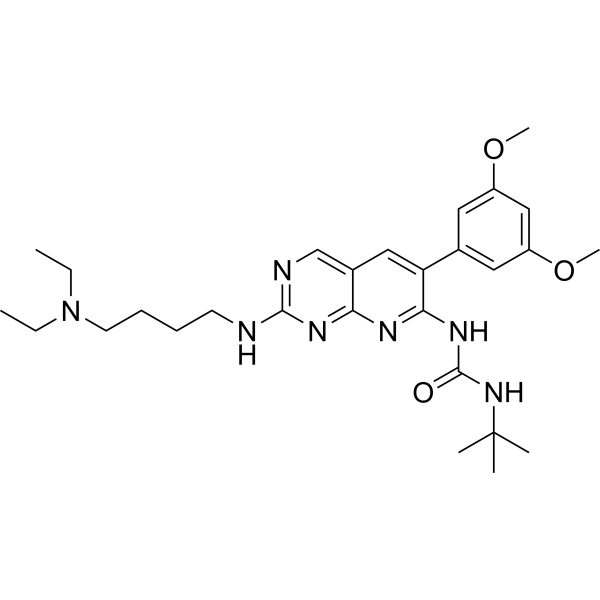
HY-10321
PD173074FGFR1 Inhibitor -
HY-13941
1-Naphthyl PP1Src Inhibitor -
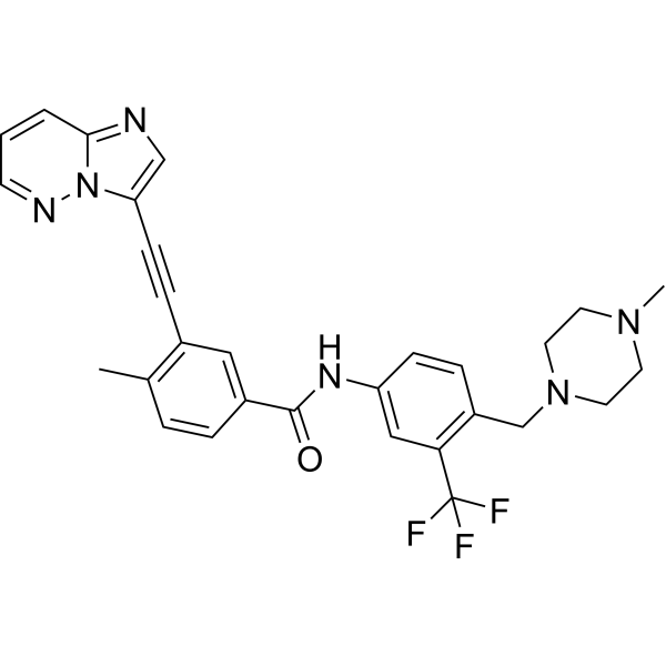
HY-12047
PonatinibMulti-targeted Kinase Inhibitor -
HY-101053
Src Inhibitor 1Src Inhibitor
Publications Citing Use of MedChemExpress (MCE) AMG-47a
More Customer Validation & Images

-
WB
All VEGFR Isoforms
More

Biological Activity
Description
IC50 & Target
IC50: 0.2 nM (Lck), 1 nM (VEGF2), 3 nM (p38α), 72 nM (Jak3), 30 nM (MLR), 21 nM (IL-2)[2]
Cellular Effect
|
Cell Line

|
Type | Value | Description | References |
|---|---|---|---|---|
| T-cell | IC50 |
21 nM
Compound: 47
|
Inhibition of anti CD3/CD28-induced IL2 secretion in human T cells
Inhibition of anti CD3/CD28-induced IL2 secretion in human T cells
|
[PMID: 16970394] |
| T-cell | IC50 |
30 nM
Compound: 47
|
Inhibition of human T cell proliferation by mixed lymphocyte reaction
Inhibition of human T cell proliferation by mixed lymphocyte reaction
|
[PMID: 16970394] |
In Vitro
AMG-47a (100 nM-5 μM; 48 h) significantly reduces the level of EGFP-KRASG12V protein in HeLa cells, without significant effect on EGFP protein[1].
AMG-47a (0.2 nM; 5 min) effectively inhibits the phosphorylation of Lck in CD4+ T cells, wtih comparable function to that of Nintedanib (HY-50904)[3].
MedChemExpress (MCE) has not independently confirmed the accuracy of these methods. They are for reference only. Further protocols information, click here.
In Vivo
MedChemExpress (MCE) has not independently confirmed the accuracy of these methods. They are for reference only.


Chemical Information
-
CAS No. 882663-88-9
-
Appearance Solid
-
Molecular Weight 535.56
-
Formula C29H28F3N5O2
-
Color Light yellow to yellow
-
SMILES
O=C(NC1=CC=CC(C(F)(F)F)=C1)C2=CC=C(C)C(C3=CC4=CN=C(NCCN5CCOCC5)N=C4C=C3)=C2
-
Shipping
Room temperature in continental US; may vary elsewhere.
-
Storage
Powder -20°C 3 years 4°C 2 years In solvent -80°C 6 months -20°C 1 month


Publications (3)
-
Journal Impact Factor
-
Most Recent
-
Cell Death Dis
The Lck inhibitor, AMG-47a, blocks necroptosis and implicates RIPK1 in signalling downstream of MLKL. [Abstract]2022 Apr 1;13(4):291. PMID: 35365636 -
Oncogene
2018 Aug;37(31):4226-4238. PMID: 29717260
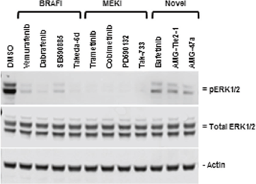
AMG-47a purchased from MedChemExpress. Usage Cited in: Oncogene. 2018 Aug;37(31):4226-4238. [Abstract]
AMG-47 results in decreased ERK phosphorylation.
-
Arch Pharm (Weinheim)
Discovery of potent CSK inhibitors through integrated virtual screening and molecular dynamic simulation. [Abstract]2024 Sep;357(9):e2400066. PMID: 38809025


Solvent & Solubility
In Vitro:
DMSO : ≥ 34 mg/mL (63.48 mM; Hygroscopic DMSO has a significant impact on the solubility of product, please use newly opened DMSO)
* "≥" means soluble, but saturation unknown.
Please refer to the solubility information to select the appropriate solvent. Once prepared, please aliquot and store the solution to prevent product inactivation from repeated freeze-thaw cycles.
Storage method and period of stock solution: -80°C, 6 months; -20°C, 1 month. When stored at -80°C, please use it within 6 months. When stored at -20°C, please use it within 1 month.
Please refer to the solubility information to select the appropriate solvent. Once prepared, please aliquot and store the solution to prevent product inactivation from repeated freeze-thaw cycles.
Storage method and period of stock solution: -80°C, 6 months; -20°C, 1 month. When stored at -80°C, please use it within 6 months. When stored at -20°C, please use it within 1 month.
Concentration (start) × Volume (start) = Concentration (final) × Volume (final)

Protocols
-
Fibrosis/Collagen Morphometry
Fibrosis and collagen morphometry is based on the quantitative visualization of fibrillar collagen deposition in tissue sections using histochemical stains such as Sirius Red (Picrosirius Red) or Masson's trichrome, followed by image-based or polarization-enhanced analysis to estimate collagen proportional area as a surrogate of extracellular matrix accumulation during fibrotic remodeling. Sirius Red combined with polarized light microscopy enhances detection of collagen fibers due to birefringence properties, enabling more specific visualization of collagen type I and III fibrils compared to conventional bright-field histology, while whole-section or region-restricted digital morphometry reduces field-selection bias in fibrosis assessment. Alternative quantitative approaches include second harmonic generation (SHG) and two-photon excited fluorescence microscopy, which enable label-free detection of fibrillar collagen and have been validated against histological staining and biochemica
-
Collagen: Sirius Red Staining
Sirius Red or picrosirius red staining is a histochemical method for visualizing collagen-rich extracellular matrix in tissue sections, and collagen fibers are detected as red-stained structures under bright-field microscopy with enhanced birefringence under polarized light. Picrosirius red is useful for assessing total collagen organization, distribution, and fibrosis burden, but polarized color should not be interpreted as a definitive collagen type I versus type III readout because color is affected by fiber orientation, thickness, and packing.
-
Cell Cytotoxicity Assay
Cytotoxicity assays are usually based on the assessment of cell membrane damage, which can also be indirectly detected by measuring cell viability. Detection methods include MTT assay, CKK-8 assay, LDH assay and ATP assay, etc.
-
Connective Tissue: Masson's Trichrome/Collagen Trichrome Staining
Masson’s Trichrome (collagen/trichrome staining) is a histological technique that differentially stains tissue compartments using sequential acidic dyes to distinguish collagen from muscle and cytoplasmic components based on dye affinity and tissue permeability differences, enabling visualization of fibrosis and connective tissue architecture in histological sections. The classical formulation typically uses Weigert's iron hematoxylin for nuclear staining, Biebrich scarlet-acid fuchsin for cytoplasm and muscle, and aniline blue (or light green variants) for collagen, producing a characteristic blue/green collagen signal contrasted against red cytoplasm and dark nuclei. The staining principle relies on selective displacement of smaller dye molecules by larger anionic dyes in collagen-rich regions under controlled acidified conditions, which enhances collagen-specific dye retention. This property makes the method widely used for fibrosis assessment in organs such as heart, liver, lung, a
-
Research Protocol for Inflammation-related Diseases
The NLRP3 inflammasome is a cytosolic innate immune signaling platform that integrates priming signals and danger-signal activation to promote caspase-1 activation, maturation of IL-1β and IL-18, and gasdermin D-mediated pyroptotic cell death. The core experimental logic is to determine whether inflammatory disease phenotypes are driven by increased NLRP3 expression, ASC-containing inflammasome assembly, caspase-1 cleavage, GSDMD cleavage, and extracellular release of IL-1β/IL-18 rather than by nonspecific cell injury alone. The pathway is strongly linked to inflammation-related disease phenotypes because monosodium urate crystals activate NALP3/NLRP3 inflammasome signaling in gout-like crystal inflammation, cholesterol crystals activate NLRP3 inflammasomes in atherogenesis models, and DSS-induced intestinal inflammation has been reported to involve NLRP3 inflammasome activity. However, experimental colitis studies also show context-dependent protective effects of NLRP3 inflammasome co
-
Research Protocol for Cancer Immunology
Cancer immunology studies how the immune system recognizes, suppresses, edits, or fails to eliminate malignant cells through tumor antigen release, antigen presentation, T-cell priming, immune trafficking, tumor-cell killing, and feedback inhibition in the tumor microenvironment. The cancer-immunity cycle links tumor antigenicity, dendritic-cell priming, CD8+ T-cell infiltration, cytotoxic function, and immune-checkpoint regulation to tumor rejection or immune escape. Immune-checkpoint pathways such as PD-1/PD-L1 and CTLA-4 suppress antitumor T-cell activity and can be therapeutically blocked, but many tumors remain resistant because of poor antigen presentation, weak T-cell infiltration, suppressive myeloid cells, regulatory T cells, and tumor-intrinsic immune-exclusion programs. Unresolved questions include which immune-cell states predict response, how tumor-intrinsic pathways exclude immune cells, how myeloid suppression limits checkpoint blockade, and which combination strategies
-
Genotoxicity/Mutagenicity Study
The bacterial reverse mutation assay detects point mutations that restore amino-acid prototrophy in auxotrophic Salmonella typhimurium or Escherichia coli tester strains; after exposure to a test article, mutagenic activity is read out as an increased number of revertant colonies on minimal agar compared with the vehicle control. The assay uses tester strains with different mutation targets so that base-substitution and frameshift mutagens can be detected, and testing is performed with and without exogenous mammalian metabolic activation because some chemicals require biotransformation to become mutagenic.


Purity & Documentation
-
Data Sheet (272 KB)
-
SDS (393 KB)
- English - EN (393 KB)
- Français - FR (393 KB)
- Deutsch - DE (393 KB)
- Norwegian - NO (393 KB)
- Español - ES (393 KB)
- Swedish - SV (393 KB)
- Italian - IT (393 KB)
- Korean - KR (393 KB)
- Portuguese - PT (393 KB)
-
Handling Instructions (2659 KB)
References
[1]. Carver J, et al. A high-throughput assay for small molecule destabilizers of the KRAS oncoprotein. PLoS One. 2014 Aug 5;9(8):e103836. [Content Brief]
[2]. DiMauro EF, et al. Discovery of aminoquinazolines as potent, orally bioavailable inhibitors of Lck: synthesis, SAR, and in vivo anti-inflammatory activity. J Med Chem. 2006 Sep 21;49(19):5671-86. [Content Brief]
[3]. Kagawa K, et al. The lymphocyte-specific protein tyrosine kinase-specific inhibitor A-770041 attenuates lung fibrosis via the suppression of TGF-β production in regulatory T-cells. PLoS One. 2022 Oct 27;17(10):e0275987. [Content Brief]


Complete Stock Solution Preparation Table
Please refer to the solubility information to select the appropriate solvent. Once prepared, please aliquot and store the solution to prevent product inactivation from repeated freeze-thaw cycles.
Storage method and period of stock solution: -80°C, 6 months; -20°C, 1 month. When stored at -80°C, please use it within 6 months. When stored at -20°C, please use it within 1 month.
| Optional Solvent | Concentration Solvent Mass | 1 mg | 5 mg | 10 mg | 25 mg |
|---|---|---|---|---|---|
| DMSO | 1 mM | 1.8672 mL | 9.3360 mL | 18.6720 mL | 46.6801 mL |
| 5 mM | 0.3734 mL | 1.8672 mL | 3.7344 mL | 9.3360 mL | |
| 10 mM | 0.1867 mL | 0.9336 mL | 1.8672 mL | 4.6680 mL | |
| 15 mM | 0.1245 mL | 0.6224 mL | 1.2448 mL | 3.1120 mL | |
| 20 mM | 0.0934 mL | 0.4668 mL | 0.9336 mL | 2.3340 mL | |
| 25 mM | 0.0747 mL | 0.3734 mL | 0.7469 mL | 1.8672 mL | |
| 30 mM | 0.0622 mL | 0.3112 mL | 0.6224 mL | 1.5560 mL | |
| 40 mM | 0.0467 mL | 0.2334 mL | 0.4668 mL | 1.1670 mL | |
| 50 mM | 0.0373 mL | 0.1867 mL | 0.3734 mL | 0.9336 mL | |
| 60 mM | 0.0311 mL | 0.1556 mL | 0.3112 mL | 0.7780 mL |


AMG-47a Related Classifications
HY-18303 Related Classifications
 Powered by Bioz