



Avermectin B1
Based on 5 publication(s) in Google Scholar
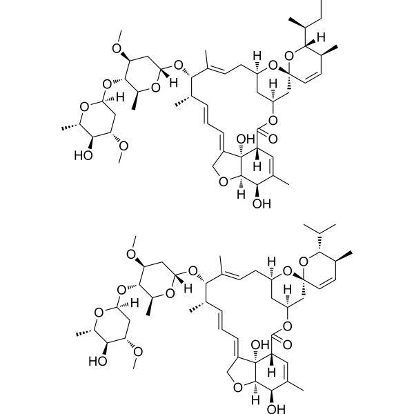
Avermectin B1 (Abamectin) is a mixture of two similar segments of avermectin. Avermectin B1 is an orally anti-infection agent, which can be used in the research of parasitic worms, insect pests, agriculture and animal husbandry. Avermectin B1 can also induce the production of ROS and induces cytotoxicity, apoptosis and autophagy.
For research use only. We do not sell to patients.
- Purity : 98.99%
- CAS No.: 71751-41-2
- Formula: C48H72O14 (for Avermectin B1a)
- Molecular Weight:873.09
-
Storage:
-20°C, sealed storage, away from moisture and light
* In solvent : -80°C, 6 months; -20°C, 1 month (sealed storage, away from moisture and light)

To place orders, for customer services and technical support, please contact: MedChemExpress USA
Tel: 609-228-6898 E-mail: [email protected] [email protected]


-
 Biological Activity
-
Chemical Information
-
Solvent & Solubility
- Protocols
- Purity & Documentation
- References
-
 Help & FAQs


-

HY-100558
Bafilomycin A1V-ATPase Inhibitor -

HY-Y0320
Dimethyl sulfoxide, meets analytical specification of Ch.P.Aprotic Solvent -
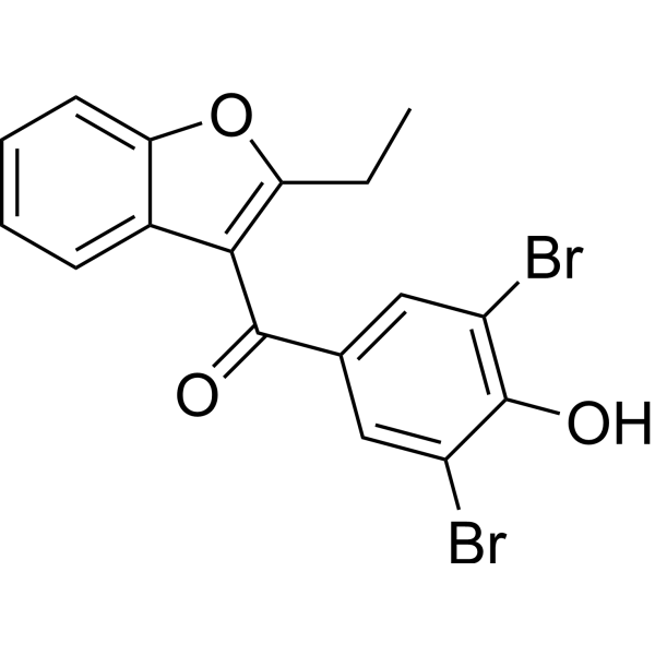
HY-B1135
BenzbromaroneApoptosis Inducer -
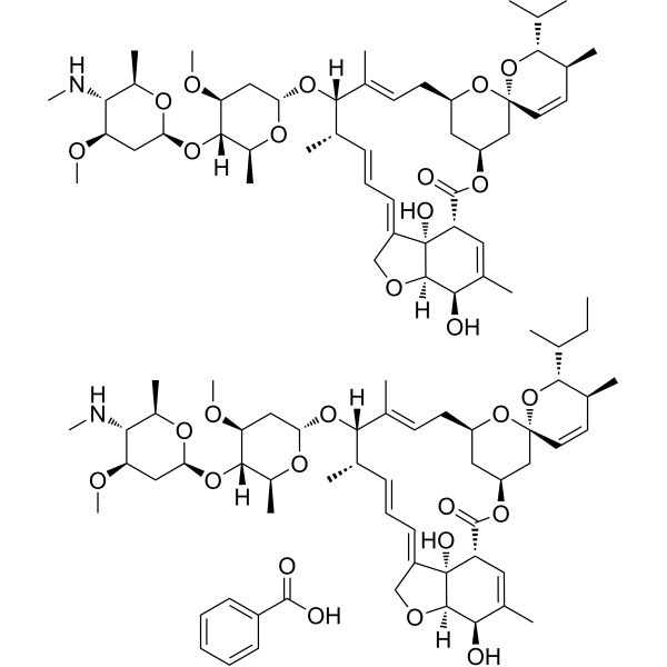
HY-B0837
Emamectin BenzoateNervoussystem Toxicant -
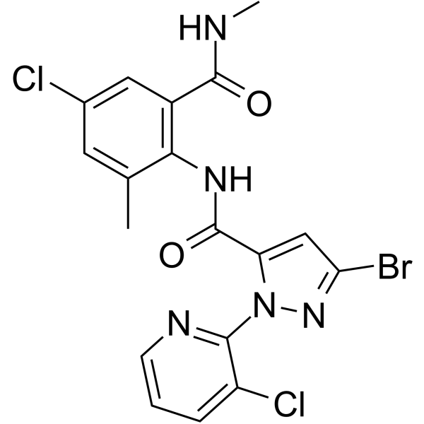
HY-112820
ChlorantraniliproleInsecticide -
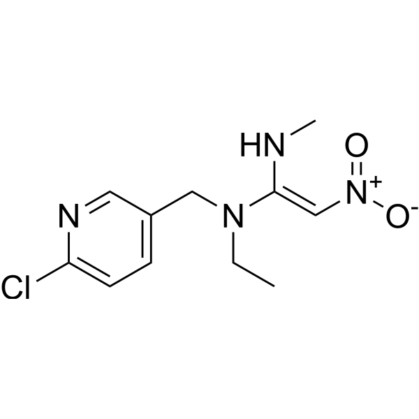
HY-B0820
NitenpyramnAChR Agonist -
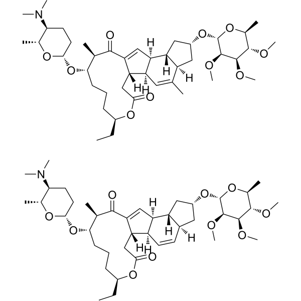
HY-138800
SpinosadAcetylcholine Agonist -

HY-15311R
Avermectin B1 (Standard)Anti-infection Agent -
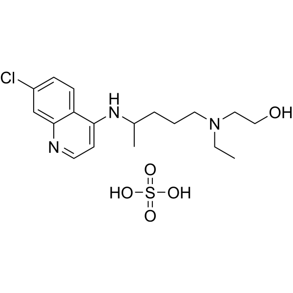
HY-B1370
Hydroxychloroquine sulfateToll-like Receptor (TLR) Inhibitor -
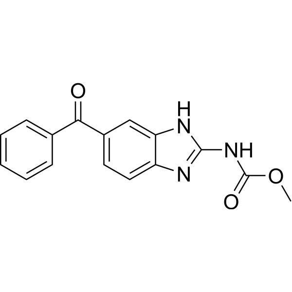
HY-17595
MebendazoleApoptosis Inducer -
HY-N0484
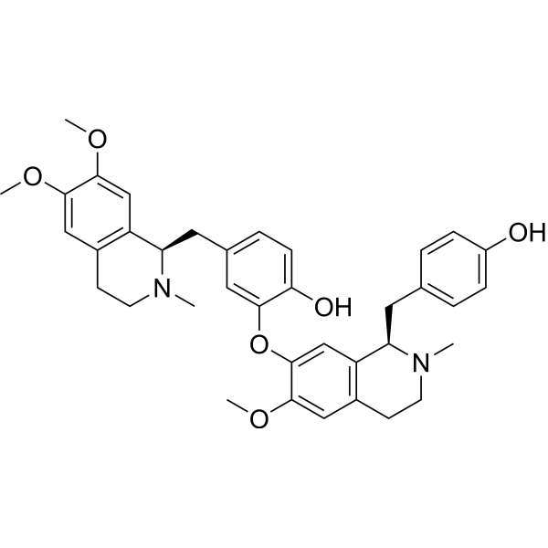
LiensininePI3K/AKT/MAPK/JAK2/STAT3 Inhibitor
Publications Citing Use of MedChemExpress (MCE) Avermectin B1
More Customer Validation & Images

-
Cell Proliferation/Viability Assay
-
Cell Imaging/Staining

-
Flow Cytometry
-
IF
-
WB
All Antibiotic Isoforms
MoreAll Parasite Isoforms
More

Biological Activity
Description
In Vitro
Avermectin B1 (0-80 μM, 12 h) induces cytotoxicity through MAPK and ATM/ATR pathway in mouse embryonic fibroblast (MEF) cells, and induces ROS-mediated DNA damage[1].
Avermectin B1 (36 μg/mL, 72 h) has strong nematicidal efect of on G. pallida in aqueous solution, and negatively influences viability and infectivity of G. pallida J2 detected in potato roots(cv. Spunta)[2].
Abamectin (10 μM, 24 h) induces significant cytotoxicity by overproduction of ROS in haemocytes of Erocheir sinensis[3].
Abamectin (4 μM, 24 h) induces apoptosis and autophagy by inhibiting ROS-mediated PI3K/AKT signaling in MGC803 cells[4].
MedChemExpress (MCE) has not independently confirmed the accuracy of these methods. They are for reference only. Further protocols information, click here.
-
Cell Line:Mouse embryonic fibroblast (MEF) cells
-
Concentration:0, 0.5, 5, 10, 20, 40, 80 μM
-
Incubation Time:12 h
-
Result:Reduced cell viability with an IC50 value of 45.6 μM.
-
Cell Line:MGC803 cells
-
Concentration:0-4 μM
-
Incubation Time:24 h
-
Result:Increased active caspase-3 and expression of Bax/Bcl-2, decreased MMP in a dose-dependent manner.
In Vivo
MedChemExpress (MCE) has not independently confirmed the accuracy of these methods. They are for reference only.
-
Animal Model:Horses (had a pruritic dermatosis)[5]
-
Dosage:0.2 mg/kg, a single dose.
-
Administration:Oral administration
-
Result:Decreased in mean strongyle egg counts 14, 28 and 42 d after treatment, and resulted in zero microfilaria counts in all horses 14 d after treatment.
-
Animal Model:Healthy adult female sheep (Pharmacokinetic assay)[6]
-
Dosage:0.2 mg/kg
-
Administration:Subcutaneous administration (in the left neck area of each sheep)
-
Result:Pharmacokinetic profiles of Avermectin B1
Parameters Mean Kcl (/day) 0.17 t1/2cl (day) 4.36 Kab (/day) 0.24 t1/2ab (day) 3.15 Cmax (ng/mL) 6.24 tmax (day) 4.20 AUC (0-27) (ng/day/mL) 80.2 AUC(0-∞) (ng/day/mL) 84.7 MRT (day) 8.80
Clinical Trial
| NCT Number | Sponsor | Condition | Start Date |
Phase

|
|---|---|---|---|---|
| NCT01329991 | Plexxikon| | 2011-05 | PHASE1 |


Chemical Information
-
CAS No. 71751-41-2
-
Appearance Solid
-
Molecular Weight 873.09
-
Formula C48H72O14 (for Avermectin B1a)
-
Color White to off-white
-
SMILES
C[C@@H](CC)[C@]1([H])[C@@H](C)C=C[C@@]2(O[C@@]3([H])C[C@]([H])(OC([C@@]4([H])[C@@](/C(CO5)=C/C=C/[C@H](C)[C@H](O[C@@]6([H])C[C@H](OC)[C@@H](O[C@]7([H])O[C@@H](C)[C@H](O)[C@@H](OC)C7)[C@H](C)O6)/C(C)=C/C3)(O)[C@@]5([H])[C@H](O)C(C)=C4)=O)C2)O1.C[C@H]8C=C[C@@]9(O[C@@]%10([H])C[C@]([H])(OC([C@@]%11([H])[C@@](/C(CO%12)=C/C=C/[C@H](C)[C@H](O[C@@]%13([H])C[C@H](OC)[C@@H](O[C@]%14([H])O[C@@H](C)[C@H](O)[C@@H](OC)C%14)[C@H](C)O%13)/C(C)=C/C%10)(O)[C@@]%12([H])[C@H](O)C(C)=C%11)=O)C9)O[C@@H]8C(C)C
-
Synonyms
Abamectin; Avermectin B1a-Avermectin B1b mixt.
-
Initial Source
Streptomyces avermitilis
-
Shipping
Room temperature in continental US; may vary elsewhere.
-
Storage
-20°C, sealed storage, away from moisture and light
* In solvent : -80°C, 6 months; -20°C, 1 month (sealed storage, away from moisture and light)


Publications (5)
-
Journal Impact Factor
-
Most Recent
-
Mol Pharm
2022 Nov 7;19(11):4320-4332. PMID: 36269563 -
J Biochem Mol Toxicol
ROS accumulation contributes to abamectin-induced apoptosis and autophagy via the inactivation of PI3K/AKT/mTOR pathway in TM3 Leydig cells. [Abstract]2020 Aug;34(8):e22505. PMID: 32275808 -
J Biochem Mol Toxicol
Abamectin induces apoptosis and autophagy by inhibiting reactive oxygen species-mediated PI3K/AKT signaling in MGC803 cells. [Abstract]2019 Jul;33(7):e22336. PMID: 30958899
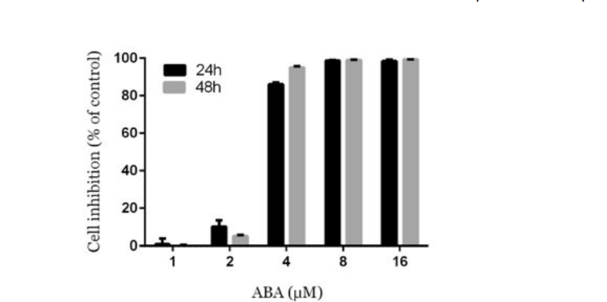
Avermectin B1 purchased from MedChemExpress. Usage Cited in: J Biochem Mol Toxicol. 2019 Jul;33(7):e22336. [Abstract]
MTS assays were carried out to assess cell viability inhibition after MGC803 cells were treated with Avermectin B1 (ABA) (1, 2, 4, 8, and 16 μM) for 24 and 48 hours.
Avermectin B1 purchased from MedChemExpress. Usage Cited in: J Biochem Mol Toxicol. 2019 Jul;33(7):e22336. [Abstract]
Dose‐dependent effect of ABA on MGC803 cells morphology. MGC803 cells were treated with Avermectin B1 (ABA) for 24 hours and examined under a light microscope at ×200 magnification.
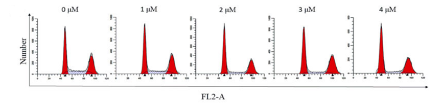
Avermectin B1 purchased from MedChemExpress. Usage Cited in: J Biochem Mol Toxicol. 2019 Jul;33(7):e22336. [Abstract]
Representative flow cytometry histograms following PI staining for cell‐cycle analysis after treatment with Avermectin B1 (ABA) (0, 1, 2, 3, and 4 μM) for 24 hours.
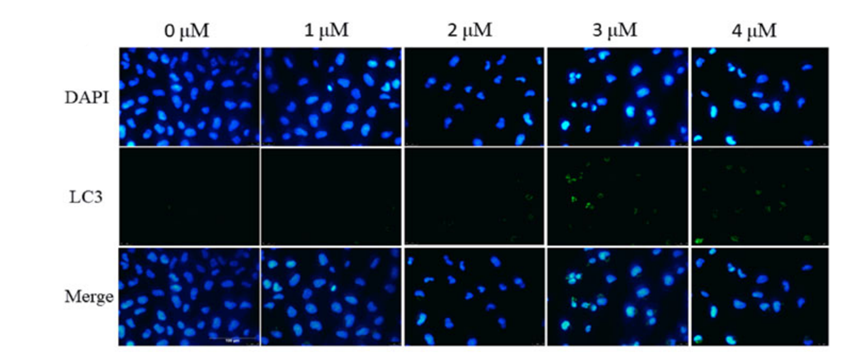
Avermectin B1 purchased from MedChemExpress. Usage Cited in: J Biochem Mol Toxicol. 2019 Jul;33(7):e22336. [Abstract]
The LC3 and DAPI staining detected MGC803 cell autophagy treated with Avermectin B1 (ABA) (0, 1, 2, 3, and 4 μM).
Avermectin B1 purchased from MedChemExpress. Usage Cited in: J Biochem Mol Toxicol. 2019 Jul;33(7):e22336. [Abstract]
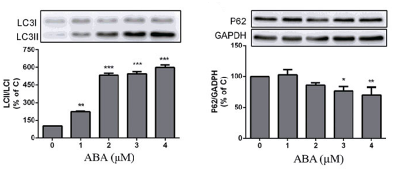
Protein expression of LC3‐Ⅰ, LC3‐II, and P62 treated with Avermectin B1 (ABA) (0, 1, 2, 3, and 4 μM).
-
J Insect Physiol
Multi-omics analysis reveals AaaaNAT1 as a critical regulator of physiological homeostasis in Aedes aegypti. [Abstract]2026 Mar:169:104956. PMID: 41722763 -
Virology
Evaluating anti-viral effect of Ivermectin on porcine epidemic diarrhea virus and analyzing the related genes and signaling pathway by RNA-seq in vitro. [Abstract]2023 Oct:587:109877. PMID: 37688922


Solvent & Solubility
In Vitro:
DMSO : 100 mg/mL (114.54 mM; Need ultrasonic; Hygroscopic DMSO has a significant impact on the solubility of product, please use newly opened DMSO)
Please refer to the solubility information to select the appropriate solvent. Once prepared, please aliquot and store the solution to prevent product inactivation from repeated freeze-thaw cycles.
Storage method and period of stock solution: -80°C, 6 months; -20°C, 1 month (sealed storage, away from moisture and light). When stored at -80°C, please use it within 6 months. When stored at -20°C, please use it within 1 month.
Please refer to the solubility information to select the appropriate solvent. Once prepared, please aliquot and store the solution to prevent product inactivation from repeated freeze-thaw cycles.
Storage method and period of stock solution: -80°C, 6 months; -20°C, 1 month (sealed storage, away from moisture and light). When stored at -80°C, please use it within 6 months. When stored at -20°C, please use it within 1 month.
Concentration (start) × Volume (start) = Concentration (final) × Volume (final)
In Vivo:
Select the appropriate dissolution method based on your experimental animal and administration route.
- For the following dissolution methods, please ensure to first prepare a clear stock solution using an In Vitro approach and then sequentially add co-solvents:
- To ensure reliable experimental results, the clarified stock solution can be appropriately stored based on storage conditions. As for the working solution for In Vivo experiments, it is recommended to prepare freshly and use it on the same day.
- The percentages shown for the solvents indicate their volumetric ratio in the final prepared solution. If precipitation or phase separation occurs during preparation, heat and/or sonication can be used to aid dissolution.
Add each solvent one by one: 10% DMSO 40% PEG300 5% Tween-80 45% Saline
Solubility: ≥ 2.08 mg/mL (2.38 mM); Clear solution
This protocol yields a clear solution of ≥ 2.08 mg/mL (saturation unknown).
Taking 1 mL working solution as an example, add 100 μL DMSO stock solution (20.8 mg/mL) to 400 μL PEG300, and mix evenly; then add 50 μL Tween-80 and mix evenly; then add 450 μL Saline to adjust the volume to 1 mL.
Preparation of Saline: Dissolve 0.9 g sodium chloride in ddH₂O and dilute to 100 mL to obtain a clear Saline solution.
Add each solvent one by one: 10% DMSO 90% (20% SBE-β-CD in Saline)
Solubility: 2.08 mg/mL (2.38 mM); Suspended solution; Need ultrasonic
This protocol yields a suspended solution of 2.08 mg/mL. Suspended solution can be used for oral and intraperitoneal injection.
Taking 1 mL working solution as an example, add 100 μL DMSO stock solution (20.8 mg/mL) to 900 μL 20% SBE-β-CD in Saline, and mix evenly.
Preparation of 20% SBE-β-CD in Saline (4°C, storage for one week): 2 g SBE-β-CD powder is dissolved in 10 mL Saline, completely dissolve until clear.
In Vivo Dissolution Calculator
Please enter the basic information of animal experiments:
-
-
-
-
Recommended: Prepare an additional quantity of animals to account for potential losses during experiments.
Please enter your animal formula composition:
-
%DMSO +
Recommended: Keep the proportion of DMSO in working solution below 2% if your animal is weak.
-
%+
-
+%Tween-80 + +
-
%Saline +
The co-solvents required include: DMSO, . All of co-solvents are available by MedChemExpress (MCE). , Tween 80. All of co-solvents are available by MedChemExpress (MCE).
Working solution concentration: 0.22 mg/mL
Method for preparing stock solution: mg drug dissolved in μL DMSO. Stock solution concentration: mg/mL. * In solvent : -80°C, 6 months; -20°C, 1 month (sealed storage, away from moisture and light)
1. Take μL DMSO stock solution;
2. Add μL .
μL , mix evenly;
3. Then add μL Tween 80, mix evenly;
4. Then add μL
Please ensure that the stock solution in the first step is dissolved to a clear state, and add co-solvents in sequence. You can use ultrasonic heating (ultrasonic cleaner, recommended frequency 20-40 kHz), vortexing, etc. to assist dissolution.

Protocols
-
Research Protocol for Infectious Diseases
Infectious-disease experiments test how pathogens interact with host barriers, innate immune receptors, inflammatory signaling, pathogen replication, and tissue injury; pattern-recognition receptors such as TLRs, RIG-I-like receptors, NOD-like receptors, and inflammasomes detect microbial molecules and activate NF-κB, interferon, and cytokine responses. The central hypothesis is that infection severity reflects the balance between pathogen burden and host response: protective inflammation restricts pathogen growth, whereas excessive or mislocalized inflammation contributes to tissue damage and disease phenotype. Unresolved questions include which host pathways are protective versus pathogenic, why some infection models fail to translate to human disease, and which combined readouts best predict clinically relevant infection outcomes.
-
Apoptosis
Apoptosis, also called programmed cell death, is generally characterized by distinct morphological characteristics.
-
TUNEL staining for apoptotic DNA fragmentation
TUNEL staining detects DNA strand breaks by using terminal deoxynucleotidyl transferase to add labeled nucleotides to exposed 3′-OH DNA termini, generating either microscopic staining in fixed cells or tissue sections, or fluorescence/cytometric signal in cell suspensions. TUNEL positivity reflects DNA fragmentation but should not be interpreted alone as definitive apoptosis, because TUNEL can also label necrotic, autolytic, mechanically damaged, or DNA-repair-associated DNA breaks.
-
Cell Cytotoxicity Assay
Cytotoxicity assays are usually based on the assessment of cell membrane damage, which can also be indirectly detected by measuring cell viability. Detection methods include MTT assay, CKK-8 assay, LDH assay and ATP assay, etc.
-
Mammalian live/dead viability and cytotoxicity staining
Live/dead viability and cytotoxicity staining assays are based on the simultaneous detection of intracellular esterase activity in metabolically active (viable) cells and membrane integrity loss in non-viable cells. In commonly used dual-staining approaches, membrane-permeant fluorogenic substrates are converted by intracellular esterases into fluorescent products in live cells, while impermeant DNA-binding dyes selectively enter cells with compromised plasma membranes and label nucleic acids in dead or dying cells, enabling discrimination between viable and non-viable populations by fluorescence microscopy or flow cytometry.
-
Annexin V plus membrane-impermeant dye apoptosis staining
Annexin V-based apoptosis assays rely on the detection of phosphatidylserine (PS) externalization from the inner leaflet of the plasma membrane to the outer leaflet, an early biochemical hallmark of apoptosis. Fluorescently labeled Annexin V binds PS in a calcium-dependent manner, enabling identification of early apoptotic cells by flow cytometry or fluorescence microscopy. When combined with a membrane-impermeant DNA-binding dye (e. g. , propidium iodide), this approach allows discrimination between viable (Annexin V−/dye−), early apoptotic (Annexin V+/dye−), and late apoptotic or necrotic (Annexin V+/dye+) cell populations by assessing membrane integrity and PS exposure.
-
ROS/oxidative-stress fluorescent staining
ROS/oxidative-stress fluorescent staining uses cell-permeant fluorogenic probes that become fluorescent after oxidation inside cells or tissues; commonly used examples include DCFH-DA/DCFDA for broad cellular oxidant detection, DHE for superoxide-related signal detection, MitoSOX for mitochondrial superoxide-related signal detection, and CellROX probes for oxidative-stress-associated fluorescence readouts. The assay detects probe oxidation rather than a single ROS species unless the probe and analysis method have been chemically validated for that species. DCFH-DA enters cells, is deacetylated by intracellular esterases to DCFH, and produces fluorescent DCF after oxidation, so the readout is used as an operational measure of total cellular oxidative stress rather than a species-specific ROS measurement. DHE and MitoSOX can report superoxide-related oxidation, but red fluorescence alone can include non-specific ethidium-like oxidation products; HPLC or optimized spectral approaches are
-
Autophagy
Autophagy is a process in which eukaryotic cells use lysosomes to degrade their own cytoplasmic proteins and damaged organelles under the regulation of autophagy related gene (Atg). Microtubule-associated proteins light chain 3 (LC3) is recognized as autophagy marker, which transfers from cytoplasmic LC3 (LC3-I) to membrane type (LC3-II). LC3-II/I ratio could be detected by Western Blot and fluorescence microscopy.
-
Apoptosis Solutions
Apoptosis is a regulated, generally non-lytic cell-death pathway that removes unwanted, damaged, infected, or abnormal cells through coordinated morphological changes, caspase activation, DNA fragmentation, and membrane remodeling. The intrinsic apoptosis pathway is controlled mainly by mitochondrial outer membrane permeabilization, BCL-2 family proteins, cytochrome c release, apoptosome formation, caspase-9 activation, and downstream executioner caspase-3/7 activation. The extrinsic apoptosis pathway is initiated by death receptors such as Fas, TNFR, and TRAIL receptors, which recruit adaptor proteins and activate caspase-8 before engaging executioner caspases or mitochondrial amplification through BID cleavage. Apoptosis is linked to many phenotypes, including cancer cell killing, tissue homeostasis, immune regulation, neurodegeneration, infection response, and treatment-induced cytotoxicity; unresolved questions include how apoptosis interacts with necroptosis, pyroptosis, ferroptos
-
Lysosome and acidic-vesicle live-cell staining
Lysosome and acidic-vesicle live-cell staining detects acidic intracellular compartments by using membrane-permeant acidotropic probes that accumulate in low-pH vesicles, including lysosomes, late endosomes, autolysosomes, and acidic phagosomes. LysoTracker staining is commonly used as an intensity-based readout of acidic lysosomal compartment abundance or enlargement, while acridine orange produces green fluorescence in less concentrated compartments and red fluorescence after concentration-dependent accumulation in acidic vesicular organelles. Loss or reduction of acridine-orange red signal can be used as a readout of lysosomal membrane permeabilization or reduced acidic-vesicle integrity. This protocol is designed for live cultured cells and can be adapted for fluorescence microscopy, high-content imaging, plate-reader readout, or flow cytometry when the selected literature supports the readout. Because these dyes report acidotropic accumulation rather than lysosome identity alone,
-
Macroautophagy Solutions
Macroautophagy is a conserved lysosome-dependent degradation pathway in which cytoplasmic material is sequestered into double-membrane autophagosomes and delivered to lysosomes for degradation and recycling. The pathway supports cellular homeostasis during nutrient limitation, organelle stress, protein-aggregate accumulation, infection, differentiation, and tissue remodeling by coupling cargo sequestration, autophagosome maturation, lysosomal fusion, and degradation of cargo-derived macromolecules. The core molecular sequence includes initiation by nutrient- and stress-regulated autophagy machinery, autophagosome nucleation, LC3/ATG8-family conjugation to autophagosomal membranes, cargo selection through receptors such as SQSTM1/p62, autophagosome-lysosome fusion, and lysosomal degradation. LC3 was identified as a mammalian homolog of yeast Atg8 that localizes to autophagosomal membranes after processing, and p62/SQSTM1 was shown to connect ubiquitinated cargo with autophagic degradati
-
Cell Viability Determination by MTT Colorimetric Assay
The following protocol uses the MTT colorimetric assay as a classic literature-established method for assessing cell viability/metabolic activity in cultured mammalian cells. MTT[3-(4,5-dimethylthiazol-2-yl)-2,5-diphenyltetrazolium bromide] is reduced by metabolically active cells to a colored formazan product; the amount of formazan is quantified spectrophotometrically and provides an indirect measure of metabolically active viable cells. Importantly, MTT reduction reflects cellular oxidoreductase/metabolic activity rather than an absolute direct count of living cells, so changes in cellular metabolism can alter the signal independently of cell number.


Purity & Documentation
-
Data Sheet (291 KB)
-
SDS (624 KB)
- English - EN (624 KB)
- Français - FR (624 KB)
- Deutsch - DE (624 KB)
- Norwegian - NO (624 KB)
- Español - ES (624 KB)
- Swedish - SV (624 KB)
- Italian - IT (624 KB)
- Korean - KR (624 KB)
- Portuguese - PT (624 KB)
-
Handling Instructions (2659 KB)
References
[1]. [1] Yiran Liang, et al. Abamectin induces cytotoxicity via the ROS, JNK, and ATM/ATR pathways. PLoS One. Environ Sci Pollut Res Int. 2020 Apr;27(12):13726-13734. [Content Brief]
[2]. Nicola Sasanelli, et al. Abamectin Efficacy on the Potato Cyst Nematode Globodera pallida. Plants (Basel). 2019 Dec 19;9(1):12. [Content Brief]
[3]. Yi Huang, et al. Cytotoxicity induced by abamectin exposure in haemocytes of Chinese mitten crab, Eriocheir sinensis. Environ Toxicol Pharmacol. 2020 Jul;77:103384. [Content Brief]
[4]. Shanshan Zhu, et al. Abamectin induces apoptosis and autophagy by inhibiting reactive oxygen species-mediated PI3K/AKT signaling in MGC803 cells. J Biochem Mol Toxicol. 2019 Jul;33(7):e22336. [Content Brief]
[5]. Mogg TD, et al. Efficacy of avermectin B1 given orally against equine intestinal strongyles and Onchocera microfilaria. Aust Vet J. 1990 Nov;67(11):399-401. [Content Brief]
[6]. Peyami Sari, et al. Pharmacokinetics of Abamectin/Levamisole Combination in a Medium Chain Mono and Diglyceride-Based Vehicle and an In Vitro Release and In Vitro In Vivo Correlation Study for Levamisole. AAPS PharmSciTech. 2017 May;18(4):1254-1260. [Content Brief]


Complete Stock Solution Preparation Table
Please refer to the solubility information to select the appropriate solvent. Once prepared, please aliquot and store the solution to prevent product inactivation from repeated freeze-thaw cycles.
Storage method and period of stock solution: -80°C, 6 months; -20°C, 1 month (sealed storage, away from moisture and light). When stored at -80°C, please use it within 6 months. When stored at -20°C, please use it within 1 month.
| Optional Solvent | Concentration Solvent Mass | 1 mg | 5 mg | 10 mg | 25 mg |
|---|---|---|---|---|---|
| DMSO | 1 mM | 1.1454 mL | 5.7268 mL | 11.4536 mL | 28.6339 mL |
| 5 mM | 0.2291 mL | 1.1454 mL | 2.2907 mL | 5.7268 mL | |
| 10 mM | 0.1145 mL | 0.5727 mL | 1.1454 mL | 2.8634 mL | |
| 15 mM | 0.0764 mL | 0.3818 mL | 0.7636 mL | 1.9089 mL | |
| 20 mM | 0.0573 mL | 0.2863 mL | 0.5727 mL | 1.4317 mL | |
| 25 mM | 0.0458 mL | 0.2291 mL | 0.4581 mL | 1.1454 mL | |
| 30 mM | 0.0382 mL | 0.1909 mL | 0.3818 mL | 0.9545 mL | |
| 40 mM | 0.0286 mL | 0.1432 mL | 0.2863 mL | 0.7158 mL | |
| 50 mM | 0.0229 mL | 0.1145 mL | 0.2291 mL | 0.5727 mL | |
| 60 mM | 0.0191 mL | 0.0954 mL | 0.1909 mL | 0.4772 mL | |
| 80 mM | 0.0143 mL | 0.0716 mL | 0.1432 mL | 0.3579 mL | |
| 100 mM | 0.0115 mL | 0.0573 mL | 0.1145 mL | 0.2863 mL |


Avermectin B1 Related Classifications
HY-15311 Related Classifications
 Powered by Bioz