



Bavachalcone
Based on 2 publication(s) in Google Scholar
Bavachalcone is a potent inducer of apoptosis. Bavachalcone exerts anticancer activity by promoting autophagy and apoptosis in HepG2 cells. Bavachalcone acts as an anti-neuroinflammatory and antidepressant through the NF-κB pathway. Bavachalcone inhibits osteoclasts by interfering with ERK and Akt signaling pathways and the expression of c-Fos and NFATc1. Bavachalcone exhibits a significant inhibitory effect on baculovirus-expressed BACE-1 in vitro.
For research use only. We do not sell to patients.
- Purity : 99.90%
- CAS No.: 28448-85-3
- Formula: C20H20O4
- Molecular Weight:324.37
-
Storage:Powder -20°C, 3 years , 4°C, 2 years ; In solvent -80°C, 2 years , -20°C, 1 year

To place orders, for customer services and technical support, please contact: MedChemExpress USA
Tel: 609-228-6898 E-mail: [email protected] [email protected]


-
 Biological Activity
-
Chemical Information
-
Solvent & Solubility
- Protocols
- Purity & Documentation
- References
-
 Help & FAQs


-
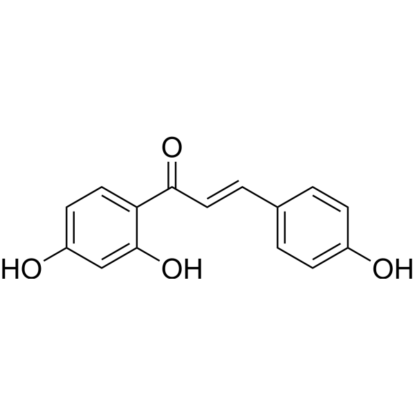
HY-N0102
IsoliquiritigeninAutophagy Inducer -
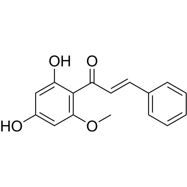
HY-N0279
CardamoninNF-κB Inhibitor -
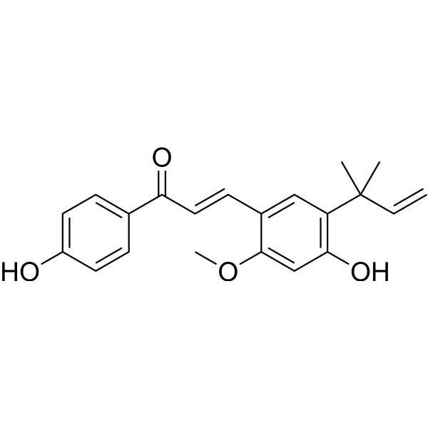
HY-N0372
Licochalcone AUGTs inhibitor -
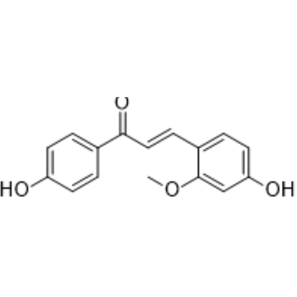
HY-N0269
EchinatinAnti-Inflammatory Agent -
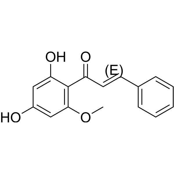
HY-N1378
(E)-CardamoninhTRPA1 Antagonist -

HY-N0231R
Bavachalcone (Standard)Bacterial Inhibitor -
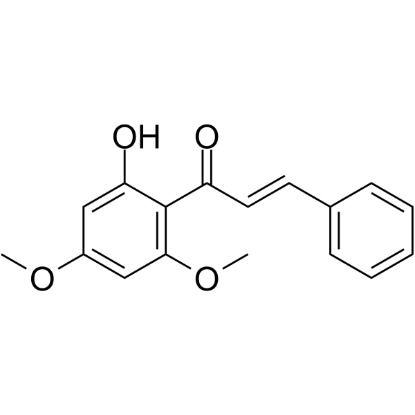
HY-N2132
Flavokawain BAnti-tumor Agent -
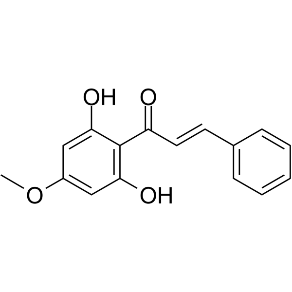
HY-N3057
Pinostrobin chalconeAnti-Cancer Agent -
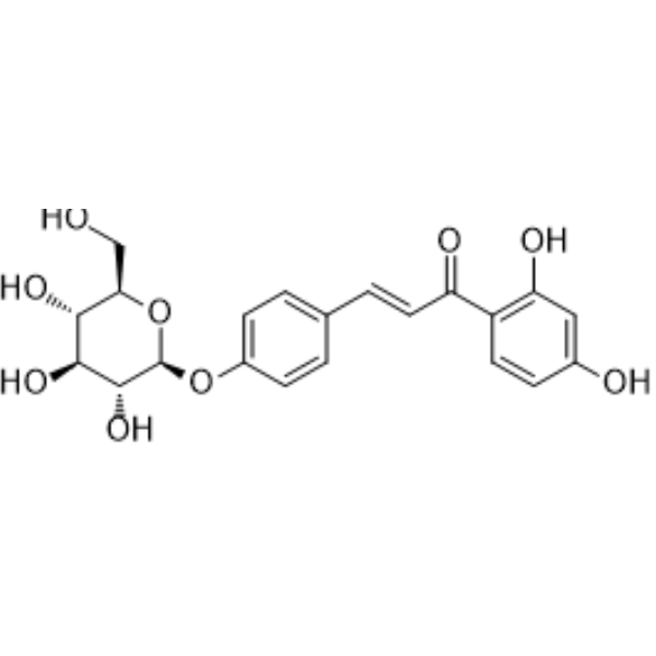
HY-N0765
IsoliquiritinFungal Inhibitor -
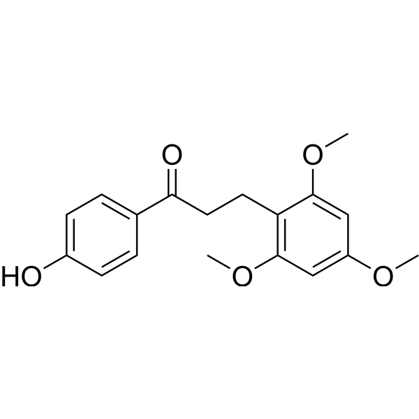
HY-N1504
Loureirin BPAI-1 Inhibitor -
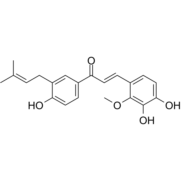
HY-N4187
Licochalcone DNF-κB p65 Inhibitor -
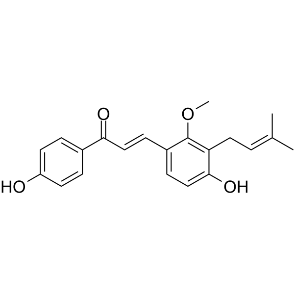
HY-N0374
Licochalcone Cα-Glucosidase Inhibitor
Publications Citing Use of MedChemExpress (MCE) Bavachalcone
MoreAll Antibiotic Isoforms
More

Biological Activity
Description
Cellular Effect
|
Cell Line

|
Type | Value | Description | References |
|---|---|---|---|---|
| A549 | IC50 |
>10 μM
Compound: 9
|
Cytotoxicity against human A549 cells after 48 hrs by MTT assay
Cytotoxicity against human A549 cells after 48 hrs by MTT assay
|
[PMID: 25710081] |
| HUVEC | IC50 |
39.73 μM
Compound: 1a
|
Toxicity against HUVEC incubated for 48 hrs by MTT assay
Toxicity against HUVEC incubated for 48 hrs by MTT assay
|
[PMID: 25590864] |
| K562 | IC50 |
>10 μM
Compound: 9
|
Cytotoxicity against human K562 cells after 48 hrs by MTT assay
Cytotoxicity against human K562 cells after 48 hrs by MTT assay
|
[PMID: 25710081] |
| K562 | IC50 |
2.77 μM
Compound: 1a
|
Antitumor activity against human K562 cells incubated for 48 hrs by MTT assay
Antitumor activity against human K562 cells incubated for 48 hrs by MTT assay
|
[PMID: 25590864] |
In Vitro
Bavachalcone (5 μg/mL; 1-48 h) inhibits osteoclasts by interfering with ERK and Akt signaling pathways and the expression of c-Fos and NFATc1[1].
.
Bavachalcone (0-100 μg/mL; 24h) is toxic to HepG2 cells with IC50 of 20 μg/mL[3].
Bavachalcone (20 μg/mL; 24 h) induces apoptosis in HepG2[3].
Bavachalcone (0-20 μg/mL; 24 h) induces cell cycle arrest in HepG2 cells[3].
Bavachalcone (0-20 μg/mL; 24 h) induces autophagy through Akt-mTOR signaling pathway[3].
Bavachalcone (5-10 μM; 2 h) inhibits the activation of NF-κB pathway in LPS (HY-D1056)-induced BV2 cells, upregulated the expression of A20 and TAX1BP1, and enhanced their interaction[4].
MedChemExpress (MCE) has not independently confirmed the accuracy of these methods. They are for reference only. Further protocols information, click here.
-
Cell Line:RANKL treated bone marrow-derived macrophages
-
Concentration:5 μg/mL
-
Incubation Time:0, 12, 24 and 48 h;
1 h -
Result:Reduced the levels of c-Fos and NFATc1.
Reduced the phosphorylation levels of ERK and Akt.
-
Cell Line:HepG2 cell
-
Concentration:20 μg/mL
-
Incubation Time:24 h
-
Result:Significantly increased the levels of caspase-3.
-
Cell Line:HepG2 cell
-
Concentration:0, 5, 10 and 20 μg/mL
-
Incubation Time:24 h
-
Result:Decreased the levels of early cell cycle regulatory proteins cdk4 and cdk2.
Increased the levels of p21 and p27.
Inhibited the phosphorylation and expression of both Akt and mTOR.
-
Cell Line:LPS (HY-D1056) treated BV2 cells
-
Concentration:5 μM and 10 μM
-
Incubation Time:2 h
-
Result:Significantly decreased the expression of TRAF6 and phosphorylation of P65 and IκBα.
Increased the expression of A20 and TAX1BP1
In Vivo
MedChemExpress (MCE) has not independently confirmed the accuracy of these methods. They are for reference only.
-
Animal Model:LPS (HY-D1056) treated male C57BL/6 mice (8 weeks, 20 ± 2 g)
-
Dosage:30 mg/kg and 60 mg/kg
-
Administration:Intraperitoneal injection (i.p.); 4 days
-
Result:Attenuated LPS-induced depression-like behaviors in mice.
Inhibited LPS-induced activation of microglia in the brain of mice.
Inhibited LPS-induced NF-κB pathway activation and upregulated the expression of A20 and TAX1BP1 in the cortex of mice.
Weakened the production of TNF-α and IL-6 in the cortex of mice.


Chemical Information
-
CAS No. 28448-85-3
-
Appearance Solid
-
Molecular Weight 324.37
-
Formula C20H20O4
-
Color Yellow to orange
-
SMILES
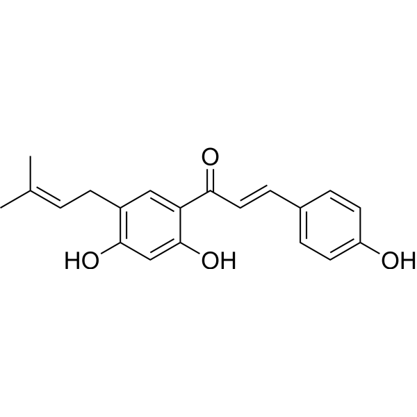
O=C(C1=CC(C/C=C(C)\C)=C(O)C=C1O)/C=C/C2=CC=C(O)C=C2
-
Synonyms
Broussochalcone B
-
Structure Classification
-
Initial Source
-
Shipping
Room temperature in continental US; may vary elsewhere.
-
Storage
Powder -20°C 3 years 4°C 2 years In solvent -80°C 2 years -20°C 1 year


Publications (2)
-
Journal Impact Factor
-
Most Recent
-
Chem Biol Interact
Bavachinin, a main compound of Psoraleae Fructus, facilitates GSDMD-mediated pyroptosis and causes hepatotoxicity in mice. [Abstract]2024 Sep 1:400:111133. PMID: 38969277 -
BMC Cancer
4'-O-Methylbroussochalcone B as a novel tubulin polymerization inhibitor suppressed the proliferation and migration of acute myeloid leukaemia cells. [Abstract]2021 Jan 22;21(1):91. PMID: 33482772


Solvent & Solubility
In Vitro:
DMSO : ≥ 34 mg/mL (104.82 mM; Hygroscopic DMSO has a significant impact on the solubility of product, please use newly opened DMSO)
* "≥" means soluble, but saturation unknown.
Please refer to the solubility information to select the appropriate solvent. Once prepared, please aliquot and store the solution to prevent product inactivation from repeated freeze-thaw cycles.
Storage method and period of stock solution: -80°C, 2 years; -20°C, 1 year. When stored at -80°C, please use it within 2 years. When stored at -20°C, please use it within 1 year.
Please refer to the solubility information to select the appropriate solvent. Once prepared, please aliquot and store the solution to prevent product inactivation from repeated freeze-thaw cycles.
Storage method and period of stock solution: -80°C, 2 years; -20°C, 1 year. When stored at -80°C, please use it within 2 years. When stored at -20°C, please use it within 1 year.
Concentration (start) × Volume (start) = Concentration (final) × Volume (final)

Protocols
-
RNA extraction experimental
By lysing cells, releasing RNA, and removing impurities such as proteins and DNA, high-purity RNA products are finally obtained. The commonly used traditional method is the guanidine isothiocyanate/phenol/chloroform method (Trizol), which is suitable for a variety of animal materials including animal tissues, microorganisms, cultured cells, etc., and most plant materials.
-
Apoptosis
Apoptosis, also called programmed cell death, is generally characterized by distinct morphological characteristics.
-
TUNEL staining for apoptotic DNA fragmentation
TUNEL staining detects DNA strand breaks by using terminal deoxynucleotidyl transferase to add labeled nucleotides to exposed 3′-OH DNA termini, generating either microscopic staining in fixed cells or tissue sections, or fluorescence/cytometric signal in cell suspensions. TUNEL positivity reflects DNA fragmentation but should not be interpreted alone as definitive apoptosis, because TUNEL can also label necrotic, autolytic, mechanically damaged, or DNA-repair-associated DNA breaks.
-
Annexin V plus membrane-impermeant dye apoptosis staining
Annexin V-based apoptosis assays rely on the detection of phosphatidylserine (PS) externalization from the inner leaflet of the plasma membrane to the outer leaflet, an early biochemical hallmark of apoptosis. Fluorescently labeled Annexin V binds PS in a calcium-dependent manner, enabling identification of early apoptotic cells by flow cytometry or fluorescence microscopy. When combined with a membrane-impermeant DNA-binding dye (e. g. , propidium iodide), this approach allows discrimination between viable (Annexin V−/dye−), early apoptotic (Annexin V+/dye−), and late apoptotic or necrotic (Annexin V+/dye+) cell populations by assessing membrane integrity and PS exposure.
-
Autophagy
Autophagy is a process in which eukaryotic cells use lysosomes to degrade their own cytoplasmic proteins and damaged organelles under the regulation of autophagy related gene (Atg). Microtubule-associated proteins light chain 3 (LC3) is recognized as autophagy marker, which transfers from cytoplasmic LC3 (LC3-I) to membrane type (LC3-II). LC3-II/I ratio could be detected by Western Blot and fluorescence microscopy.
-
Apoptosis Solutions
Apoptosis is a regulated, generally non-lytic cell-death pathway that removes unwanted, damaged, infected, or abnormal cells through coordinated morphological changes, caspase activation, DNA fragmentation, and membrane remodeling. The intrinsic apoptosis pathway is controlled mainly by mitochondrial outer membrane permeabilization, BCL-2 family proteins, cytochrome c release, apoptosome formation, caspase-9 activation, and downstream executioner caspase-3/7 activation. The extrinsic apoptosis pathway is initiated by death receptors such as Fas, TNFR, and TRAIL receptors, which recruit adaptor proteins and activate caspase-8 before engaging executioner caspases or mitochondrial amplification through BID cleavage. Apoptosis is linked to many phenotypes, including cancer cell killing, tissue homeostasis, immune regulation, neurodegeneration, infection response, and treatment-induced cytotoxicity; unresolved questions include how apoptosis interacts with necroptosis, pyroptosis, ferroptos
-
Lysosome and acidic-vesicle live-cell staining
Lysosome and acidic-vesicle live-cell staining detects acidic intracellular compartments by using membrane-permeant acidotropic probes that accumulate in low-pH vesicles, including lysosomes, late endosomes, autolysosomes, and acidic phagosomes. LysoTracker staining is commonly used as an intensity-based readout of acidic lysosomal compartment abundance or enlargement, while acridine orange produces green fluorescence in less concentrated compartments and red fluorescence after concentration-dependent accumulation in acidic vesicular organelles. Loss or reduction of acridine-orange red signal can be used as a readout of lysosomal membrane permeabilization or reduced acidic-vesicle integrity. This protocol is designed for live cultured cells and can be adapted for fluorescence microscopy, high-content imaging, plate-reader readout, or flow cytometry when the selected literature supports the readout. Because these dyes report acidotropic accumulation rather than lysosome identity alone,
-
Research Protocol for Inflammation-related Diseases
The NLRP3 inflammasome is a cytosolic innate immune signaling platform that integrates priming signals and danger-signal activation to promote caspase-1 activation, maturation of IL-1β and IL-18, and gasdermin D-mediated pyroptotic cell death. The core experimental logic is to determine whether inflammatory disease phenotypes are driven by increased NLRP3 expression, ASC-containing inflammasome assembly, caspase-1 cleavage, GSDMD cleavage, and extracellular release of IL-1β/IL-18 rather than by nonspecific cell injury alone. The pathway is strongly linked to inflammation-related disease phenotypes because monosodium urate crystals activate NALP3/NLRP3 inflammasome signaling in gout-like crystal inflammation, cholesterol crystals activate NLRP3 inflammasomes in atherogenesis models, and DSS-induced intestinal inflammation has been reported to involve NLRP3 inflammasome activity. However, experimental colitis studies also show context-dependent protective effects of NLRP3 inflammasome co
-
Macroautophagy Solutions
Macroautophagy is a conserved lysosome-dependent degradation pathway in which cytoplasmic material is sequestered into double-membrane autophagosomes and delivered to lysosomes for degradation and recycling. The pathway supports cellular homeostasis during nutrient limitation, organelle stress, protein-aggregate accumulation, infection, differentiation, and tissue remodeling by coupling cargo sequestration, autophagosome maturation, lysosomal fusion, and degradation of cargo-derived macromolecules. The core molecular sequence includes initiation by nutrient- and stress-regulated autophagy machinery, autophagosome nucleation, LC3/ATG8-family conjugation to autophagosomal membranes, cargo selection through receptors such as SQSTM1/p62, autophagosome-lysosome fusion, and lysosomal degradation. LC3 was identified as a mammalian homolog of yeast Atg8 that localizes to autophagosomal membranes after processing, and p62/SQSTM1 was shown to connect ubiquitinated cargo with autophagic degradati


Purity & Documentation
-
Data Sheet (277 KB)
-
SDS (252 KB)
- English - EN (252 KB)
- Français - FR (252 KB)
- Deutsch - DE (252 KB)
- Norwegian - NO (252 KB)
- Español - ES (252 KB)
- Swedish - SV (252 KB)
- Italian - IT (252 KB)
- Korean - KR (252 KB)
- Portuguese - PT (252 KB)
-
Handling Instructions (2659 KB)
References
[1]. Park CK, et al. Bavachalcone inhibits osteoclast differentiation through suppression of NFATc1 induction by RANKL. Biochem Pharmacol. 2008 Jun 1;75(11):2175-82. [Content Brief]
[2]. Choi YH, et al. In vitro BACE-1 inhibitory phenolic components from the seeds of Psoralea corylifolia. Planta Med. 2008 Sep;74(11):1405-8. [Content Brief]
[3]. Song HS, et al. Bavachalcone from Cullen corylifolium induces apoptosis and autophagy in HepG2 cells. Phytomedicine. 2018 Feb 1;40:37-47. [Content Brief]
[4]. Wu X, et al. Upregulation of A20 and TAX1BP1 contributes to the anti-neuroinflammatory and antidepressant effects of bavachalcone. Int Immunopharmacol. 2023 Sep;122:110552. [Content Brief]


Complete Stock Solution Preparation Table
Please refer to the solubility information to select the appropriate solvent. Once prepared, please aliquot and store the solution to prevent product inactivation from repeated freeze-thaw cycles.
Storage method and period of stock solution: -80°C, 2 years; -20°C, 1 year. When stored at -80°C, please use it within 2 years. When stored at -20°C, please use it within 1 year.
| Optional Solvent | Concentration Solvent Mass | 1 mg | 5 mg | 10 mg | 25 mg |
|---|---|---|---|---|---|
| DMSO | 1 mM | 3.0829 mL | 15.4145 mL | 30.8290 mL | 77.0725 mL |
| 5 mM | 0.6166 mL | 3.0829 mL | 6.1658 mL | 15.4145 mL | |
| 10 mM | 0.3083 mL | 1.5414 mL | 3.0829 mL | 7.7072 mL | |
| 15 mM | 0.2055 mL | 1.0276 mL | 2.0553 mL | 5.1382 mL | |
| 20 mM | 0.1541 mL | 0.7707 mL | 1.5414 mL | 3.8536 mL | |
| 25 mM | 0.1233 mL | 0.6166 mL | 1.2332 mL | 3.0829 mL | |
| 30 mM | 0.1028 mL | 0.5138 mL | 1.0276 mL | 2.5691 mL | |
| 40 mM | 0.0771 mL | 0.3854 mL | 0.7707 mL | 1.9268 mL | |
| 50 mM | 0.0617 mL | 0.3083 mL | 0.6166 mL | 1.5414 mL | |
| 60 mM | 0.0514 mL | 0.2569 mL | 0.5138 mL | 1.2845 mL | |
| 80 mM | 0.0385 mL | 0.1927 mL | 0.3854 mL | 0.9634 mL | |
| 100 mM | 0.0308 mL | 0.1541 mL | 0.3083 mL | 0.7707 mL |


Bavachalcone Related Classifications
HY-N0231 Related Classifications
 Powered by Bioz