



Gluconate Calcium
Based on 1 publication(s) in Google Scholar
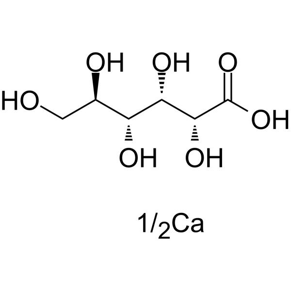
Gluconate (D-Gluconic acid) Calcium is an orally active glucose derivative. Gluconate Calcium reduces nitric oxide and inflammatory cytokines (IL-1β and IL-6). Gluconate Calcium inhibits ERK phosphorylation. Gluconate Calcium has antioxidant and antiplatelet activation activities. Gluconate Calcium has antitumor activity against colorectal cancer. Gluconate Calcium improves osteoarthritis, intestinal damage and acute lung injury.
Nur für Forschungszwecke. Wir verkaufen nicht an Patienten.
- Reinheit : 98.0%
- CAS. Nr.: 299-28-5
- Formel: C6H12O7.1/2Ca
- Molecular Weight:216.20
-
Speicherung:
4°C, sealed storage, away from moisture
* In solvent : -80°C, 6 months; -20°C, 1 month (sealed storage, away from moisture)

To place orders, for customer services and technical support, please contact: MedChemExpress USA
Tel: 609-228-6898 E-mail: [email protected] [email protected]


-
 Biologische Aktivität
-
Chemical Information
-
Lösungsmittel & Löslichkeit
- Protokoll
- Reinheit & Dokumentation
- Verweise
-
 Help & FAQs


-
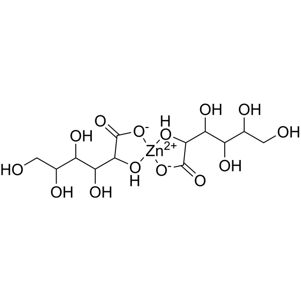
HY-W145499
Zinc GluconateZinc Supplement -
HY-Y1310A
Sodium alginate (Viscosity 200±20mpa.s)Biochemical Assay Reagent -
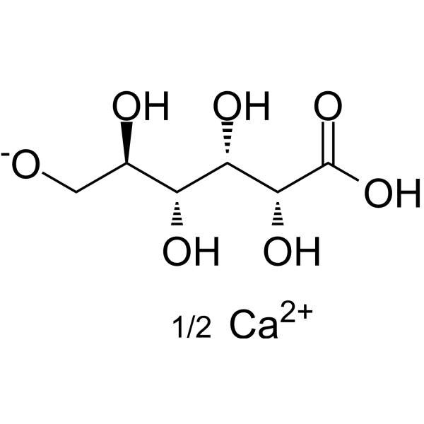
HY-W145486
Calcium gluconateCalcium Salt Supplement -
HY-N1938
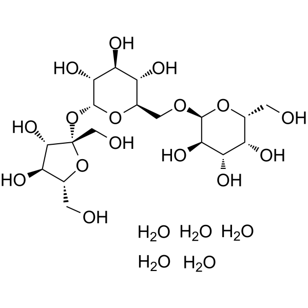
D(+)-Raffinose pentahydrateBiofilm-inhibitory Agent -

HY-B1092R
Gluconate Calcium (Standard)Analytical Standard -

HY-W145486R
Calcium gluconate (Standard)Biochemical Assay Reagent
Publications Citing Use of MedChemExpress (MCE) Gluconate Calcium
MoreAlle Endogenous Metabolite Isoform-spezifische Produkte anzeigen
More

Biologische Aktivität
Beschreibung
IC50 & Target
|
Human Endogenous Metabolite |
In Vitro
Gluconate Calcium (0.5-3 mM; 5 min) inhibits thrombin-induced arachidonic acid peroxidation and superoxide anion production in platelets, reduces platelet protein oxidation/nitration, and decreases carbonyl group generation, thiol oxidation, and nitrotyrosine formation[1].
MedChemExpress (MCE) has not independently confirmed the accuracy of these methods. They are for reference only.
In Vivo
Gluconate Calcium (50 mg/kg body weight; once a day for 5 weeks) regulates the changes in hematological parameters induced by Dimethylhydrazine (HY-W460407) in mice, reduces the levels of lipid peroxidation and nitric oxide (NO) in colon tissues, and improves the pathological changes of colon tissues[3].
Gluconate sodium (2500 ppm; in diet; p.o.; 15 days) increases the average daily gain of weaned piglets, reduces the feed-to-gain ratio, alleviates the intestinal injury induced by recombinant Escherichia coli, and improves the intestinal morphology[4].
Gluconate sodium (Calcium gluconate; 50 mg/kg; p.o.; 84 days) alleviates the symptoms of osteoarthritis induced by anterior cruciate ligament transection and partial medial meniscectomy in Sprague-Dawley rats, and inhibits joint stiffness and cartilage damage[5].
Gluconate sodium (Calcium gluconate; 12.5-50 mg/kg; i.p.; 30 min after modeling) alleviates lipopolysaccharide (LPS)-induced acute lung injury in mice, and inhibits the infiltration of inflammatory cells and the release of inflammatory cytokines[6].
MedChemExpress (MCE) has not independently confirmed the accuracy of these methods. They are for reference only.


Chemical Information
-
CAS. Nr. 299-28-5
-
Appearance Solid
-
Molecular Weight 216.20
-
Formel C6H12O7.1/2Ca
-
Color White to off-white
-
SMILES
O[C@H]([C@H]([C@@H]([C@@H](CO)O)O)O)C(O)=O.[1/2Ca]
-
Synonyms
Calcium D-gluconate; Gluconic acid hemicalcium salt
-
Structure Classification
-
Initial Source
-
Versand
Room temperature in continental US; may vary elsewhere.
-
Speicherung
4°C, sealed storage, away from moisture
* In solvent : -80°C, 6 months; -20°C, 1 month (sealed storage, away from moisture)


Publications (1)
-
Journal Impact Factor
-
Most Recent


Lösungsmittel & Löslichkeit
In Vitro:
H2O : 20 mg/mL (92.51 mM; Need ultrasonic)
DMSO : 1 mg/mL (4.63 mM; ultrasonic and warming and heat to 80°C; Hygroscopic DMSO has a significant impact on the solubility of product, please use newly opened DMSO)
Please refer to the solubility information to select the appropriate solvent. Once prepared, please aliquot and store the solution to prevent product inactivation from repeated freeze-thaw cycles.
Storage method and period of stock solution: -80°C, 6 months; -20°C, 1 month (sealed storage, away from moisture). When stored at -80°C, please use it within 6 months. When stored at -20°C, please use it within 1 month.
* Note: If you choose water as the stock solution, please dilute it to the working solution, then filter and sterilize it with a 0.22 μm filter before use.
Please refer to the solubility information to select the appropriate solvent. Once prepared, please aliquot and store the solution to prevent product inactivation from repeated freeze-thaw cycles.
Storage method and period of stock solution: -80°C, 6 months; -20°C, 1 month (sealed storage, away from moisture). When stored at -80°C, please use it within 6 months. When stored at -20°C, please use it within 1 month.
* Note: If you choose water as the stock solution, please dilute it to the working solution, then filter and sterilize it with a 0.22 μm filter before use.
Konzentration (Stammlösung) × Volumen (Stammlösung) = Konzentration (Ziellösung) × Volumen (Ziellösung)


Reinheit & Dokumentation
-
Data Sheet (278 KB)
-
SDS (254 KB)
- English - EN (254 KB)
- Français - FR (254 KB)
- Deutsch - DE (254 KB)
- Norwegian - NO (254 KB)
- Español - ES (254 KB)
- Swedish - SV (254 KB)
- Italian - IT (254 KB)
- Korean - KR (254 KB)
- Portuguese - PT (254 KB)
-
Handling Instructions (2659 KB)
Verweise
[1]. Saluk-Juszczak J. A comparative study of antioxidative activity of calcium-D-glucarate, sodium-D-gluconate and D-glucono-1,4-lactone in a human blood platelet model. Platelets. 2010;21(8):632-40. [Content Brief]
[2]. Kameue C, et al. Dietary sodium gluconate protects rats from large bowel cancer by stimulating butyrate production. J Nutr. 2004 Apr;134(4):940-4. [Content Brief]
[3]. Saleem TH, et al. Possible Protective Effects of Quercetin and Sodium Gluconate Against Colon Cancer Induction by Dimethylhydrazine in Mice. Asian Pac J Cancer Prev. 2015;16(14):5823-8. [Content Brief]
[4]. Zhang Y, et al. Dietary supplementation with sodium gluconate improves the growth performance and intestinal function in weaned pigs challenged with a recombinant Escherichia coli strain. BMC Vet Res. 2022 Aug 6;18(1):303. [Content Brief]
[5]. Kang SJ, et al. Protective effects of calcium gluconate on osteoarthritis induced by anterior cruciate ligament transection and partial medial meniscectomy in Sprague-Dawley rats. J Orthop Surg Res. 2014 Mar 7;9(1):14. [Content Brief]
[6]. Liu L, et al. Effects of calcium gluconate on lipopolysaccharide-induced acute lung injury in mice. Biochem Biophys Res Commun. 2018 Sep 18;503(4):2931-2935. [Content Brief]


Complete Stock Solution Preparation Table
Please refer to the solubility information to select the appropriate solvent. Once prepared, please aliquot and store the solution to prevent product inactivation from repeated freeze-thaw cycles.
Storage method and period of stock solution: -80°C, 6 months; -20°C, 1 month (sealed storage, away from moisture). When stored at -80°C, please use it within 6 months. When stored at -20°C, please use it within 1 month.
| Optional Solvent | Concentration Solvent Mass | 1 mg | 5 mg | 10 mg | 25 mg |
|---|---|---|---|---|---|
| DMSO / H2O | 1 mM | 4.6253 mL | 23.1267 mL | 46.2535 mL | 115.6337 mL |
| H2O | 5 mM | 0.9251 mL | 4.6253 mL | 9.2507 mL | 23.1267 mL |
| 10 mM | 0.4625 mL | 2.3127 mL | 4.6253 mL | 11.5634 mL | |
| 15 mM | 0.3084 mL | 1.5418 mL | 3.0836 mL | 7.7089 mL | |
| 20 mM | 0.2313 mL | 1.1563 mL | 2.3127 mL | 5.7817 mL | |
| 25 mM | 0.1850 mL | 0.9251 mL | 1.8501 mL | 4.6253 mL | |
| 30 mM | 0.1542 mL | 0.7709 mL | 1.5418 mL | 3.8545 mL | |
| 40 mM | 0.1156 mL | 0.5782 mL | 1.1563 mL | 2.8908 mL | |
| 50 mM | 0.0925 mL | 0.4625 mL | 0.9251 mL | 2.3127 mL | |
| 60 mM | 0.0771 mL | 0.3854 mL | 0.7709 mL | 1.9272 mL | |
| 80 mM | 0.0578 mL | 0.2891 mL | 0.5782 mL | 1.4454 mL |
* Note: If you choose water as the stock solution, please dilute it to the working solution, then filter and sterilize it with a 0.22 μm filter before use.


Gluconate Calcium Related Classifications
HY-B1092 Related Classifications
 Powered by Bioz