



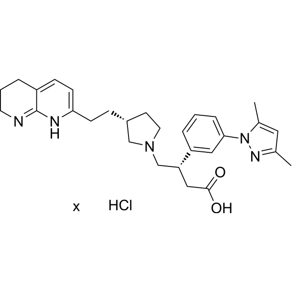
GSK 3008348 hydrochloride
Based on 2 publication(s) in Google Scholar
GSK 3008348 hydrochloride is a small molecule antagonist of integrin αvβ6 with IC50 values for αvβ6, αvβ1, αvβ3, αvβ5, and αvβ8 are 1.50, 2.83, 12.53, 4.00, and 2.26 nM, respectively. GSK 3008348 hydrochloride can target the αvβ6 integrin, inhibit TGF-β activation, and thereby alleviate the fibrotic process. GSK 3008348 hydrochloride prevents the binding of the foot-and-mouth disease virus (FMDV) to the αvβ6 integrin receptor on the surface of host cells, thereby inhibiting the entry of the virus. GSK 3008348 hydrochloride can be used for research on pulmonary fibrosis and various types of cancer.
For research use only. We do not sell to patients.
- Purity : 99.86%
- CAS No.: 1629249-40-6
- Formula: C29H38ClN5O2
-
Storage:
4°C, sealed storage, away from moisture
* In solvent : -80°C, 6 months; -20°C, 1 month (sealed storage, away from moisture)

To place orders, for customer services and technical support, please contact: MedChemExpress USA
Tel: 609-228-6898 E-mail: [email protected] [email protected]


-
 Biological Activity
-
Chemical Information
-
Solvent & Solubility
- Protocols
- Purity & Documentation
- References
-
 Help & FAQs


-
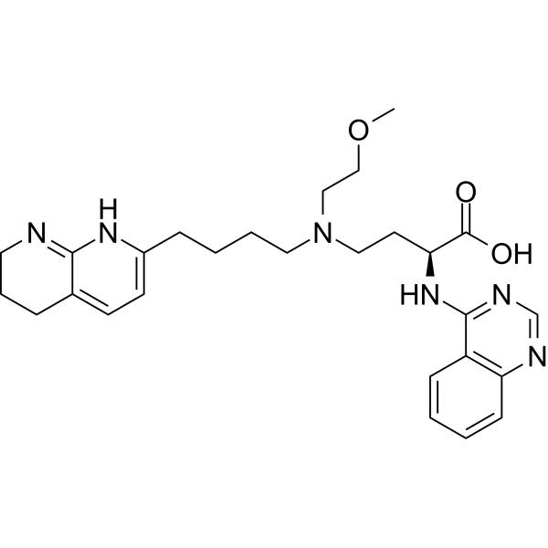
HY-137561
Bexotegrastαvβ6/αvβ1 Integrin Inhibitor -
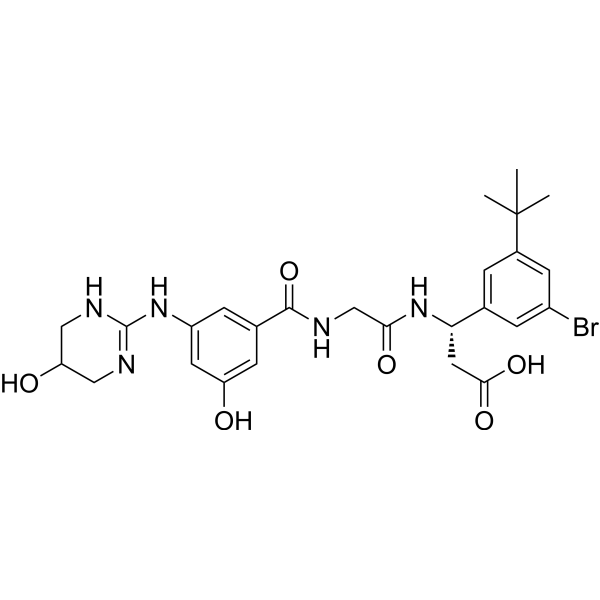
HY-18644
CWHM-12αV Integrins Inhibitor -
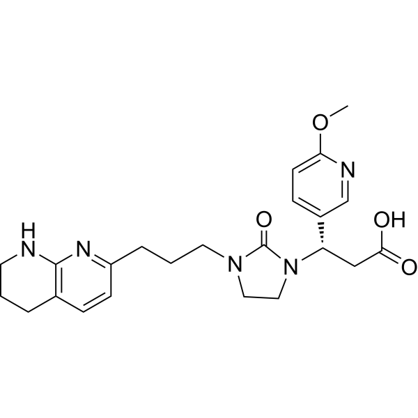
HY-15102
MK-0429αvβ3 Integrin Inhibitor -
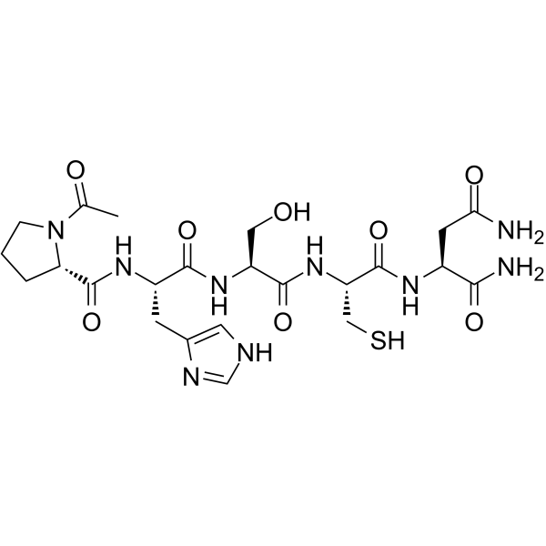
HY-13535
ATN-161α5β1/αvβ3 Integrin Binder -
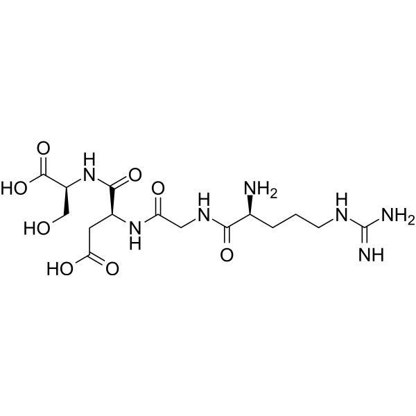
HY-12290
Arg-Gly-Asp-SerIntegrin Inhibitor -
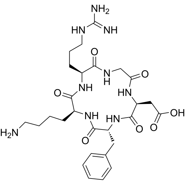
HY-P0023
Cyclo(-RGDfK)αvβ3 Integrin Inhibitor -
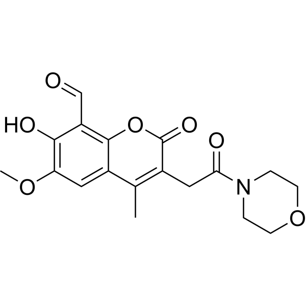
HY-104040
MKC8866IRE1α RNase Inhibitor -
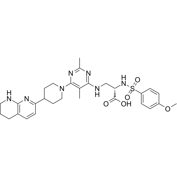
HY-100506
GLPG0187Integrin Receptor Antagonist -
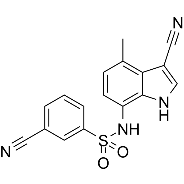
HY-14571
E7820Angiogenesis Inhibitor -

HY-P9911
Vedolizumab -
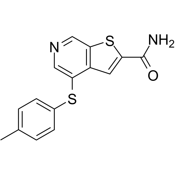
HY-100226
A-205804E-selectin/ICAM-1 Inhibitor -
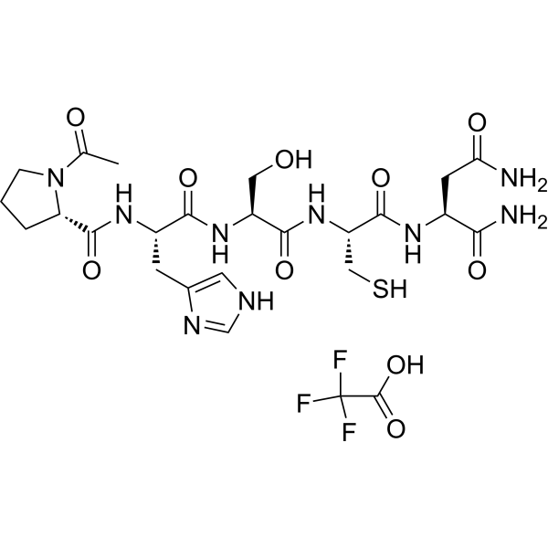
HY-13535A
ATN-161 trifluoroacetate saltIntegrin Antagonist
Publications Citing Use of MedChemExpress (MCE) GSK 3008348 hydrochloride
More

Biological Activity
Description
IC50 & Target
[3]|
αvβ6 1.50 nM (IC50) |
αvβ1 2.83 nM (IC50) |
αvβ3 12.53 nM (IC50) |
αvβ5 4.00 nM (IC50) |
αvβ8 2.26 nM (IC50) |
In Vitro
GSK 3008348 hydrochloride antagonizes integrin αvβ6 with an affinity (pIC50) value of 8.1 in the fluorescence polarisation assay, whereas its affinity in the cell adhesion assays (K562 cells) is for:αvβ6 (pIC50 = 8.4); αvβ3 (pIC50 = 6); αvβ5 (pIC50 = 6.9); αvβ8 (pIC50 = 7.7)[1].
GSK 3008348 hydrochloride (8 h) suppresses FMDV RNA (vRNA) replication and virus production when added prior to or concurrently with virus infection in PK-15 cells[2].
GSK 3008348 hydrochloride (10 μM, 1 h) decreases TGF-β1-activated procollagen I production in primary human hepatic stellate cells, fails to inhibit TGF-β1 induced SMAD3 and SMAD2 phosphorylation, but inhibits TGF-β-induced phosphorylation of ERK1/2 and STAT3[3].
MedChemExpress (MCE) has not independently confirmed the accuracy of these methods. They are for reference only. Further protocols information, click here.
-
Cell Line:Hepatic stellate cells
-
Concentration:10 μM
-
Incubation Time:1 h
-
Result:Reversed TGF-β1 induced p-SMAD3 nuclear translocation.
Significantly inhibited TGFβ1 induced ERK1/2 phosphorylation levels.
Significantly inhibited STAT3 phosphorylation in HSCs.
Did not show effects on TGF-β1-induced SMAD3 phosphorylation.


Chemical Information
-
CAS No. 1629249-40-6
-
Appearance Solid
-
Formula C29H38ClN5O2
-
Color Light yellow to khaki
-
SMILES
O=C(O)C[C@@H](C1=CC(N2N=C(C)C=C2C)=CC=C1)CN3C[C@H](CCC(N4)=CC=C5C4=NCCC5)CC3.[x].Cl
-
Shipping
Room temperature in continental US; may vary elsewhere.
-
Storage
4°C, sealed storage, away from moisture
* In solvent : -80°C, 6 months; -20°C, 1 month (sealed storage, away from moisture)


Publications (2)
-
Journal Impact Factor
-
Most Recent
-
mBio
Heat Shock Protein 60 Is Involved in Viral Replication Complex Formation and Facilitates Foot and Mouth Virus Replication by Stabilizing Viral Nonstructural Proteins 3A and 2C. [Abstract]2022 Oct 26;13(5):e0143422. PMID: 36106732 -
Biochem J
Integrin αVβ1 regulates procollagen I production through a non-canonical transforming growth factor β signaling pathway in human hepatic stellate cells. [Abstract]2021 May 14;478(9):1689-1703. PMID: 33876829


Solvent & Solubility
In Vitro:
H2O : 100 mg/mL (Need ultrasonic)
DMSO : 11.66 mg/mL (Need ultrasonic and warming; Hygroscopic DMSO has a significant impact on the solubility of product, please use newly opened DMSO)

Protocols
-
Fibrosis/Collagen Morphometry
Fibrosis and collagen morphometry is based on the quantitative visualization of fibrillar collagen deposition in tissue sections using histochemical stains such as Sirius Red (Picrosirius Red) or Masson's trichrome, followed by image-based or polarization-enhanced analysis to estimate collagen proportional area as a surrogate of extracellular matrix accumulation during fibrotic remodeling. Sirius Red combined with polarized light microscopy enhances detection of collagen fibers due to birefringence properties, enabling more specific visualization of collagen type I and III fibrils compared to conventional bright-field histology, while whole-section or region-restricted digital morphometry reduces field-selection bias in fibrosis assessment. Alternative quantitative approaches include second harmonic generation (SHG) and two-photon excited fluorescence microscopy, which enable label-free detection of fibrillar collagen and have been validated against histological staining and biochemica
-
Collagen: Sirius Red Staining
Sirius Red or picrosirius red staining is a histochemical method for visualizing collagen-rich extracellular matrix in tissue sections, and collagen fibers are detected as red-stained structures under bright-field microscopy with enhanced birefringence under polarized light. Picrosirius red is useful for assessing total collagen organization, distribution, and fibrosis burden, but polarized color should not be interpreted as a definitive collagen type I versus type III readout because color is affected by fiber orientation, thickness, and packing.
-
Cell Cytotoxicity Assay
Cytotoxicity assays are usually based on the assessment of cell membrane damage, which can also be indirectly detected by measuring cell viability. Detection methods include MTT assay, CKK-8 assay, LDH assay and ATP assay, etc.
-
Connective Tissue: Masson's Trichrome/Collagen Trichrome Staining
Masson’s Trichrome (collagen/trichrome staining) is a histological technique that differentially stains tissue compartments using sequential acidic dyes to distinguish collagen from muscle and cytoplasmic components based on dye affinity and tissue permeability differences, enabling visualization of fibrosis and connective tissue architecture in histological sections. The classical formulation typically uses Weigert's iron hematoxylin for nuclear staining, Biebrich scarlet-acid fuchsin for cytoplasm and muscle, and aniline blue (or light green variants) for collagen, producing a characteristic blue/green collagen signal contrasted against red cytoplasm and dark nuclei. The staining principle relies on selective displacement of smaller dye molecules by larger anionic dyes in collagen-rich regions under controlled acidified conditions, which enhances collagen-specific dye retention. This property makes the method widely used for fibrosis assessment in organs such as heart, liver, lung, a


Purity & Documentation
-
Data Sheet (276 KB)
-
SDS (394 KB)
- English - EN (394 KB)
- Français - FR (394 KB)
- Deutsch - DE (394 KB)
- Norwegian - NO (394 KB)
- Español - ES (394 KB)
- Swedish - SV (394 KB)
- Italian - IT (394 KB)
- Korean - KR (394 KB)
- Portuguese - PT (394 KB)
-
Handling Instructions (2659 KB)
References
[2]. Tang J, et al. Heat Shock Protein 60 Is Involved in Viral Replication Complex Formation and Facilitates Foot and Mouth Virus Replication by Stabilizing Viral Nonstructural Proteins 3A and 2C. mBio. 2022 Oct 26;13(5):e0143422. [Content Brief]
[3]. Han Z, et al. Integrin αVβ1 regulates procollagen I production through a non-canonical transforming growth factor β signaling pathway in human hepatic stellate cells. Biochem J. 2021 May 14;478(9):1689-1703. [Content Brief]

Calculators
Concentration (start) × Volume (start) = Concentration (final) × Volume (final)


GSK 3008348 hydrochloride Related Classifications
HY-19767A Related Classifications
 Powered by Bioz