



Sanguinarine chloride
Based on 16 publication(s) in Google Scholar
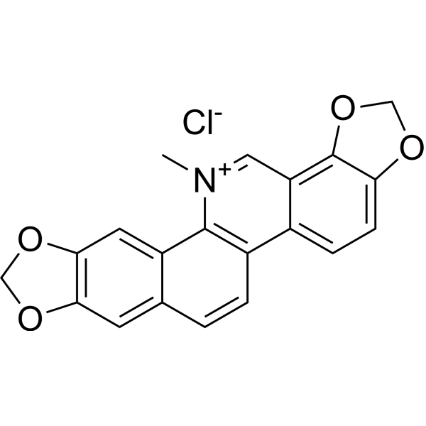
Sanguinarine (Sanguinarin) chloride, a benzophenanthridine alkaloid derived from the root of Sanguinaria Canadensis, can stimulate apoptosis via activating the production of reactive oxygen species (ROS). Sanguinarine-induced apoptosis is associated with the activation of JNK and NF-κB.
For research use only. We do not sell to patients.
- Purity : 98.71%
- CAS No.: 5578-73-4
- Formula: C20H14ClNO4
- Molecular Weight:367.78
-
Storage:
4°C, sealed storage, away from moisture and light
* In solvent : -80°C, 1 year; -20°C, 6 months (sealed storage, away from moisture and light)

To place orders, for customer services and technical support, please contact: MedChemExpress USA
Tel: 609-228-6898 E-mail: [email protected] [email protected]


-
 Biological Activity
-
Chemical Information
-
Solvent & Solubility
- Protocols
- Purity & Documentation
- References
-
 Help & FAQs


-
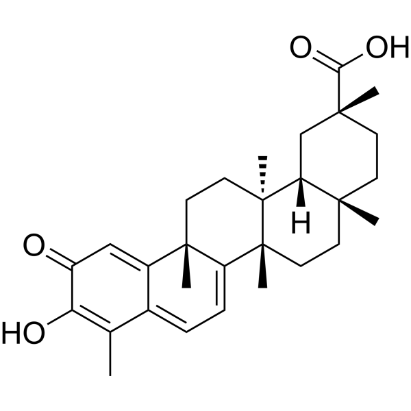
HY-13067
CelastrolAutophagy Inducer -
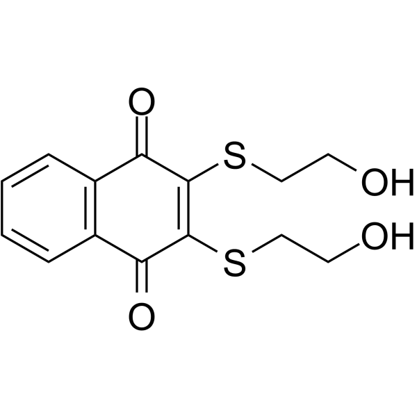
HY-108543
NSC 95397Cdc25 Phosphatase Inhibitor -
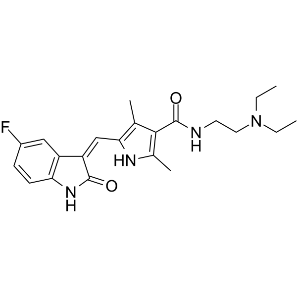
HY-10255A
SunitinibMulti-targeted RTK Inhibitor -
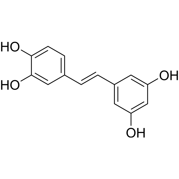
HY-13518
PiceatannolSyk Inhibitor -
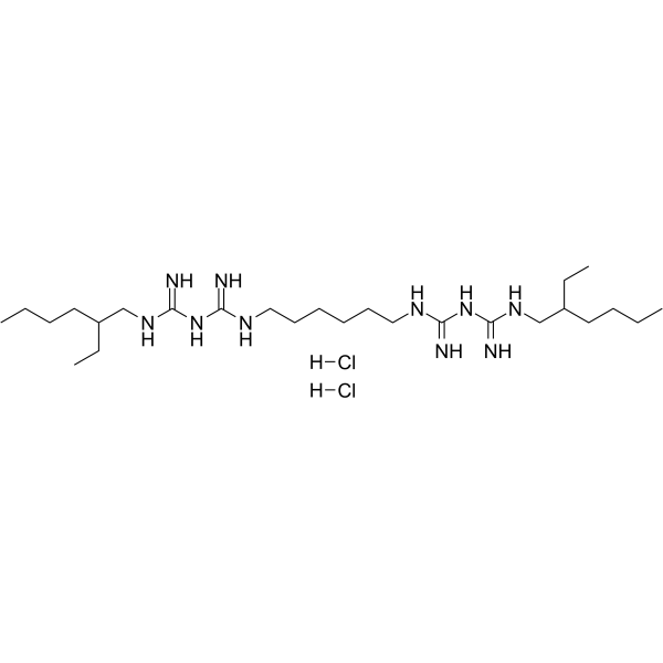
HY-108547
Alexidine dihydrochlorideAntifungal Agent -

HY-13259
MG-132Proteasome Inhibitor -
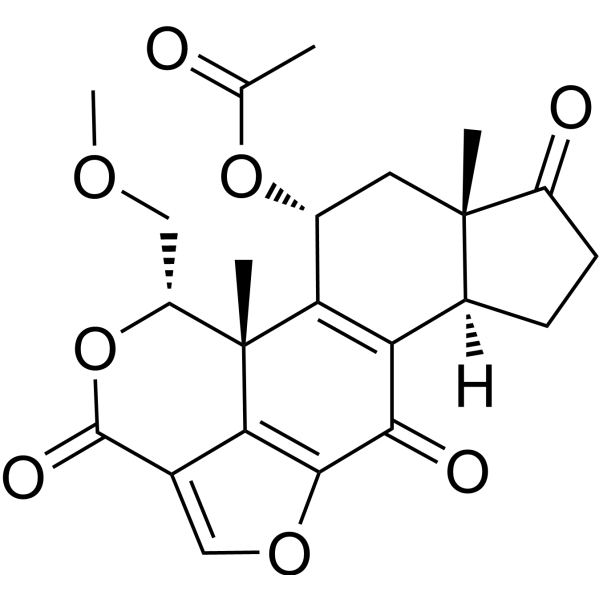
HY-10197
WortmanninPI3K Inhibitor -
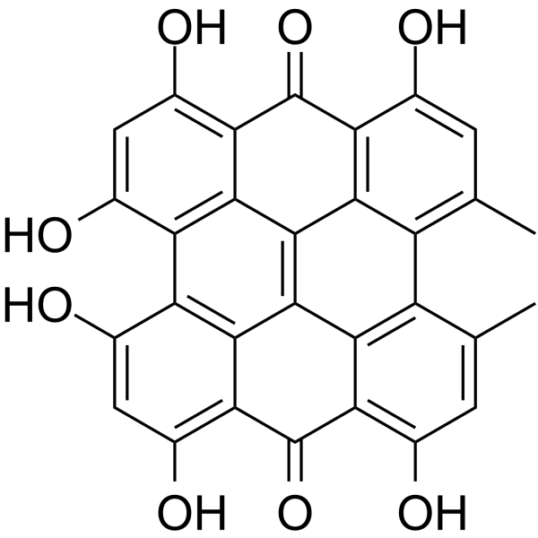
HY-N0453
HypericinPKC Inhibitor -
HY-N0184
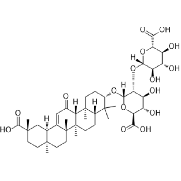
Glycyrrhizic acidHMGB1 Antagonist -
HY-13453
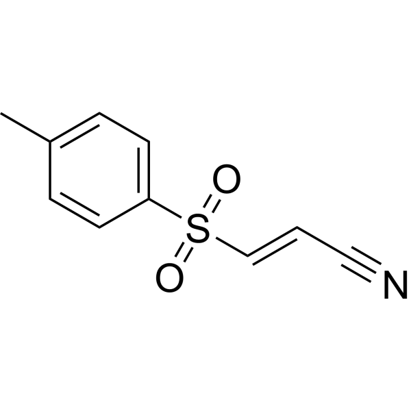
BAY 11-7082IκBα/NF-κB Inhibitor -
HY-13756
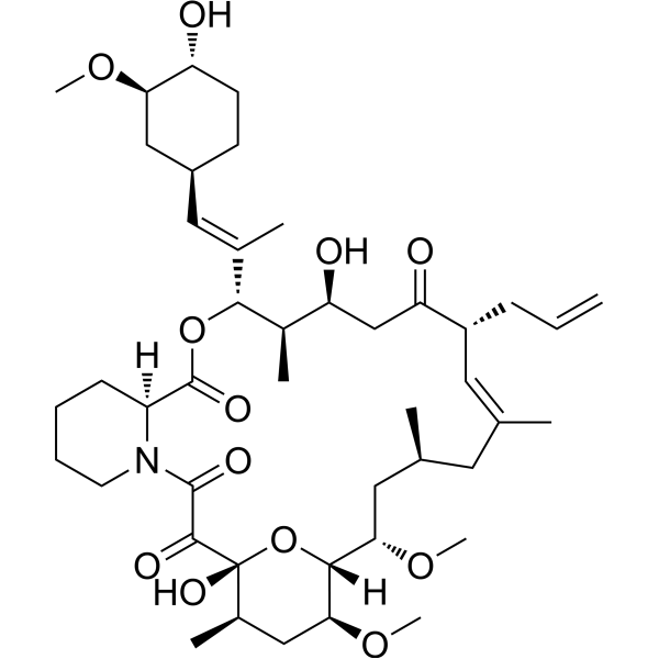
TacrolimusCalcineurin Inhibitor -

HY-100558
Bafilomycin A1V-ATPase Inhibitor
Publications Citing Use of MedChemExpress (MCE) Sanguinarine chloride
More- J Adv Res. 2026 Feb 21:S2090-1232(26)00176-1. [Abstract]
- Adv Sci (Weinh). 2024 Sep;11(34):e2403451. [Abstract]
- Oncogene. 2025 May;44(16):1078-1092. [Abstract]
- Phytother Res. 2023 Oct;37(10):4771-4790. [Abstract]
- Am J Chin Med. 2025;53(5):1545-1571. [Abstract]
- PLoS Pathog. 2026 Mar 3;22(3):e1014032. [Abstract]
- PLoS Pathog. 2023 Dec 7;19(12):e1011796. [Abstract]
- Pestic Biochem Physiol. 2022 Nov:188:105259. [Abstract]
- Comp Biochem Physiol C Toxicol Pharmacol. 2022 Feb:252:109228. [Abstract]
- Antiviral Res. 2026 Jun:250:106417. [Abstract]
- Brain Behav Immun Health. 2022 Oct 29:26:100546. [Abstract]
- Toxicol Lett. 2021 Oct 10:350:71-80. [Abstract]
- Fishes. 2022, 7(5), 229.
- Planta Med. 2024 Jun;90(7-08):523-533. [Abstract]
- bioRxiv. 2024 Feb 5.
- Research Square Print. 2022 Jun.
 Customer Validation & Images

-
Cell Proliferation/Viability Assay
-
Cell Proliferation/Viability Assay
-
Bio/Physico-chemical Assay
-
Bio/Physico-chemical Assay
-
WB
All Parasite Isoforms
More

Biological Activity
Description
IC50 & Target
Apoptosis[1]
Cellular Effect
|
Cell Line

|
Type | Value | Description | References |
|---|---|---|---|---|
| A549 | IC50 |
1.59 μM
Compound: 1; SA
|
Antiproliferative activity against human A549 cells assessed as inhibition of cell growth incubated for 72 hrs by MTT assay
Antiproliferative activity against human A549 cells assessed as inhibition of cell growth incubated for 72 hrs by MTT assay
|
[PMID: 33479636] |
| HeLa | IC50 |
0.16 μg/mL
Compound: 6
|
Growth inhibition of human HeLa S3 cells after 72 hrs
Growth inhibition of human HeLa S3 cells after 72 hrs
|
[PMID: 10395504] |
| HeLa | IC50 |
2.43 μM
Compound: 1; SA
|
Antiproliferative activity against human HeLa cells assessed as reduction in cell viability after 24 hrs by MTT assay
Antiproliferative activity against human HeLa cells assessed as reduction in cell viability after 24 hrs by MTT assay
|
[PMID: 33479636] |
| NCI-H1975 | IC50 |
1.32 μM
Compound: 1; SA
|
Antiproliferative activity against human H1975 cells assessed as inhibition of cell growth incubated for 72 hrs by MTT assay
Antiproliferative activity against human H1975 cells assessed as inhibition of cell growth incubated for 72 hrs by MTT assay
|
[PMID: 33479636] |
In Vitro
Sanguinarine (SANG)-induced apoptosis is associated with the activation of JNK and NF-κB signal pathways.To determine the effects of Sanguinarine on cell viability, 22B-cFluc cells are stimulated with different concentrations of Sanguinarine for 24 h, and then a CKK-8 assay is performed. The treatment with Sanguinarine decreases the proliferation of 22B cells in a dose-dependent manner. Meanwhile, the cytosolic extracts of 22B-cFluc cells treated with different dose of Sanguinarine are measured to detect cellular caspase-3 activity using Ac-DEVD-pNA, which is a validated caspase-3 substrate. The absorbance at 450 nm increases in a dose-dependent manner, indicating increased caspase-3 activity stimulated by Sanguinarine[1].
MedChemExpress (MCE) has not independently confirmed the accuracy of these methods. They are for reference only. Further protocols information, click here.
In Vivo
MedChemExpress (MCE) has not independently confirmed the accuracy of these methods. They are for reference only.


Chemical Information
-
CAS No. 5578-73-4
-
Appearance Solid
-
Molecular Weight 367.78
-
Formula C20H14ClNO4
-
Color Orange to red
-
SMILES
C[N+]1=CC2=C3C(OCO3)=CC=C2C(C=CC4=C5)=C1C4=CC6=C5OCO6.[Cl-]
-
Synonyms
Sanguinarin chloride; Sanguinarium chloride; Pseudochelerythrine chloride
-
Structure Classification
-
Initial Source
-
Shipping
Room temperature in continental US; may vary elsewhere.
-
Storage
4°C, sealed storage, away from moisture and light
* In solvent : -80°C, 1 year; -20°C, 6 months (sealed storage, away from moisture and light)


Publications (16)
-
Journal Impact Factor
-
Most Recent
-
J Adv Res
Identification of a novel EphB4 inhibitor, Sanguinarine, which attenuates β-catenin signaling to inhibit tumor proliferation and migration in lung cancer. [Abstract]2026 Feb 21:S2090-1232(26)00176-1. PMID: 41730417 -
Adv Sci (Weinh)
Short-Term Statin Therapy Induces Hepatic Insulin Resistance Through HNF4α/PAQR9/PPM1α Axis Regulated AKT Phosphorylation. [Abstract]2024 Sep;11(34):e2403451. PMID: 38970167 -
Oncogene
The protection of UCK2 protein stability by GART maintains pyrimidine salvage synthesis for HCC growth under glucose limitation. [Abstract]2025 May;44(16):1078-1092. PMID: 39865175 -
Phytother Res
NPLC0393 from Gynostemma pentaphyllum ameliorates Alzheimer's disease-like pathology in mice by targeting protein phosphatase magnesium-dependent 1A phosphatase. [Abstract]2023 Oct;37(10):4771-4790. PMID: 37434441 -
Am J Chin Med
Sanguinarine Inhibits Gastric Cancer Progression by Targeting the NOS2/SOD1 Axis to Promote Ferroptosis. [Abstract]2025;53(5):1545-1571. PMID: 40663435
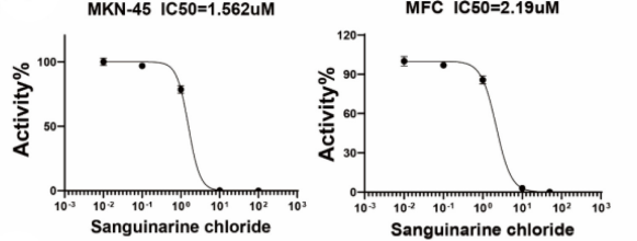
Sanguinarine chloride purchased from MedChemExpress. Usage Cited in: Am J Chin Med. 2025;53(5):1545-1571. [Abstract]
MKN-45 and MFC cells were treated with different concentration of Sanguinarin chloride (0, 0.01, 0.1, 10, 100 μM) for 24h, and cell viability was measured by CCK-8 assays.
Sanguinarine chloride purchased from MedChemExpress. Usage Cited in: Am J Chin Med. 2025;53(5):1545-1571. [Abstract]
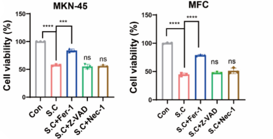
Cell viability was measured in MKN-45 and MFC cells treated with Sanguinarin chloride (2 μM) with Fer-1 (10 μM), Z-VAD (10 μM), or Nec-1 (10 μM) for 24h.
Sanguinarine chloride purchased from MedChemExpress. Usage Cited in: Am J Chin Med. 2025;53(5):1545-1571. [Abstract]
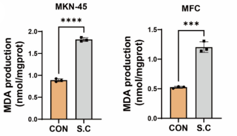
MKN-45 and MFC cells were treated with Sanguinarin chloride (2 μM) for 24 h. MDA level was detected by MDA assay kit.
Sanguinarine chloride purchased from MedChemExpress. Usage Cited in: Am J Chin Med. 2025;53(5):1545-1571. [Abstract]
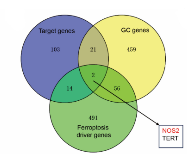
Prediction of Sanguinarin chloride-targeted genes, GC-related proteins, and ferroptosis driver genes, respectively. Two overlapping proteins are displayed.
Sanguinarine chloride purchased from MedChemExpress. Usage Cited in: Am J Chin Med. 2025;53(5):1545-1571. [Abstract]
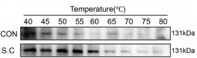
GETSA was performed after 24h of Sanguinarin chloride (2 μM) action, and the protein stability of NOS2 was examined by western at 40-80℃.
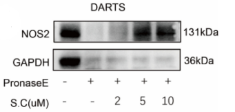
Sanguinarine chloride purchased from MedChemExpress. Usage Cited in: Am J Chin Med. 2025;53(5):1545-1571. [Abstract]
Cell lysates were incubated with Sanguinarin chloride (2 μM, 5 μM, 10 μM), digested with PronaseE, the reaction was terminated by the addition of allozyme inhibitor, and the protein level of NOS2 was detected by western.
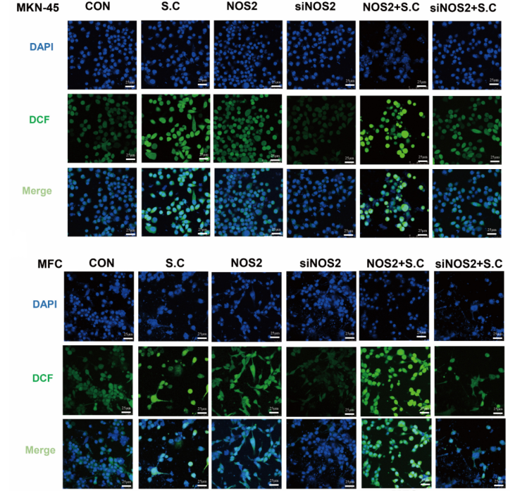
Sanguinarine chloride purchased from MedChemExpress. Usage Cited in: Am J Chin Med. 2025;53(5):1545-1571. [Abstract]
ROS level in MKN-45 and MFC cells transfected with NOS2 or siNOS2 incubated with Sanguinarin chloride was detected by DCFH-DA.
-
PLoS Pathog
The CD97-PPM1G axis dampens antiviral immunity by dephosphorylating IRF7 in type I interferon pathway. [Abstract]2026 Mar 3;22(3):e1014032. PMID: 41774756 -
PLoS Pathog
G-quadruplex in the TMV Genome Regulates Viral Proliferation and Acts as Antiviral Target of Photodynamic Therapy. [Abstract]2023 Dec 7;19(12):e1011796. PMID: 38060599 -
Pestic Biochem Physiol
Neurotoxicity of sanguinarine via inhibiting mitophagy and activating apoptosis in zebrafish and PC12 cells. [Abstract]2022 Nov:188:105259. PMID: 36464364 -
Comp Biochem Physiol C Toxicol Pharmacol
Cardiotoxicity of sanguinarine via regulating apoptosis and MAPK pathways in zebrafish and HL1 cardiomyocytes. [Abstract]2022 Feb:252:109228. PMID: 34744004 -
Antiviral Res
Repurposing screen using a robust human rhinovirus infectious clone identifies pyrvinium pamoate with antiviral activity. [Abstract]2026 Jun:250:106417. PMID: 42025967 -
Brain Behav Immun Health
Miltefosine as a PPM1A activator improves AD-like pathology in mice by alleviating tauopathy via microglia/neurons crosstalk. [Abstract]2022 Oct 29:26:100546. PMID: 36388134 -
Toxicol Lett
Developmental toxicity caused by sanguinarine in zebrafish embryos via regulating oxidative stress, apoptosis and wnt pathways. [Abstract]2021 Oct 10:350:71-80. PMID: 34252508 -
-
Planta Med
Novel Approaches for the Analysis and Isolation of Benzylisoquinoline Alkaloids in Chelidonium majus. [Abstract]2024 Jun;90(7-08):523-533. PMID: 38843792 -
-


Solvent & Solubility
In Vitro:
DMSO : 3.33 mg/mL (9.05 mM; ultrasonic and warming and heat to 60°C; Hygroscopic DMSO has a significant impact on the solubility of product, please use newly opened DMSO)
H2O : < 0.1 mg/mL (insoluble)
Please refer to the solubility information to select the appropriate solvent. Once prepared, please aliquot and store the solution to prevent product inactivation from repeated freeze-thaw cycles.
Storage method and period of stock solution: -80°C, 1 year; -20°C, 6 months (sealed storage, away from moisture and light). When stored at -80°C, please use it within 1 year. When stored at -20°C, please use it within 6 months.
Please refer to the solubility information to select the appropriate solvent. Once prepared, please aliquot and store the solution to prevent product inactivation from repeated freeze-thaw cycles.
Storage method and period of stock solution: -80°C, 1 year; -20°C, 6 months (sealed storage, away from moisture and light). When stored at -80°C, please use it within 1 year. When stored at -20°C, please use it within 6 months.
Concentration (start) × Volume (start) = Concentration (final) × Volume (final)
In Vivo:
Select the appropriate dissolution method based on your experimental animal and administration route.
- For the following dissolution methods, please ensure to first prepare a clear stock solution using an In Vitro approach and then sequentially add co-solvents:
- To ensure reliable experimental results, the clarified stock solution can be appropriately stored based on storage conditions. As for the working solution for In Vivo experiments, it is recommended to prepare freshly and use it on the same day.
- The percentages shown for the solvents indicate their volumetric ratio in the final prepared solution. If precipitation or phase separation occurs during preparation, heat and/or sonication can be used to aid dissolution.
Add each solvent one by one: 10% DMSO 40% PEG300 5% Tween-80 45% Saline
Solubility: ≥ 0.33 mg/mL (0.90 mM); Clear solution
This protocol yields a clear solution of ≥ 0.33 mg/mL (saturation unknown).
Taking 1 mL working solution as an example, add 100 μL DMSO stock solution (3.3 mg/mL) to 400 μL PEG300, and mix evenly; then add 50 μL Tween-80 and mix evenly; then add 450 μL Saline to adjust the volume to 1 mL.
Preparation of Saline: Dissolve 0.9 g sodium chloride in ddH₂O and dilute to 100 mL to obtain a clear Saline solution.
Add each solvent one by one: 10% DMSO 90% (20% SBE-β-CD in Saline)
Solubility: ≥ 0.33 mg/mL (0.90 mM); Clear solution
This protocol yields a clear solution of ≥ 0.33 mg/mL (saturation unknown).
Taking 1 mL working solution as an example, add 100 μL DMSO stock solution (3.3 mg/mL) to 900 μL 20% SBE-β-CD in Saline, and mix evenly.
Preparation of 20% SBE-β-CD in Saline (4°C, storage for one week): 2 g SBE-β-CD powder is dissolved in 10 mL Saline, completely dissolve until clear.
For the following dissolution methods, please prepare the working solution directly:
It is recommended to prepare fresh solutions and use them promptly within a short period of time.
The percentages shown for the solvents indicate their volumetric ratio in the final prepared solution. If precipitation or phase separation occurs during preparation, heat and/or sonication can be used to aid dissolution.
Add each solvent one by one: 50% PEG300 50% Saline
Solubility: 25 mg/mL (67.98 mM); Suspended solution; Need ultrasonic
In Vivo Dissolution Calculator
Please enter the basic information of animal experiments:
-
-
-
-
Recommended: Prepare an additional quantity of animals to account for potential losses during experiments.
Please enter your animal formula composition:
-
%DMSO +
Recommended: Keep the proportion of DMSO in working solution below 2% if your animal is weak.
-
%+
-
+%Tween-80 + +
-
%Saline +
The co-solvents required include: DMSO, . All of co-solvents are available by MedChemExpress (MCE). , Tween 80. All of co-solvents are available by MedChemExpress (MCE).
Working solution concentration: 0.22 mg/mL
Method for preparing stock solution: mg drug dissolved in μL DMSO. Stock solution concentration: mg/mL. * In solvent : -80°C, 1 year; -20°C, 6 months (sealed storage, away from moisture and light)
1. Take μL DMSO stock solution;
2. Add μL .
μL , mix evenly;
3. Then add μL Tween 80, mix evenly;
4. Then add μL
Please ensure that the stock solution in the first step is dissolved to a clear state, and add co-solvents in sequence. You can use ultrasonic heating (ultrasonic cleaner, recommended frequency 20-40 kHz), vortexing, etc. to assist dissolution.

Protocols
-
Apoptosis
Apoptosis, also called programmed cell death, is generally characterized by distinct morphological characteristics.
-
TUNEL staining for apoptotic DNA fragmentation
TUNEL staining detects DNA strand breaks by using terminal deoxynucleotidyl transferase to add labeled nucleotides to exposed 3′-OH DNA termini, generating either microscopic staining in fixed cells or tissue sections, or fluorescence/cytometric signal in cell suspensions. TUNEL positivity reflects DNA fragmentation but should not be interpreted alone as definitive apoptosis, because TUNEL can also label necrotic, autolytic, mechanically damaged, or DNA-repair-associated DNA breaks.
-
Annexin V plus membrane-impermeant dye apoptosis staining
Annexin V-based apoptosis assays rely on the detection of phosphatidylserine (PS) externalization from the inner leaflet of the plasma membrane to the outer leaflet, an early biochemical hallmark of apoptosis. Fluorescently labeled Annexin V binds PS in a calcium-dependent manner, enabling identification of early apoptotic cells by flow cytometry or fluorescence microscopy. When combined with a membrane-impermeant DNA-binding dye (e. g. , propidium iodide), this approach allows discrimination between viable (Annexin V−/dye−), early apoptotic (Annexin V+/dye−), and late apoptotic or necrotic (Annexin V+/dye+) cell populations by assessing membrane integrity and PS exposure.
-
ROS/oxidative-stress fluorescent staining
ROS/oxidative-stress fluorescent staining uses cell-permeant fluorogenic probes that become fluorescent after oxidation inside cells or tissues; commonly used examples include DCFH-DA/DCFDA for broad cellular oxidant detection, DHE for superoxide-related signal detection, MitoSOX for mitochondrial superoxide-related signal detection, and CellROX probes for oxidative-stress-associated fluorescence readouts. The assay detects probe oxidation rather than a single ROS species unless the probe and analysis method have been chemically validated for that species. DCFH-DA enters cells, is deacetylated by intracellular esterases to DCFH, and produces fluorescent DCF after oxidation, so the readout is used as an operational measure of total cellular oxidative stress rather than a species-specific ROS measurement. DHE and MitoSOX can report superoxide-related oxidation, but red fluorescence alone can include non-specific ethidium-like oxidation products; HPLC or optimized spectral approaches are
-
Apoptosis Solutions
Apoptosis is a regulated, generally non-lytic cell-death pathway that removes unwanted, damaged, infected, or abnormal cells through coordinated morphological changes, caspase activation, DNA fragmentation, and membrane remodeling. The intrinsic apoptosis pathway is controlled mainly by mitochondrial outer membrane permeabilization, BCL-2 family proteins, cytochrome c release, apoptosome formation, caspase-9 activation, and downstream executioner caspase-3/7 activation. The extrinsic apoptosis pathway is initiated by death receptors such as Fas, TNFR, and TRAIL receptors, which recruit adaptor proteins and activate caspase-8 before engaging executioner caspases or mitochondrial amplification through BID cleavage. Apoptosis is linked to many phenotypes, including cancer cell killing, tissue homeostasis, immune regulation, neurodegeneration, infection response, and treatment-induced cytotoxicity; unresolved questions include how apoptosis interacts with necroptosis, pyroptosis, ferroptos
-
Research Protocol for Inflammation-related Diseases
The NLRP3 inflammasome is a cytosolic innate immune signaling platform that integrates priming signals and danger-signal activation to promote caspase-1 activation, maturation of IL-1β and IL-18, and gasdermin D-mediated pyroptotic cell death. The core experimental logic is to determine whether inflammatory disease phenotypes are driven by increased NLRP3 expression, ASC-containing inflammasome assembly, caspase-1 cleavage, GSDMD cleavage, and extracellular release of IL-1β/IL-18 rather than by nonspecific cell injury alone. The pathway is strongly linked to inflammation-related disease phenotypes because monosodium urate crystals activate NALP3/NLRP3 inflammasome signaling in gout-like crystal inflammation, cholesterol crystals activate NLRP3 inflammasomes in atherogenesis models, and DSS-induced intestinal inflammation has been reported to involve NLRP3 inflammasome activity. However, experimental colitis studies also show context-dependent protective effects of NLRP3 inflammasome co


Purity & Documentation
-
Data Sheet (282 KB)
-
SDS (397 KB)
- English - EN (397 KB)
- Français - FR (397 KB)
- Deutsch - DE (397 KB)
- Norwegian - NO (397 KB)
- Español - ES (397 KB)
- Swedish - SV (397 KB)
- Italian - IT (397 KB)
- Korean - KR (397 KB)
- Portuguese - PT (397 KB)
-
Handling Instructions (2659 KB)
References


Complete Stock Solution Preparation Table
Please refer to the solubility information to select the appropriate solvent. Once prepared, please aliquot and store the solution to prevent product inactivation from repeated freeze-thaw cycles.
Storage method and period of stock solution: -80°C, 1 year; -20°C, 6 months (sealed storage, away from moisture and light). When stored at -80°C, please use it within 1 year. When stored at -20°C, please use it within 6 months.
| Optional Solvent | Concentration Solvent Mass | 1 mg | 5 mg | 10 mg | 25 mg |
|---|---|---|---|---|---|
| DMSO | 1 mM | 2.7190 mL | 13.5951 mL | 27.1902 mL | 67.9754 mL |
| 5 mM | 0.5438 mL | 2.7190 mL | 5.4380 mL | 13.5951 mL |


Sanguinarine chloride Related Classifications
HY-N0052A Related Classifications
 Powered by Bioz