



ABH hydrochloride
Based on 1 publication(s) in Google Scholar
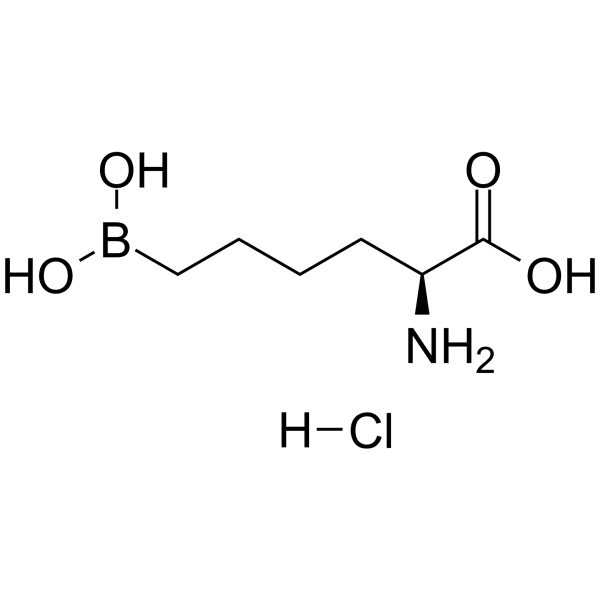
ABH (hydrochloride) is an orally active arginase inhibitor (Ki = 8.5 nM). ABH hydrochloride promotes NO production and reduces the expression of inflammatory response-related molecules (ICAM-1, VCAM-1, MCP-1). ABH hydrochloride improves erectile function, reduces lung damage, promotes wound healing, reduces arterial blood pressure, and improves vascular fibrosis.
For research use only. We do not sell to patients.
- Purity : 99.90%
- CAS No.: 194656-75-2
- Formula: C6H15BClNO4
- Molecular Weight:211.45
-
Storage:
4°C, sealed storage, away from moisture
* In solvent : -80°C, 6 months; -20°C, 1 month (sealed storage, away from moisture)

To place orders, for customer services and technical support, please contact: MedChemExpress USA
Tel: 609-228-6898 E-mail: [email protected] [email protected]


-
 Biological Activity
-
Chemical Information
-
Solvent & Solubility
- Protocols
- Purity & Documentation
- References
-
 Help & FAQs


Publications Citing Use of MedChemExpress (MCE) ABH hydrochloride
More

Biological Activity
Description
In Vitro
ABH (100 μM; pretreatment for 1 h, hypoxia for 12-24 h) hydrochloride partially prevents the hypoxia-induced reduction in in-vitro tube-like structure formation in bovine aortic endothelial cells and increased vascular endothelial growth factor (VEGF) expression[2].
ABH (0-10 μM; 3 h) hydrochloride effectively inhibits arginase from multiple Bacillus anthracis spore strains[3].
ABH (0.01-1 mM; pretreatment for 1 h) hydrochloride significantly reduces arginase activity in mouse dermal fibroblasts, with 0.1 mM ABH reducing arginase activity by 35% and 1 mM ABH reducing arginase activity by 54%[5].
ABH (100 μM; pretreatment for 1 h) hydrochloride can inhibit the increase in arginase 1 (A1) expression and activity in mouse aortic endothelial cells induced by high glucose/palmitic acid, maintain NO production, reduce the expression of inflammatory response-related molecules (ICAM-1, VCAM-1, MCP-1), and inhibit monocyte adhesion[7].
ABH (100 µM; 2 h) hydrochloride inhibits Ang II-induced proliferation of rat aortic smooth muscle cells, ODC activity and expression, and the increase in type I collagen and hydroxyproline levels[10].
MedChemExpress (MCE) has not independently confirmed the accuracy of these methods. They are for reference only. Further protocols information, click here.
In Vivo
ABH (0.1 mM, 0.1 mL; topical application; once every 8 hours; 14 days) hydrochloride can significantly promote wound healing in mice[5].
ABH (5 mg/kg; s.c.; once 1 hour before surgery; single administration) hydrochloride can increase plasma nitrite levels in rats with pneumoperitoneum, maintain nitric oxide synthase (NOS) activity, reduce oxidative stress and inflammatory response, and reduce the severity of lung injury[6].
ABH (10 mg/kg; p.o., drinking water; once daily) hydrochloride can inhibit the increase of arginase activity and A1 expression in vascular endothelial cells of obese mice induced by a high-fat and high-sugar diet, reduce inflammation and pathological remodeling of visceral adipose tissue (VAT), including reducing inflammatory monocyte infiltration, macrophage polarization to M1 type, improving adipocyte size, fibrosis and capillary density[7].
ABH (100 mg; i.p.; once at 24, 48, 60, 70 h after infection or continuously administered until day 3 after infection) hydrochloride can increase NO production in the lungs of mice infected with Pseudomonas aeruginosa, improve L-arginine availability, reduce L-ornithine concentration, and do not increase the levels of lung inflammatory markers[8].
ABH (400 µg/kg/day; subcutaneous osmotic pump injection; once/day; 21 days) hydrochloride can reduce the mean arterial blood pressure and improve the vascular reactivity of the carotid and femoral arteries in a rat model of hypertension induced by chronic intermittent hypoxia (CIH)[9].
ABH (10 mg/kg/day; drinking water administration; continuous administration; 6 months) hydrochloride can improve endothelial function, reduce aortic stiffness and fibrosis, and reduce plasma and aortic arginase activities in a mouse model of obesity-related type 2 diabetes induced by a high-fat and high-sugar diet[10].
ABH (400 µg/day; subcutaneous osmotic pump administration; continuous administration) hydrochloride can preserve the total volume of the fetal lung, prevent excessive proliferation of pulmonary vascular smooth muscle cells, and improve the number of pulmonary vessels and lung morphology in a rat model of congenital diaphragmatic hernia (CDH) induced by Nitrofen (HY-B1877)[11].
MedChemExpress (MCE) has not independently confirmed the accuracy of these methods. They are for reference only.
-
Animal Model:9-11 weeks old, male C57BL/6 mice, full-thickness dorsal excisional wound model[5]
-
Dosage:0.1 mM in saline
-
Administration:Topical application, every 8 hours for 14 days
-
Result:Accelerated wound closure 7-14 days post-surgery.
Increased wound granulation tissue and decreased connective tissue on postoperative day 3.
Enhanced re-epithelialization and changed the localization of myofibroblasts on postoperative day 7. Increased NOx levels in the wound on day 3.


Chemical Information
-
CAS No. 194656-75-2
-
Appearance Solid
-
Molecular Weight 211.45
-
Formula C6H15BClNO4
-
Color White to off-white
-
SMILES
O=C([C@H](CCCCB(O)O)N)O.[H]Cl
-
Shipping
Room temperature in continental US; may vary elsewhere.
-
Storage
4°C, sealed storage, away from moisture
* In solvent : -80°C, 6 months; -20°C, 1 month (sealed storage, away from moisture)


Publications (1)
-
Journal Impact Factor
-
Most Recent
-
Adv Sci (Weinh)
Time-Controlled Refrigerated Stem Cell Therapy Mitigates Scleroderma Fibrosis via Modulation of Mitochondrial Autophagy and Gut Metabolism. [Abstract]2026 Mar 31:e15505. PMID: 41915839


Solvent & Solubility
In Vitro:
H2O : 100 mg/mL (472.93 mM; Need ultrasonic)
DMSO : 100 mg/mL (472.93 mM; Need ultrasonic; Hygroscopic DMSO has a significant impact on the solubility of product, please use newly opened DMSO)
Please refer to the solubility information to select the appropriate solvent. Once prepared, please aliquot and store the solution to prevent product inactivation from repeated freeze-thaw cycles.
Storage method and period of stock solution: -80°C, 6 months; -20°C, 1 month (sealed storage, away from moisture). When stored at -80°C, please use it within 6 months. When stored at -20°C, please use it within 1 month.
* Note: If you choose water as the stock solution, please dilute it to the working solution, then filter and sterilize it with a 0.22 μm filter before use.
Please refer to the solubility information to select the appropriate solvent. Once prepared, please aliquot and store the solution to prevent product inactivation from repeated freeze-thaw cycles.
Storage method and period of stock solution: -80°C, 6 months; -20°C, 1 month (sealed storage, away from moisture). When stored at -80°C, please use it within 6 months. When stored at -20°C, please use it within 1 month.
* Note: If you choose water as the stock solution, please dilute it to the working solution, then filter and sterilize it with a 0.22 μm filter before use.
Concentration (start) × Volume (start) = Concentration (final) × Volume (final)
In Vivo:
Select the appropriate dissolution method based on your experimental animal and administration route.
- For the following dissolution methods, please ensure to first prepare a clear stock solution using an In Vitro approach and then sequentially add co-solvents:
- To ensure reliable experimental results, the clarified stock solution can be appropriately stored based on storage conditions. As for the working solution for In Vivo experiments, it is recommended to prepare freshly and use it on the same day.
- The percentages shown for the solvents indicate their volumetric ratio in the final prepared solution. If precipitation or phase separation occurs during preparation, heat and/or sonication can be used to aid dissolution.
Add each solvent one by one: 10% DMSO 40% PEG300 5% Tween-80 45% Saline
Solubility: 2.5 mg/mL (11.82 mM); Clear solution; Need ultrasonic
This protocol yields a clear solution of 2.5 mg/mL.
Taking 1 mL working solution as an example, add 100 μL DMSO stock solution (25.0 mg/mL) to 400 μL PEG300, and mix evenly; then add 50 μL Tween-80 and mix evenly; then add 450 μL Saline to adjust the volume to 1 mL.
Preparation of Saline: Dissolve 0.9 g sodium chloride in ddH₂O and dilute to 100 mL to obtain a clear Saline solution.
Add each solvent one by one: 10% DMSO 90% (20% SBE-β-CD in Saline)
Solubility: 2.5 mg/mL (11.82 mM); Clear solution; Need ultrasonic
This protocol yields a clear solution of 2.5 mg/mL.
Taking 1 mL working solution as an example, add 100 μL DMSO stock solution (25.0 mg/mL) to 900 μL 20% SBE-β-CD in Saline, and mix evenly.
Preparation of 20% SBE-β-CD in Saline (4°C, storage for one week): 2 g SBE-β-CD powder is dissolved in 10 mL Saline, completely dissolve until clear.
In Vivo Dissolution Calculator
Please enter the basic information of animal experiments:
-
-
-
-
Recommended: Prepare an additional quantity of animals to account for potential losses during experiments.
Working solution concentration: 0.22 mg/mL
This product has good water solubility, please refer to the measured solubility data in water/PBS/Saline for details.

Protocols
-
Fibrosis/Collagen Morphometry
Fibrosis and collagen morphometry is based on the quantitative visualization of fibrillar collagen deposition in tissue sections using histochemical stains such as Sirius Red (Picrosirius Red) or Masson's trichrome, followed by image-based or polarization-enhanced analysis to estimate collagen proportional area as a surrogate of extracellular matrix accumulation during fibrotic remodeling. Sirius Red combined with polarized light microscopy enhances detection of collagen fibers due to birefringence properties, enabling more specific visualization of collagen type I and III fibrils compared to conventional bright-field histology, while whole-section or region-restricted digital morphometry reduces field-selection bias in fibrosis assessment. Alternative quantitative approaches include second harmonic generation (SHG) and two-photon excited fluorescence microscopy, which enable label-free detection of fibrillar collagen and have been validated against histological staining and biochemica
-
RNA extraction experimental
By lysing cells, releasing RNA, and removing impurities such as proteins and DNA, high-purity RNA products are finally obtained. The commonly used traditional method is the guanidine isothiocyanate/phenol/chloroform method (Trizol), which is suitable for a variety of animal materials including animal tissues, microorganisms, cultured cells, etc., and most plant materials.
-
Collagen: Sirius Red Staining
Sirius Red or picrosirius red staining is a histochemical method for visualizing collagen-rich extracellular matrix in tissue sections, and collagen fibers are detected as red-stained structures under bright-field microscopy with enhanced birefringence under polarized light. Picrosirius red is useful for assessing total collagen organization, distribution, and fibrosis burden, but polarized color should not be interpreted as a definitive collagen type I versus type III readout because color is affected by fiber orientation, thickness, and packing.
-
Cell migration
Cell migration is a method that plays an important role in wound healing, cell differentiation, embryonic development, etc.
-
Connective Tissue: Masson's Trichrome/Collagen Trichrome Staining
Masson’s Trichrome (collagen/trichrome staining) is a histological technique that differentially stains tissue compartments using sequential acidic dyes to distinguish collagen from muscle and cytoplasmic components based on dye affinity and tissue permeability differences, enabling visualization of fibrosis and connective tissue architecture in histological sections. The classical formulation typically uses Weigert's iron hematoxylin for nuclear staining, Biebrich scarlet-acid fuchsin for cytoplasm and muscle, and aniline blue (or light green variants) for collagen, producing a characteristic blue/green collagen signal contrasted against red cytoplasm and dark nuclei. The staining principle relies on selective displacement of smaller dye molecules by larger anionic dyes in collagen-rich regions under controlled acidified conditions, which enhances collagen-specific dye retention. This property makes the method widely used for fibrosis assessment in organs such as heart, liver, lung, a
-
Research Protocol for Inflammation-related Diseases
The NLRP3 inflammasome is a cytosolic innate immune signaling platform that integrates priming signals and danger-signal activation to promote caspase-1 activation, maturation of IL-1β and IL-18, and gasdermin D-mediated pyroptotic cell death. The core experimental logic is to determine whether inflammatory disease phenotypes are driven by increased NLRP3 expression, ASC-containing inflammasome assembly, caspase-1 cleavage, GSDMD cleavage, and extracellular release of IL-1β/IL-18 rather than by nonspecific cell injury alone. The pathway is strongly linked to inflammation-related disease phenotypes because monosodium urate crystals activate NALP3/NLRP3 inflammasome signaling in gout-like crystal inflammation, cholesterol crystals activate NLRP3 inflammasomes in atherogenesis models, and DSS-induced intestinal inflammation has been reported to involve NLRP3 inflammasome activity. However, experimental colitis studies also show context-dependent protective effects of NLRP3 inflammasome co
-
Cell-Exclusion Zone Migration Assay
The Cell-Exclusion Zone (CEZ) migration assay is an in vitro 2D cell migration method in which a defined cell-free area is created using removable physical barriers such as silicone stoppers, allowing cells to be seeded around the barrier and subsequently migrate into the cleared zone after barrier removal. This approach enables quantification of collective cell migration by monitoring repopulation of the initially cell-free region over time using microscopy-based imaging. Compared with scratch-based wound healing assays, barrier-based exclusion methods are designed to avoid mechanical damage to the extracellular matrix and reduce injury-induced effects on boundary cells, thereby improving interpretability of migration behavior in vitro. The assay readout is typically the progressive reduction in the cell-free area or the number of cells invading the exclusion zone, reflecting coordinated cell motility relevant to physiological processes such as wound healing, epithelial repair, and ca


Purity & Documentation
-
Data Sheet (279 KB)
-
SDS (394 KB)
- English - EN (394 KB)
- Français - FR (394 KB)
- Deutsch - DE (394 KB)
- Norwegian - NO (394 KB)
- Español - ES (394 KB)
- Swedish - SV (394 KB)
- Italian - IT (394 KB)
- Korean - KR (394 KB)
- Portuguese - PT (394 KB)
-
Handling Instructions (2659 KB)
References
[2]. Wang L, et al. Arginase inhibition enhances angiogenesis in endothelial cells exposed to hypoxia. Microvasc Res. 2015 Mar;98:1-8. [Content Brief]
[3]. Tsai P, et al. Effect of the mammalian arginase inhibitor 2(S)-amino-6-boronohexanoic acid on Bacillus anthracis arginase. Curr Microbiol. 2012 Apr;64(4):379-84. [Content Brief]
[4]. Segal R, et al. Chronic oral administration of the arginase inhibitor 2(S)-amino-6-boronohexanoic acid (ABH) improves erectile function in aged rats. J Androl. 2012 Nov-Dec;33(6):1169-75. [Content Brief]
[5]. Kavalukas SL, et al. Arginase inhibition promotes wound healing in mice. Surgery. 2012 Feb;151(2):287-95. [Content Brief]
[6]. Cho JS, et al. The effects of arginase inhibitor on lung oxidative stress and inflammation caused by pneumoperitoneum in rats. BMC Anesthesiol. 2015 Sep 28;15:129. [Content Brief]
[7]. Yao L, et al. Obesity-induced vascular inflammation involves elevated arginase activity. Am J Physiol Regul Integr Comp Physiol. 2017 Nov 1;313(5):R560-R571. [Content Brief]
[8]. Mehl A, et al. Effect of arginase inhibition on pulmonary L-arginine metabolism in murine Pseudomonas pneumonia. PLoS One. 2014 Mar 3;9(3):e90232. [Content Brief]
[9]. Krause BJ, et al. Chronic Intermittent Hypoxia-Induced Vascular Dysfunction in Rats is Reverted by N-Acetylcysteine Supplementation and Arginase Inhibition. Front Physiol. 2018 Jul 24;9:901. [Content Brief]
[11]. Toso A, et al. Congenital diaphragmatic hernia: phosphodiesterase-5 and Arginase inhibitors prevent pulmonary vascular hypoplasia in rat lungs. Pediatr Res. 2024 Mar;95(4):941-948. [Content Brief]


Complete Stock Solution Preparation Table
Please refer to the solubility information to select the appropriate solvent. Once prepared, please aliquot and store the solution to prevent product inactivation from repeated freeze-thaw cycles.
Storage method and period of stock solution: -80°C, 6 months; -20°C, 1 month (sealed storage, away from moisture). When stored at -80°C, please use it within 6 months. When stored at -20°C, please use it within 1 month.
| Optional Solvent | Concentration Solvent Mass | 1 mg | 5 mg | 10 mg | 25 mg |
|---|---|---|---|---|---|
| H2O / DMSO | 1 mM | 4.7293 mL | 23.6463 mL | 47.2925 mL | 118.2313 mL |
| 5 mM | 0.9459 mL | 4.7293 mL | 9.4585 mL | 23.6463 mL | |
| 10 mM | 0.4729 mL | 2.3646 mL | 4.7293 mL | 11.8231 mL | |
| 15 mM | 0.3153 mL | 1.5764 mL | 3.1528 mL | 7.8821 mL | |
| 20 mM | 0.2365 mL | 1.1823 mL | 2.3646 mL | 5.9116 mL | |
| 25 mM | 0.1892 mL | 0.9459 mL | 1.8917 mL | 4.7293 mL | |
| 30 mM | 0.1576 mL | 0.7882 mL | 1.5764 mL | 3.9410 mL | |
| 40 mM | 0.1182 mL | 0.5912 mL | 1.1823 mL | 2.9558 mL | |
| 50 mM | 0.0946 mL | 0.4729 mL | 0.9459 mL | 2.3646 mL | |
| 60 mM | 0.0788 mL | 0.3941 mL | 0.7882 mL | 1.9705 mL | |
| 80 mM | 0.0591 mL | 0.2956 mL | 0.5912 mL | 1.4779 mL | |
| 100 mM | 0.0473 mL | 0.2365 mL | 0.4729 mL | 1.1823 mL |
* Note: If you choose water as the stock solution, please dilute it to the working solution, then filter and sterilize it with a 0.22 μm filter before use.


ABH hydrochloride Related Classifications
HY-112868B Related Classifications
 Powered by Bioz