



(E)-Ferulic acid
Based on 1 publication(s) in Google Scholar
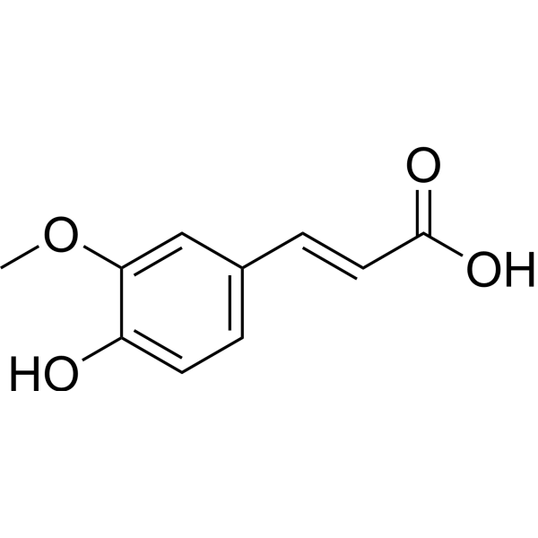
(E)-Ferulic acid is an isomer of ferulic acid, an aromatic compound abundant in plant cell walls. (E)-Ferulic acid causes phosphorylation of β-catenin (β-catenin), leading to proteasome degradation, increasing the expression of pro-apoptotic factor Bax and reducing pro-apoptotic factor Expression of the survival factor survivin. (E)-Ferulic acid can effectively remove reactive oxygen species (ROS) and inhibit lipid peroxidation. (E)-Ferulic acid exerts antiproliferative and antimigratory effects in the human lung cancer cell line H1299.
Nos produits utilisent uniquement pour la recherche. Nous ne vendons pas aux patients.
- Pureté : 99.82%
- CAS No.: 537-98-4
- Formule: C10H10O4
- Masse moléculaire:194.19
-
Stockage:
Store at room temperature 3 years.
In solvent -80°C, 2 years , -20°C, 1 year

To place orders, for customer services and technical support, please contact: MedChemExpress USA
Tel: 609-228-6898 E-mail: [email protected] [email protected]


-
 Activité biologique
-
Chemical Information
-
Solvant et solubilité
- Protocole
- Pureté et documentation
- Références
-
 Help & FAQs


-
HY-N0060
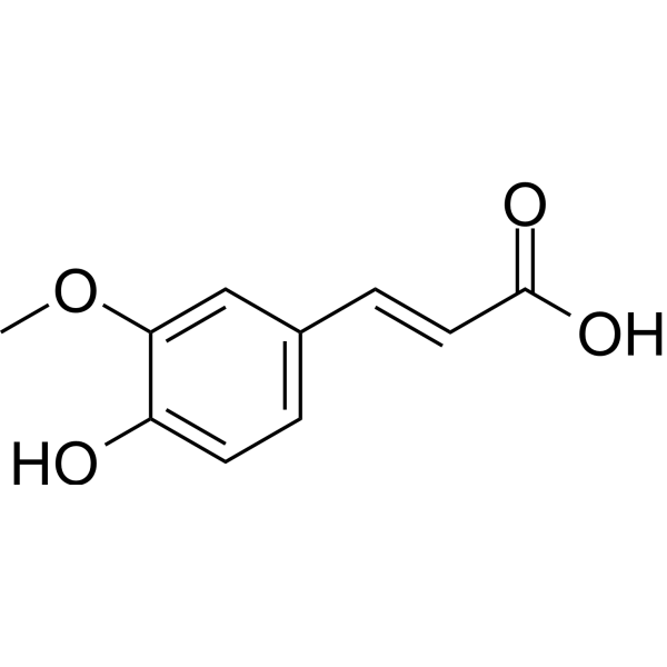
Ferulic acidFGFR1 Inhibitor -
HY-14590
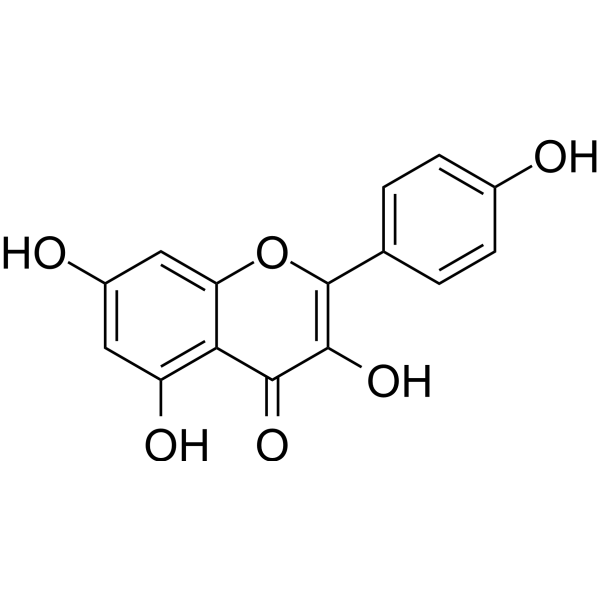
KaempferolEstrogen Receptor α Inhibitor -

HY-13259
MG-132Proteasome Inhibitor -
HY-N0172
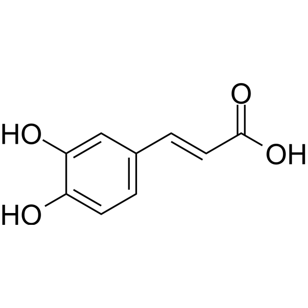
Caffeic acidEndogenous Metabolite -
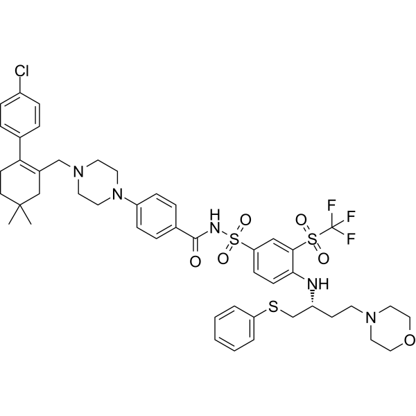
HY-10087
NavitoclaxBcl-2 Inhibitor -

HY-N0060BR
(E)-Ferulic acid (Standard)Bcl-2 Family Activator -
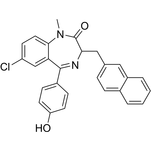
HY-13108
Bz 423Bcl-2 Family Activator -
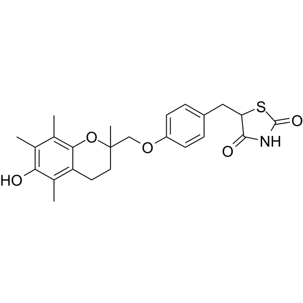
HY-50935
TroglitazonePPARγ Agonist -
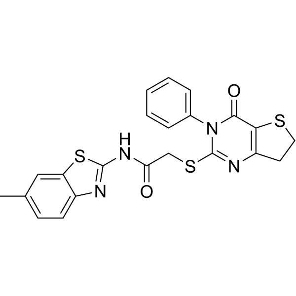
HY-13912
IWP-2Wnt Production Inhibitor -
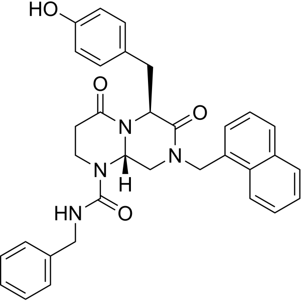
HY-14428
ICG-001Apoptosis Inducer -
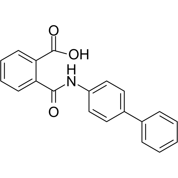
HY-16268
KartogeninChondroprotective Agent -
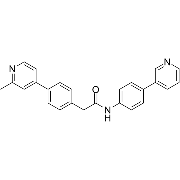
HY-15659
Wnt-C59PORCN Inhibitor
Publications Citing Use of MedChemExpress (MCE) (E)-Ferulic acid
MoreVoir tous les produits spécifiques à Isoform Endogenous Metabolite
More

Activité biologique
Description
IC50 & Target
[1]|
Bax |
Microbial Metabolite |
Cellular Effect
|
Cell Line

|
Type | Value | Description | References |
|---|---|---|---|---|
| A549 | IC50 |
>100 μM
Compound: 13
|
Cytotoxicity against human A549 cells after 72 hrs by MTT assay
Cytotoxicity against human A549 cells after 72 hrs by MTT assay
|
[PMID: 21696954] |
| A549 | IC50 |
2.6 mM
Compound: Ferulic acid
|
Cytotoxicity against human A549 cells assessed as reduction in cell viability measured after 48 hrs by FMCA assay
Cytotoxicity against human A549 cells assessed as reduction in cell viability measured after 48 hrs by FMCA assay
|
[PMID: 27162124] |
| A549 | IC50 |
3.1 mM
Compound: Ferulic acid
|
Cytotoxicity against human A549 cells assessed as reduction in cell viability measured after 48 hrs by MTT assay
Cytotoxicity against human A549 cells assessed as reduction in cell viability measured after 48 hrs by MTT assay
|
[PMID: 27162124] |
| A549 | IC50 |
3.2 mM
Compound: Ferulic acid
|
Cytotoxicity against human A549 cells assessed as reduction in cell viability measured after 48 hrs by luminescence-based ATP assay
Cytotoxicity against human A549 cells assessed as reduction in cell viability measured after 48 hrs by luminescence-based ATP assay
|
[PMID: 27162124] |
| A549 | IC50 |
>10 μM
Compound: 7
|
Cytotoxicity against human A549 cells after 48 hrs by SRB assay
Cytotoxicity against human A549 cells after 48 hrs by SRB assay
|
[PMID: 27700070] |
| BT-549 | IC50 |
>10 μM
Compound: 7
|
Cytotoxicity against human BT549 cells after 48 hrs by SRB assay
Cytotoxicity against human BT549 cells after 48 hrs by SRB assay
|
[PMID: 27700070] |
| BT-549 | IC50 |
1.2 μM
Compound: 1
|
Antiproliferative activity against human BT549 cells after 24 hrs by MTT assay
Antiproliferative activity against human BT549 cells after 24 hrs by MTT assay
|
[PMID: 29144746] |
| BV-2 | IC50 |
>50 μM
Compound: 7
|
Antineuroinflammatory activity in mouse BV2 cells assessed as inhibition of LPS-induced nitric oxide production after 24 hrs by Griess assay
Antineuroinflammatory activity in mouse BV2 cells assessed as inhibition of LPS-induced nitric oxide production after 24 hrs by Griess assay
|
[PMID: 27700070] |
| BV-2 | EC50 |
>10 μM
Compound: 2
|
Antiinflammatory activity in mouse BV2 cells assessed as inhibition of LPS-induced microglial activation after 24 hrs by Griess reagent based assay
Antiinflammatory activity in mouse BV2 cells assessed as inhibition of LPS-induced microglial activation after 24 hrs by Griess reagent based assay
|
[PMID: 29407994] |
| C2BBe1 | IC50 |
>200 μM
Compound: 2; FA
|
Cytotoxicity against human C2BBe1 cells assessed as reduction in cell proliferation after 24 hrs by sulforhodamine B assay
Cytotoxicity against human C2BBe1 cells assessed as reduction in cell proliferation after 24 hrs by sulforhodamine B assay
|
[PMID: 27290693] |
| C2BBe1 | IC50 |
>200 μM
Compound: 2; FA
|
Cytotoxicity against human C2BBe1 cells assessed as reduction in cell proliferation after 48 hrs by sulforhodamine B assay
Cytotoxicity against human C2BBe1 cells assessed as reduction in cell proliferation after 48 hrs by sulforhodamine B assay
|
[PMID: 27290693] |
| C2BBe1 | IC50 |
>200 μM
Compound: 2; FA
|
Cytotoxicity against human C2BBe1 cells assessed as reduction in cell proliferation after 72 hrs by sulforhodamine B assay
Cytotoxicity against human C2BBe1 cells assessed as reduction in cell proliferation after 72 hrs by sulforhodamine B assay
|
[PMID: 27290693] |
| C2BBe1 | IC50 |
>200 μM
Compound: 2; FA
|
Cytotoxicity against human C2BBe1 cells assessed as reduction in cell proliferation after 96 hrs by sulforhodamine B assay
Cytotoxicity against human C2BBe1 cells assessed as reduction in cell proliferation after 96 hrs by sulforhodamine B assay
|
[PMID: 27290693] |
| HT-22 | EC50 |
>10 μM
Compound: 2
|
Neuroprotective activity against IAA-induced ischemia in mouse HT22 cells assessed as increase in cell viability cotreated with IAA for 2 hrs followed by IAA wash out measured after 24 hrs by MTT assay
Neuroprotective activity against IAA-induced ischemia in mouse HT22 cells assessed as increase in cell viability cotreated with IAA for 2 hrs followed by IAA wash out measured after 24 hrs by MTT assay
|
[PMID: 29407994] |
| K562 | IC50 |
>1000 μM
Compound: Ferulic acid
|
Cytotoxicity against human K562 cells after 5 days by XTT assay
Cytotoxicity against human K562 cells after 5 days by XTT assay
|
[PMID: 18076140] |
| LoVo | IC50 |
95 μM
Compound: 13
|
Cytotoxicity against human LoVo cells after 72 hrs by MTT assay
Cytotoxicity against human LoVo cells after 72 hrs by MTT assay
|
[PMID: 21696954] |
| MCF7 | IC50 |
0.36 μM
Compound: FerA
|
Antiproliferative activity against human MCF7 cells assessed as cell viability after 24 hrs
Antiproliferative activity against human MCF7 cells assessed as cell viability after 24 hrs
|
[PMID: 33139111] |
| MDA-MB-231 | IC50 |
0.5 μM
Compound: 1
|
Antiproliferative activity against human MDA-MB-231 cells after 24 hrs by MTT assay
Antiproliferative activity against human MDA-MB-231 cells after 24 hrs by MTT assay
|
[PMID: 29144746] |
| MDA-MB-231 | IC50 |
15 μM
Compound: FerA
|
Antiproliferative activity against human MDA-MB-231 cells assessed as cell viability after 24 hrs
Antiproliferative activity against human MDA-MB-231 cells assessed as cell viability after 24 hrs
|
[PMID: 33139111] |
| MT4 | CC50 |
>10 μM
Compound: 8
|
Cytotoxicity against human MT4 cells assessed as reduction in cell viability after 3 days by CytoTox-Glo assay
Cytotoxicity against human MT4 cells assessed as reduction in cell viability after 3 days by CytoTox-Glo assay
|
[PMID: 26756779] |
| MT4 | EC50 |
>10 μM
Compound: 8
|
Antiviral activity against HIV1 NL4-3 infected in human MT4 cells assessed as reduction in viral replication measured on day 3 post infection by luciferase reporter gene assay
Antiviral activity against HIV1 NL4-3 infected in human MT4 cells assessed as reduction in viral replication measured on day 3 post infection by luciferase reporter gene assay
|
[PMID: 26756779] |
| Neutrophil | IC50 |
200 μM
Compound: 2; FA
|
Inhibition of phorbol myristate acetate-induced human neutrophils oxidative burst assessed as reduction in luminol oxidation preincubated for 5 mins followed by PMA addition measured after 15 mins by chemiluminescence assay
Inhibition of phorbol myristate acetate-induced human neutrophils oxidative burst assessed as reduction in luminol oxidation preincubated for 5 mins followed by PMA addition measured after 15 mins by chemiluminescence assay
|
[PMID: 27290693] |
| PC-3 | IC50 |
>100 μM
Compound: 13
|
Cytotoxicity against human PC3 cells after 72 hrs by MTT assay
Cytotoxicity against human PC3 cells after 72 hrs by MTT assay
|
[PMID: 21696954] |
| RAW264.7 | IC50 |
>100 μM
Compound: 12
|
Antiinflammatory activity in mouse RAW264.7 cells assessed as decrease in LPS-induced NO production after 24 hrs by Griess assay
Antiinflammatory activity in mouse RAW264.7 cells assessed as decrease in LPS-induced NO production after 24 hrs by Griess assay
|
[PMID: 25666824] |
| SH-SY5Y | IC50 |
11.82 μM
Compound: ferulic acid
|
Antioxidant activity in human SH-SY5Y cells assessed as reduction in H2O2-induced reactive oxygen species formation measured after 24 hrs by DCFH-DA probe based fluorescence assay
Antioxidant activity in human SH-SY5Y cells assessed as reduction in H2O2-induced reactive oxygen species formation measured after 24 hrs by DCFH-DA probe based fluorescence assay
|
[PMID: 28282613] |
| SK-MEL-2 | IC50 |
>10 μM
Compound: 7
|
Cytotoxicity against human SK-MEL-2 cells after 48 hrs by SRB assay
Cytotoxicity against human SK-MEL-2 cells after 48 hrs by SRB assay
|
[PMID: 27700070] |
| SK-MEL-28 | IC50 |
>100 μM
Compound: 13
|
Cytotoxicity against human SK-MEL-28 cells after 72 hrs by MTT assay
Cytotoxicity against human SK-MEL-28 cells after 72 hrs by MTT assay
|
[PMID: 21696954] |
| SK-OV-3 | IC50 |
>10 μM
Compound: 7
|
Cytotoxicity against human SKOV3 cells after 48 hrs by SRB assay
Cytotoxicity against human SKOV3 cells after 48 hrs by SRB assay
|
[PMID: 27700070] |
| TT | IC50 |
150 μM
Compound: Ferulic acid
|
Cytotoxicity against human TT cells by trypan blue dye-based assay
Cytotoxicity against human TT cells by trypan blue dye-based assay
|
[PMID: 31336310] |
| U-373MG ATCC | IC50 |
>100 μM
Compound: 13
|
Cytotoxicity against human U373 cells after 72 hrs by MTT assay
Cytotoxicity against human U373 cells after 72 hrs by MTT assay
|
[PMID: 21696954] |
| WiDr | IC50 |
>200 μM
Compound: 2; FA
|
Cytotoxicity against human WiDr cells assessed as reduction in cell proliferation after 24 hrs by sulforhodamine B assay
Cytotoxicity against human WiDr cells assessed as reduction in cell proliferation after 24 hrs by sulforhodamine B assay
|
[PMID: 27290693] |
| WiDr | IC50 |
>200 μM
Compound: 2; FA
|
Cytotoxicity against human WiDr cells assessed as reduction in cell proliferation after 48 hrs by sulforhodamine B assay
Cytotoxicity against human WiDr cells assessed as reduction in cell proliferation after 48 hrs by sulforhodamine B assay
|
[PMID: 27290693] |
| WiDr | IC50 |
>200 μM
Compound: 2; FA
|
Cytotoxicity against human WiDr cells assessed as reduction in cell proliferation after 72 hrs by sulforhodamine B assay
Cytotoxicity against human WiDr cells assessed as reduction in cell proliferation after 72 hrs by sulforhodamine B assay
|
[PMID: 27290693] |
| WiDr | IC50 |
>200 μM
Compound: 2; FA
|
Cytotoxicity against human WiDr cells assessed as reduction in cell proliferation after 96 hrs by sulforhodamine B assay
Cytotoxicity against human WiDr cells assessed as reduction in cell proliferation after 96 hrs by sulforhodamine B assay
|
[PMID: 27290693] |
In Vitro
(E)-Ferulic acid (0.03-0.6 mM, 24 h, 48 h) has anti-proliferation and anti-migration effects in human lung cancer cell line H1299 [1].
MedChemExpress (MCE) has not independently confirmed the accuracy of these methods. They are for reference only.
-
Cell Line:H1299
-
Concentration:0.03-0.6 mM
-
Incubation Time:24 h, 48 h
-
Result:Low dose had no significant cytotoxic effect, but cytotoxicity was observed with 0.3 and 0.6 mM for 48 h.
-
Cell Line:H1299
-
Concentration:0.03-0.6 mM
-
Incubation Time:48 h
-
Result:Caused the arrest of the cell cycle at G0/G1 and a decrease in the percentage of the G2/M phase. And induced a significant increase in apoptotic populations. with 0.6 mM
-
Cell Line:H1299
-
Concentration:0.03-0.6 mM
-
Incubation Time:16 h, (48 h)
-
Result:Inhibited cell migration and invasion. by reducing the activity of MMP‑2 and MMP‑9 and increased β-catenin phosphorylation at Thr41/Ser45 but did not affect β-catenin protein levels.
In Vivo
MedChemExpress (MCE) has not independently confirmed the accuracy of these methods. They are for reference only.
-
Animal Model:Male Wister Albino Rat model[2]
-
Dosage:100 mg/kg
-
Administration:i.g, once a day for 21 days
-
Result:Decreased tamoxifen-induced elevated enzyme activities of AST, ALT, and ALP and ameliorated the decrease in the hepatic enzyme activities.
- Kinase activity and phosphorylation assays
- Western Blot
- RNA extraction experimental
- Detection of Apoptosis
- ROS/oxidative-stress fluorescent staining
- Apoptosis Solutions
- Protocol for Kinase activity and phosphorylation assays
- Ferroptosis Solutions
- Cell Viability Determination by MTT Colorimetric Assay


Chemical Information
-
CAS No. 537-98-4
-
Appearance Solid
-
Masse moléculaire 194.19
-
Formule C10H10O4
-
Color White to light yellow
-
SMILES
O=C(O)/C=C/C1=CC=C(O)C(OC)=C1
-
Synonyms
(E)-Coniferic acid
-
Structure Classification
-
Livraison
Room temperature in continental US; may vary elsewhere.
-
Stockage
Store at room temperature 3 years
In solvent -80°C 2 years -20°C 1 year


Publications (1)
-
Journal Impact Factor
-
Most Recent
-
Adv Sci (Weinh)
Targeting Fibrotic Scarring by Mechanoregulation of Il11ra1+/Itga11+ Fibroblast Patterning Promotes Axon Growth after Spinal Cord Injury. [Abstract]2025 Sep 9:e13476. PMID: 40923460


Solvant et solubilité
In Vitro:
DMSO : 100 mg/mL (514.97 mM; Need ultrasonic; Hygroscopic DMSO has a significant impact on the solubility of product, please use newly opened DMSO)
Please refer to the solubility information to select the appropriate solvent. Once prepared, please aliquot and store the solution to prevent product inactivation from repeated freeze-thaw cycles.
Storage method and period of stock solution: -80°C, 2 years; -20°C, 1 year. When stored at -80°C, please use it within 2 years. When stored at -20°C, please use it within 1 year.
Please refer to the solubility information to select the appropriate solvent. Once prepared, please aliquot and store the solution to prevent product inactivation from repeated freeze-thaw cycles.
Storage method and period of stock solution: -80°C, 2 years; -20°C, 1 year. When stored at -80°C, please use it within 2 years. When stored at -20°C, please use it within 1 year.
Concentration (start) × Volume (start) = Concentration (final) × Volume (final)
In Vivo:
Select the appropriate dissolution method based on your experimental animal and administration route.
- For the following dissolution methods, please ensure to first prepare a clear stock solution using an In Vitro approach and then sequentially add co-solvents:
- To ensure reliable experimental results, the clarified stock solution can be appropriately stored based on storage conditions. As for the working solution for In Vivo experiments, it is recommended to prepare freshly and use it on the same day.
- The percentages shown for the solvents indicate their volumetric ratio in the final prepared solution. If precipitation or phase separation occurs during preparation, heat and/or sonication can be used to aid dissolution.
Add each solvent one by one: 10% DMSO 40% PEG300 5% Tween-80 45% Saline
Solubility: ≥ 2.5 mg/mL (12.87 mM); Clear solution
This protocol yields a clear solution of ≥ 2.5 mg/mL (saturation unknown).
Taking 1 mL working solution as an example, add 100 μL DMSO stock solution (25.0 mg/mL) to 400 μL PEG300, and mix evenly; then add 50 μL Tween-80 and mix evenly; then add 450 μL Saline to adjust the volume to 1 mL.
Preparation of Saline: Dissolve 0.9 g sodium chloride in ddH₂O and dilute to 100 mL to obtain a clear Saline solution.
Add each solvent one by one: 10% DMSO 90% (20% SBE-β-CD in Saline)
Solubility: ≥ 2.5 mg/mL (12.87 mM); Clear solution
This protocol yields a clear solution of ≥ 2.5 mg/mL (saturation unknown).
Taking 1 mL working solution as an example, add 100 μL DMSO stock solution (25.0 mg/mL) to 900 μL 20% SBE-β-CD in Saline, and mix evenly.
Preparation of 20% SBE-β-CD in Saline (4°C, storage for one week): 2 g SBE-β-CD powder is dissolved in 10 mL Saline, completely dissolve until clear.
In Vivo Dissolution Calculator
Please enter the basic information of animal experiments:
-
-
-
-
Recommended: Prepare an additional quantity of animals to account for potential losses during experiments.
Please enter your animal formula composition:
-
%DMSO +
Recommended: Keep the proportion of DMSO in working solution below 2% if your animal is weak.
-
%+
-
+%Tween-80 + +
-
%Saline +
The co-solvents required include: DMSO, . All of co-solvents are available by MedChemExpress (MCE). , Tween 80. All of co-solvents are available by MedChemExpress (MCE).
Working solution concentration: 0.22 mg/mL
Method for preparing stock solution: mg drug dissolved in μL DMSO. Stock solution concentration: mg/mL.
1. Take μL DMSO stock solution;
2. Add μL .
μL , mix evenly;
3. Then add μL Tween 80, mix evenly;
4. Then add μL
Please ensure that the stock solution in the first step is dissolved to a clear state, and add co-solvents in sequence. You can use ultrasonic heating (ultrasonic cleaner, recommended frequency 20-40 kHz), vortexing, etc. to assist dissolution.


Pureté et documentation
-
Fiche technique (277 KB)
-
SDS (394 KB)
- English - EN (394 KB)
- Français - FR (394 KB)
- Deutsch - DE (394 KB)
- Norwegian - NO (394 KB)
- Español - ES (394 KB)
- Swedish - SV (394 KB)
- Italian - IT (394 KB)
- Korean - KR (394 KB)
- Portuguese - PT (394 KB)
-
Instruction de manipulation (2659 KB)
Références


Complete Stock Solution Preparation Table
Please refer to the solubility information to select the appropriate solvent. Once prepared, please aliquot and store the solution to prevent product inactivation from repeated freeze-thaw cycles.
Storage method and period of stock solution: -80°C, 2 years; -20°C, 1 year. When stored at -80°C, please use it within 2 years. When stored at -20°C, please use it within 1 year.
| Optional Solvent | Concentration Solvent Mass | 1 mg | 5 mg | 10 mg | 25 mg |
|---|---|---|---|---|---|
| DMSO | 1 mM | 5.1497 mL | 25.7485 mL | 51.4970 mL | 128.7425 mL |
| 5 mM | 1.0299 mL | 5.1497 mL | 10.2994 mL | 25.7485 mL | |
| 10 mM | 0.5150 mL | 2.5749 mL | 5.1497 mL | 12.8743 mL | |
| 15 mM | 0.3433 mL | 1.7166 mL | 3.4331 mL | 8.5828 mL | |
| 20 mM | 0.2575 mL | 1.2874 mL | 2.5749 mL | 6.4371 mL | |
| 25 mM | 0.2060 mL | 1.0299 mL | 2.0599 mL | 5.1497 mL | |
| 30 mM | 0.1717 mL | 0.8583 mL | 1.7166 mL | 4.2914 mL | |
| 40 mM | 0.1287 mL | 0.6437 mL | 1.2874 mL | 3.2186 mL | |
| 50 mM | 0.1030 mL | 0.5150 mL | 1.0299 mL | 2.5749 mL | |
| 60 mM | 0.0858 mL | 0.4291 mL | 0.8583 mL | 2.1457 mL | |
| 80 mM | 0.0644 mL | 0.3219 mL | 0.6437 mL | 1.6093 mL | |
| 100 mM | 0.0515 mL | 0.2575 mL | 0.5150 mL | 1.2874 mL |


(E)-Ferulic acid Related Classifications
HY-N0060B Related Classifications
 Powered by Bioz