



Columbamine
Based on 3 publication(s) in Google Scholar
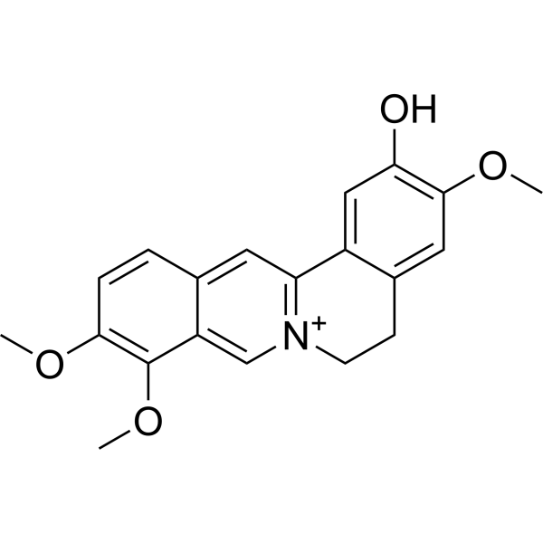
Columbamine (Columbamin; Dehydroisocorypalmine) is an organic heterotetracyclic alkaloid extracted from plants. Columbamine is a metabolite of Berberine (HY-N0716). Columbamine inhibits the cytochrome P450 (CYP) isoform CYP3A4 (IC50 = 30.6 µM). Columbamine induces apoptosis in cancer cells. Columbamine can be used for antioxidant, anti-inflammatory, antitumor, antifungal, antiparasite, hepatoprotective, neuroprotective, hypolipidemic and hypoglycemic study.
For research use only. We do not sell to patients.
- Purity : 98.38%
- CAS No.: 3621-36-1
- Formula: C20H20NO4
- Molecular Weight:338.38
-
Storage:
4°C, protect from light
* In solvent : -80°C, 6 months; -20°C, 1 month (protect from light)

To place orders, for customer services and technical support, please contact: MedChemExpress USA
Tel: 609-228-6898 E-mail: [email protected] [email protected]


-
 Biological Activity
-
Chemical Information
-
Solvent & Solubility
- Protocols
- Purity & Documentation
- References
-
 Help & FAQs


Publications Citing Use of MedChemExpress (MCE) Columbamine
MoreAll Parasite Isoforms
More

Biological Activity
Description
IC50 & Target
|
CYP3A4 30.6 μM (IC50) |
In Vitro
Columbamine (M2) (15 μM, 24 h) decreases the intracellular triglyceride (TG) contents in Hep G2 cells[2].
Columbamine (15 μM, 24 h) induces a reduction of lipid synthesis and increases in fatty acid oxidation mediated via AMPK activation in Hep G2 cells[2].
Columbamine (10-50 μM, 24-72 h) inhibits the proliferation of colon cancer cells via suppressing Wnt/β-catenin signaling pathway[4].
Columbamine (20-40 μM, 24 h) inhibits the invasion and migration of colon cancer cells[4].
Columbamine (20-40 μM, 48 h) triggers apoptosis by inhibiting a caspase-dependent mechanism in colon cancer cells[4].
MedChemExpress (MCE) has not independently confirmed the accuracy of these methods. They are for reference only.
-
Cell Line:LoVo, HCT-116, SW480 (Human colorectal cancer cell lines)
-
Concentration:10, 20, 30, 40, 50 μM
-
Incubation Time:24, 48, 72 h
-
Result:Inhibited the growth of the colon cells, which were proportional to treatment time and dosage.
Suppressed clone formation of HCT116 cells in a concentration and time-dependent manner.
-
Cell Line:LoVo, HCT-116
-
Concentration:20, 30, 40 μM
-
Incubation Time:24 h
-
Result:Significantly inhibited the migration and invasion of HCT116 and Lovo cells compared with the control cells.
Greatly impaired the invasion and migration of HCT116 cells in dose-dependent manner.
Suppressed the expression of N-Cadherin and promoted the expression of E-cadherin, resulting in the inhibition of epithelial-mesenchymal transition (EMT) of colon cancer cells.
Attenuated expressions of MMP2, MMP7 and MMP9 proteins.
-
Cell Line:HCT-116
-
Concentration:20, 30, 40 μM
-
Incubation Time:48 h
-
Result:Significantly prevented cell growth and promoted cell death at concentration from 20 μM to 40 μM.
Increased the population of Annexin V-positive cells.
The cleaved form of caspase-3 and PARP were increased without affecting the expression of caspase-3 and PARP.
Promoted pro-apoptosis factor BAD expression and repressed anti-apoptosis factor Bcl-2 expression.
-
Cell Line:Hep G2 cells
-
Concentration:15 μM
-
Incubation Time:24 h
-
Result:Inhibited the expression of genes, such as FAS, HMGR, ACC and GPAT, which are associated with fat synthesis.
Increased the gene expression of mCAD, but had no effects on CPT-1.
In Vivo
MedChemExpress (MCE) has not independently confirmed the accuracy of these methods. They are for reference only.
-
Animal Model:Six-weeks-old male BALB/c nude mice (subcutaneously injected with HCT116 and control cells on the opposite flanks of the same mouse)[4]
-
Dosage:5, 10, 20 mg/kg
-
Administration:Administered to experimental mice for 23 days
-
Result:Decreased the tumor volumes in a dose-dependent manner.


Chemical Information
-
CAS No. 3621-36-1
-
Appearance Solid
-
Molecular Weight 338.38
-
Formula C20H20NO4
-
Color Light yellow to orange
-
SMILES
COC1=C(OC)C2=C[N+]3=C(C4=CC(O)=C(OC)C=C4CC3)C=C2C=C1
-
Synonyms
Columbamin; Dehydroisocorypalmine
-
Initial Source
-
Shipping
Room temperature in continental US; may vary elsewhere.
-
Storage
4°C, protect from light
* In solvent : -80°C, 6 months; -20°C, 1 month (protect from light)


Publications (3)
-
Journal Impact Factor
-
Most Recent
-
-
Molecules
Discovery of Nine Dipeptidyl Peptidase-4 Inhibitors from Coptis chinensis Using Virtual Screening, Bioactivity Evaluation, and Binding Studies. [Abstract]2024 May 14;29(10):2304. PMID: 38792165 -


Solvent & Solubility
In Vitro:
DMSO : 5 mg/mL (14.78 mM; Need ultrasonic; Hygroscopic DMSO has a significant impact on the solubility of product, please use newly opened DMSO)
Please refer to the solubility information to select the appropriate solvent. Once prepared, please aliquot and store the solution to prevent product inactivation from repeated freeze-thaw cycles.
Storage method and period of stock solution: -80°C, 6 months; -20°C, 1 month (protect from light). When stored at -80°C, please use it within 6 months. When stored at -20°C, please use it within 1 month.
Please refer to the solubility information to select the appropriate solvent. Once prepared, please aliquot and store the solution to prevent product inactivation from repeated freeze-thaw cycles.
Storage method and period of stock solution: -80°C, 6 months; -20°C, 1 month (protect from light). When stored at -80°C, please use it within 6 months. When stored at -20°C, please use it within 1 month.
Concentration (start) × Volume (start) = Concentration (final) × Volume (final)


Purity & Documentation
-
Data Sheet (281 KB)
-
SDS (480 KB)
- English - EN (480 KB)
- Français - FR (480 KB)
- Deutsch - DE (480 KB)
- Norwegian - NO (480 KB)
- Español - ES (480 KB)
- Swedish - SV (480 KB)
- Italian - IT (480 KB)
- Korean - KR (480 KB)
- Portuguese - PT (480 KB)
-
Handling Instructions (2659 KB)
References
[1]. Wright CW, et al. In vitro antiplasmodial, antiamoebic, and cytotoxic activities of some monomeric isoquinoline alkaloids. J Nat Prod. 2000 Dec;63(12):1638-40. [Content Brief]
[2]. Cao S, et al. Berberine metabolites exhibit triglyceride-lowering effects via activation of AMP-activated protein kinase in Hep G2 cells. J Ethnopharmacol. 2013 Sep 16;149(2):576-82. [Content Brief]
[3]. Su CR, et al. Cytochrome P3A4 inhibitors and other constituents of Fibraurea tinctoria. J Nat Prod. 2007 Dec;70(12):1930-3. [Content Brief]
[4]. Lei C, et al. Columbamine suppresses the proliferation and malignization of colon cancer cells via abolishing Wnt/β-catenin signaling pathway. Cancer Manag Res. 2019 Sep 23;11:8635-8645. [Content Brief]


Complete Stock Solution Preparation Table
Please refer to the solubility information to select the appropriate solvent. Once prepared, please aliquot and store the solution to prevent product inactivation from repeated freeze-thaw cycles.
Storage method and period of stock solution: -80°C, 6 months; -20°C, 1 month (protect from light). When stored at -80°C, please use it within 6 months. When stored at -20°C, please use it within 1 month.
| Optional Solvent | Concentration Solvent Mass | 1 mg | 5 mg | 10 mg | 25 mg |
|---|---|---|---|---|---|
| DMSO | 1 mM | 2.9553 mL | 14.7763 mL | 29.5526 mL | 73.8814 mL |
| 5 mM | 0.5911 mL | 2.9553 mL | 5.9105 mL | 14.7763 mL | |
| 10 mM | 0.2955 mL | 1.4776 mL | 2.9553 mL | 7.3881 mL |


Columbamine Related Classifications
HY-N0926 Related Classifications
 Powered by Bioz