



DL-Tryptophan

Based on 1 Customer Validation
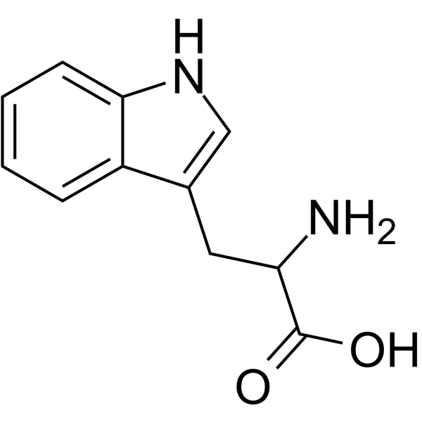
DL-Tryptophan ((±)-Tryptophan) is an orally effective aryl hydrocarbon receptor (AhR) agonist. DL-Tryptophan reduces the expression of pro-inflammatory cytokines IL-6, TNF-α, IL-1β, as well as liver injury markers aspartate aminotransferase and alanine aminotransferase, and alleviates hepatocyte apoptosis (apoptosis) in sepsis induced by cecal ligation and puncture. DL-Tryptophan can be used in the research of sepsis-related acute liver injury.
연구목적의 판매만을 진행합니다. 환자를 대상으로 한 판매는 하지 않습니다.
- Purity : 99.96%
- CAS No.: 54-12-6
- 화학식: C11H12N2O2
- 분자량:204.23
-
보관:Powder -20°C, 3 years , 4°C, 2 years ; In solvent -80°C, 6 months , -20°C, 1 month

To place orders, for customer services and technical support, please contact: MedChemExpress USA
Tel: 609-228-6898 E-mail: [email protected] [email protected]


-
 Biological Activity
-
Chemical Information
-
용액&용해도
- Protocol
- 순도&문서
- References
-
 Help & FAQs


-
HY-N0623
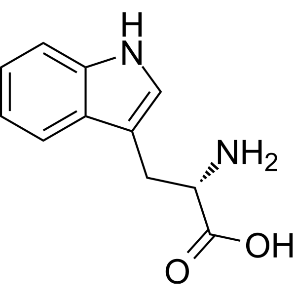
L-TryptophanEssential Amino Acid -
HY-104026
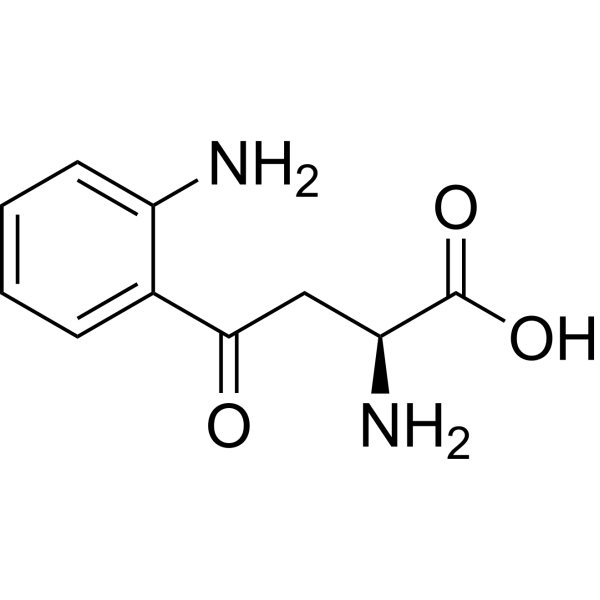
L-KynurenineAryl Hydrocarbon Receptor Agonist -
HY-100807
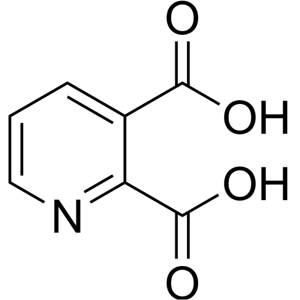
Quinolinic acidNMDA Receptor Agonist -
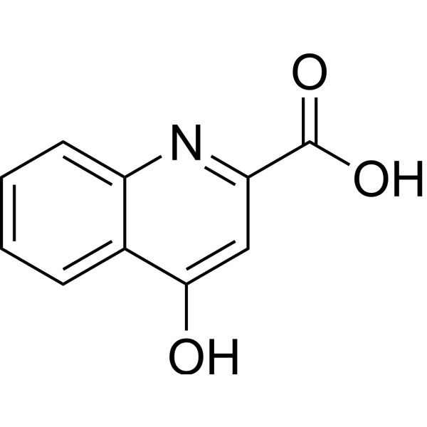
HY-100806
Kynurenic acidiGluR/α7 nAChR Antagonist -
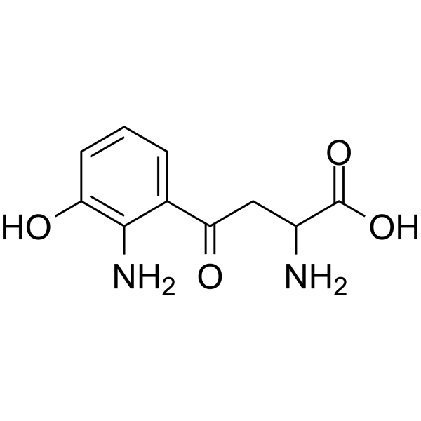
HY-113294
3-HydroxykynurenineEndogenous Neurotoxin -
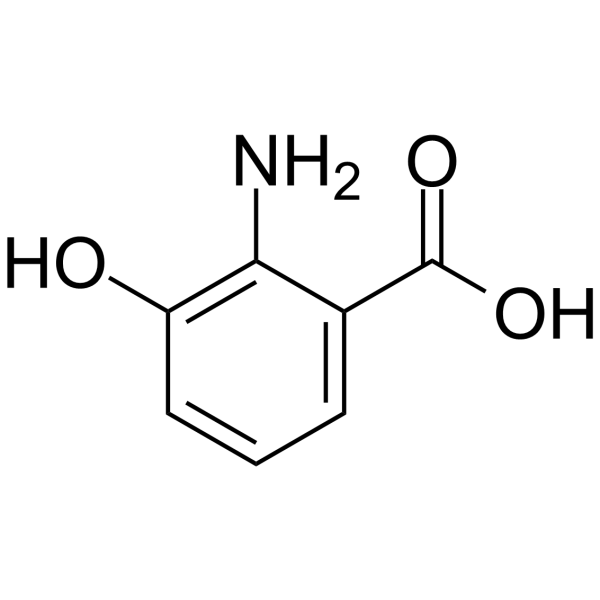
HY-W001171
3-Hydroxyanthranilic acidTryptophan Metabolite -
HY-W014666
Xanthurenic acid -
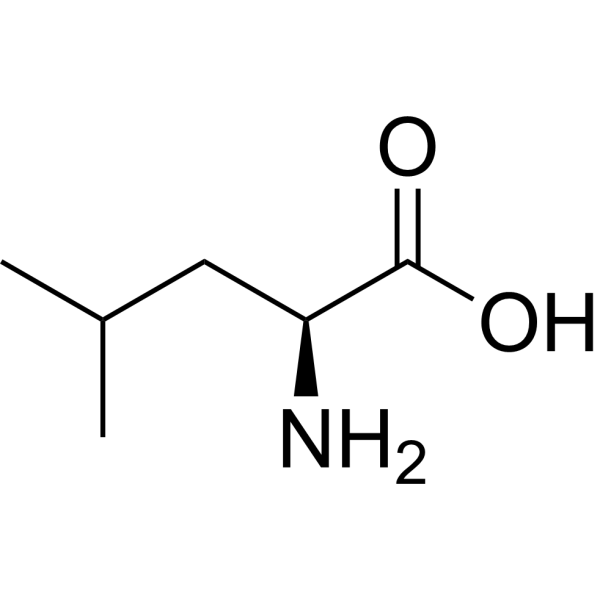
HY-N0486
L-LeucinemTOR activator -
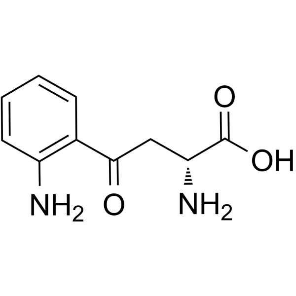
HY-W014502
D-KynurenineGPR109B Agonist -
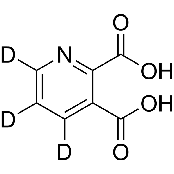
HY-100807S
Quinolinic acid-d3Stable Isotope -

HY-N0623R
L-Tryptophan (standard)Essential Amino Acid -

HY-W012480R
DL-Tryptophan (Standard)Standard
All Endogenous Metabolite Isoforms
More

Biological Activity
제품 설명
IC50 & Target
[1]|
Human Endogenous Metabolite |
IL-6 |
IL-1β |
In Vitro
MedChemExpress (MCE) has not independently confirmed the accuracy of these methods. They are for reference only.
In Vivo
MedChemExpress (MCE) has not independently confirmed the accuracy of these methods. They are for reference only.
-
Animal Model:C57BL/6 (8-week-old male or female, 20-25 g; CLP surgery-induced sepsis)[1]
-
Dosage:200 mg/kg
-
Administration:p.o.; daily; 7 consecutive days
-
Result:Significantly reduced hepatic mRNA expression of pro-inflammatory cytokines IL-6, TNF-α, and IL-1β
Decreased serum levels of liver damage markers AST and ALT.
Mitigated liver lobular structure destruction, edema, and inflammatory cell infiltration compared to the CLP group.
Reduced F4/80+ macrophage infiltration and hepatocyte apoptosis in liver tissue.
Restored hepatic expression of AhR/GAPDH and CYP1A1/GAPDH to levels not significantly different from sham-operated mice.


Chemical Information
-
CAS No. 54-12-6
-
Appearance Solid
-
분자량 204.23
-
화학식 C11H12N2O2
-
Color White to light yellow
-
SMILES
NC(CC1=CNC2=CC=CC=C12)C(O)=O
-
Synonyms
(±)-Tryptophan
-
Structure Classification
-
Initial Source
-
선적
Room temperature in continental US; may vary elsewhere.
-
보관
Powder -20°C 3 years 4°C 2 years In solvent -80°C 6 months -20°C 1 month


용액&용해도
In Vitro:
DMSO : 2.86 mg/mL (14.00 mM; ultrasonic and warming and heat to 60°C; Hygroscopic DMSO has a significant impact on the solubility of product, please use newly opened DMSO)
Please refer to the solubility information to select the appropriate solvent. Once prepared, please aliquot and store the solution to prevent product inactivation from repeated freeze-thaw cycles.
Storage method and period of stock solution: -80°C, 6 months; -20°C, 1 month. When stored at -80°C, please use it within 6 months. When stored at -20°C, please use it within 1 month.
Please refer to the solubility information to select the appropriate solvent. Once prepared, please aliquot and store the solution to prevent product inactivation from repeated freeze-thaw cycles.
Storage method and period of stock solution: -80°C, 6 months; -20°C, 1 month. When stored at -80°C, please use it within 6 months. When stored at -20°C, please use it within 1 month.
Concentration (start) × Volume (start) = Concentration (final) × Volume (final)


순도&문서
-
Data Sheet (290 KB)
-
SDS (396 KB)
- English - EN (396 KB)
- Français - FR (396 KB)
- Deutsch - DE (396 KB)
- Norwegian - NO (396 KB)
- Español - ES (396 KB)
- Swedish - SV (396 KB)
- Italian - IT (396 KB)
- Korean - KR (396 KB)
- Portuguese - PT (396 KB)
-
Handling Instructions (2659 KB)
References


Complete Stock Solution Preparation Table
Please refer to the solubility information to select the appropriate solvent. Once prepared, please aliquot and store the solution to prevent product inactivation from repeated freeze-thaw cycles.
Storage method and period of stock solution: -80°C, 6 months; -20°C, 1 month. When stored at -80°C, please use it within 6 months. When stored at -20°C, please use it within 1 month.
| Optional Solvent | Concentration Solvent Mass | 1 mg | 5 mg | 10 mg | 25 mg |
|---|---|---|---|---|---|
| DMSO | 1 mM | 4.8964 mL | 24.4822 mL | 48.9644 mL | 122.4110 mL |
| 5 mM | 0.9793 mL | 4.8964 mL | 9.7929 mL | 24.4822 mL | |
| 10 mM | 0.4896 mL | 2.4482 mL | 4.8964 mL | 12.2411 mL |


DL-Tryptophan Related Classifications
HY-W012480 Related Classifications
 Powered by Bioz
Keywords
- DL-Tryptophan
- 54-12-6
- (±)-Tryptophan
- Endogenous Metabolite
- Aryl Hydrocarbon Receptor
- Interleukin Related
- TNF Receptor
- Apoptosis
- macrophage infiltration
- inflammatory cell infiltration
- AhR
- sepsis-related acute liver injury
- pro-inflammatory cytokine
- aryl hydrocarbon receptor
- liver lobular architecture
- CLP-induced sepsis-related acute liver injury
- hepatocyte apoptosis
- Inhibitor
- inhibitor
- inhibit