



ARP-100
Based on 3 publication(s) in Google Scholar
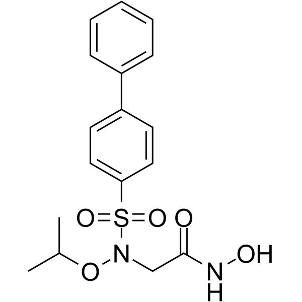
ARP-100 is a potent and selective matrix metalloproteinase MMP-2 inhibitor (IC50=12 nM). ARP-100 interacts with S1' pocket of MMP-2 and shows anti-invasive properties in an in vitro model of invasion on matrigel. ARP-100 shows the less inhibitory activity towards MMP-1 (>50 μM), MMP-3 (4.5 μM), MMP-7 (>50 μM), and MMP-9 (0.2 μM).
For research use only. We do not sell to patients.
- Purity : 98.07%
- CAS No.: 704888-90-4
- Formula: C17H20N2O5S
- Molecular Weight:364.42
-
Storage:Powder -20°C, 3 years ; In solvent -80°C, 6 months , -20°C, 1 month

To place orders, for customer services and technical support, please contact: MedChemExpress USA
Tel: 609-228-6898 E-mail: [email protected] [email protected]


-
 Biological Activity
-
Chemical Information
-
Solvent & Solubility
- Protocols
- Purity & Documentation
- References
-
 Help & FAQs


-
HY-12354
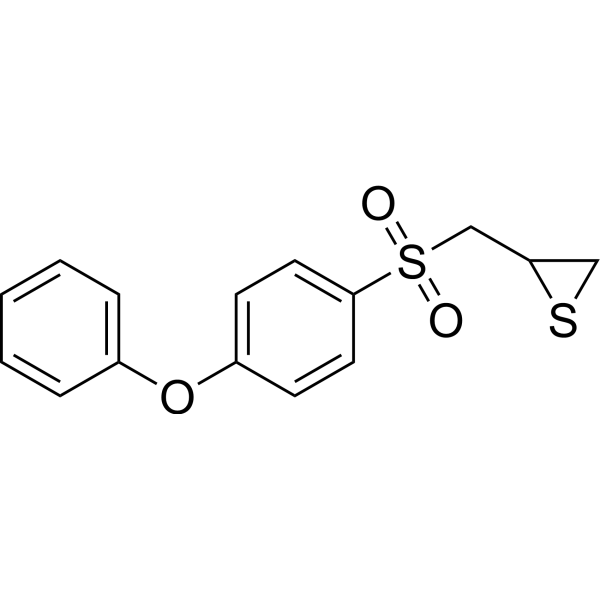
SB-3CTMMP2/9 Inhibitor -
HY-15768
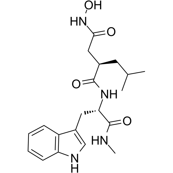
IlomastatMMP Inhibitor -
HY-12169
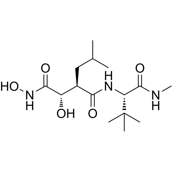
MarimastatMMP Inhibitor -
HY-13564
BatimastatMMP Inhibitor -
HY-15827
NSC 405020MMP14 Inhibitor -
HY-135232
MMP-9-IN-1MMP-9 Inhibitor -
HY-100359
CL-82198MMP-13 Inhibitor -
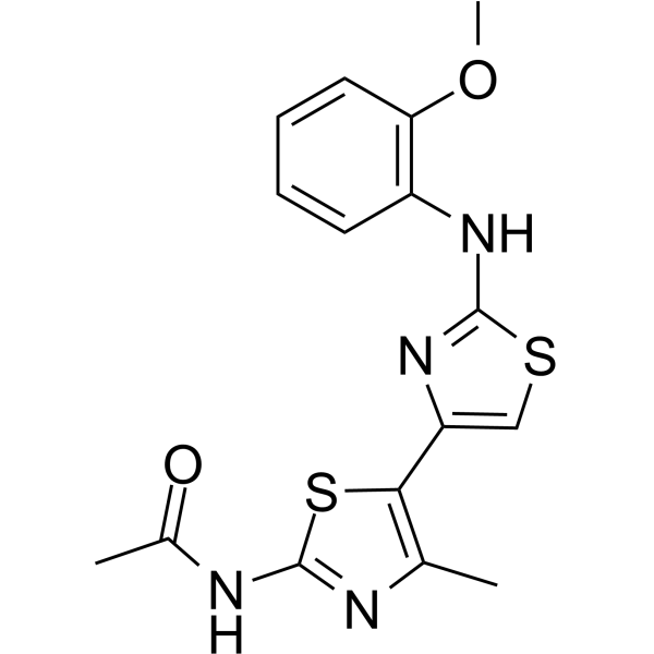
HY-103482
JNJ0966MMP-9 Inhibitor -
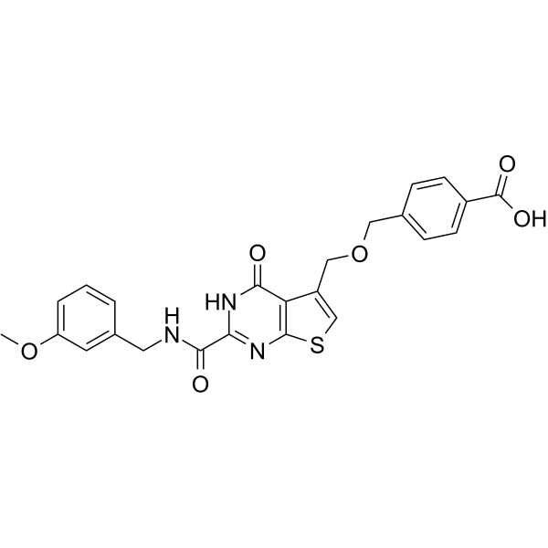
HY-100518
T-26cMMP-13 Inhibitor -
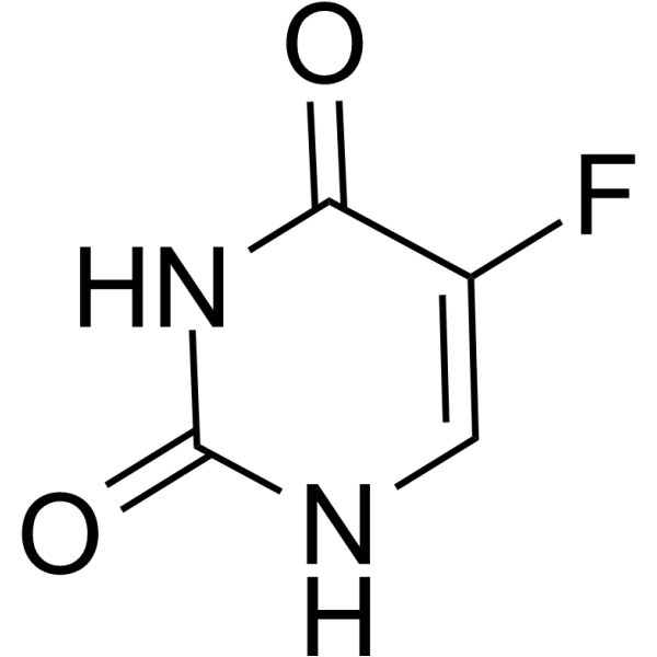
HY-90006
5-FluorouracilThymidylate Synthase Inhibitor -
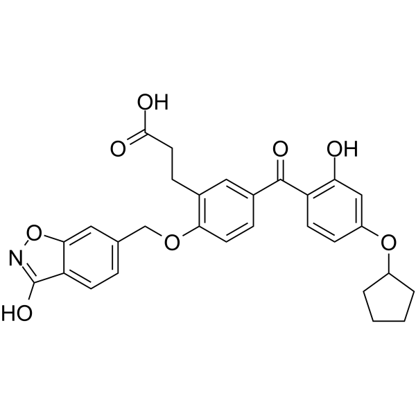
HY-12270
T-5224c-Fos/AP-1 Inhibitor -
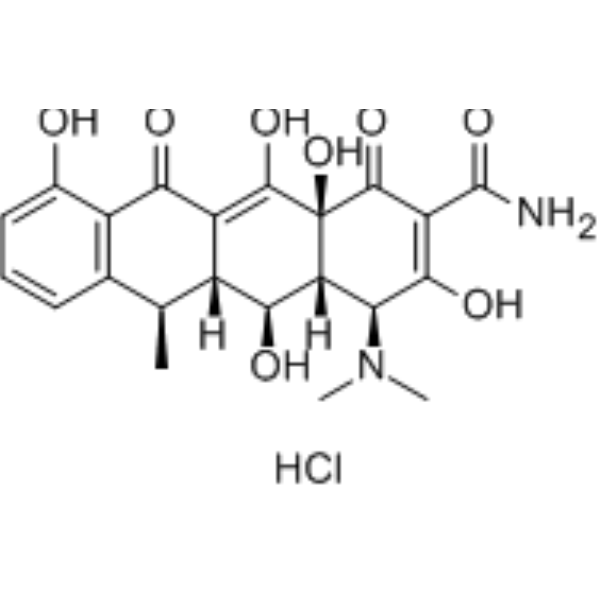
HY-N0565A
Doxycycline hydrochlorideMMP Inhibitor
Publications Citing Use of MedChemExpress (MCE) ARP-100
More Customer Validation & Images

-
IF
-
RT-PCR
-
IF
-
WB
-
In Vivo Efficacy Study


Biological Activity
Description
IC50 & Target
[1]|
MMP-2 12 nM (IC50) |
MMP-9 0.2 μM (IC50) |
MMP-3 4.5 μM (IC50) |
MMP-1 >50 μM (IC50) |
MMP-7 >50 μM (IC50) |
In Vitro
ARP-100 (50 nM) shows a significant reduction in the total number of invasive elongations[1].
MedChemExpress (MCE) has not independently confirmed the accuracy of these methods. They are for reference only.


Chemical Information
-
CAS No. 704888-90-4
-
Appearance Solid
-
Molecular Weight 364.42
-
Formula C17H20N2O5S
-
Color Off-white to light yellow
-
SMILES
O=C(NO)CN(S(=O)(C1=CC=C(C2=CC=CC=C2)C=C1)=O)OC(C)C
-
Shipping
Room temperature in continental US; may vary elsewhere.
-
Storage
Powder -20°C 3 years In solvent -80°C 6 months -20°C 1 month


Publications (3)
-
Journal Impact Factor
-
Most Recent
-
Cancer Lett
Vesicular QSOX1-MMP2 from inflammatory cancer-associated fibroblasts degrades the extracellular matrix to drive colorectal cancer pulmonary dissemination. [Abstract]2026 Sep 28:656:218645. PMID: 42217558 -
Food Res Int
δ-Tocotrienol re-sensitizes vemurafenib-resistant melanoma cells to BRAF inhibition via modulation of AKT signaling. [Abstract]2026 Aug 31:238:119457. PMID: 42215124 -
Invest Ophthalmol Vis Sci
Regulatory Effects of the Wnt7b/β-Catenin/MMP-2 Signaling Pathway on Scleral Stiffness in Guinea Pigs With Form-Deprivation Myopia. [Abstract]2025 May 1;66(5):19. PMID: 40338179
ARP-100 purchased from MedChemExpress. Usage Cited in: Invest Ophthalmol Vis Sci. 2025 May 1;66(5):19. [Abstract]
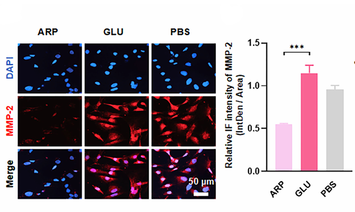
The fluorescence intensity of MMP-2 in the HFSFs treated with glutathione oxidized (GLU, 2.48 mg/mL, 24 h) was 2.09 times greater than that in the HFSFs treated with ARP-100 (ARP, 100 μM, 24 h).
ARP-100 purchased from MedChemExpress. Usage Cited in: Invest Ophthalmol Vis Sci. 2025 May 1;66(5):19. [Abstract]
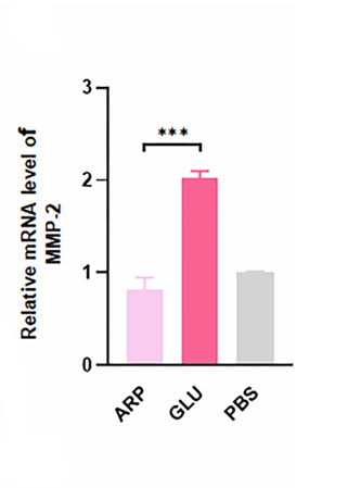
The MMP-2 mRNA level in the HFSFs treated with glutathione oxidized (GLU, 2.48 mg/mL, 24 h) was 2.46 times greater than that in the ARP-100 (ARP, 100 μM, 24 h)-treated HFSFs.
ARP-100 purchased from MedChemExpress. Usage Cited in: Invest Ophthalmol Vis Sci. 2025 May 1;66(5):19. [Abstract]
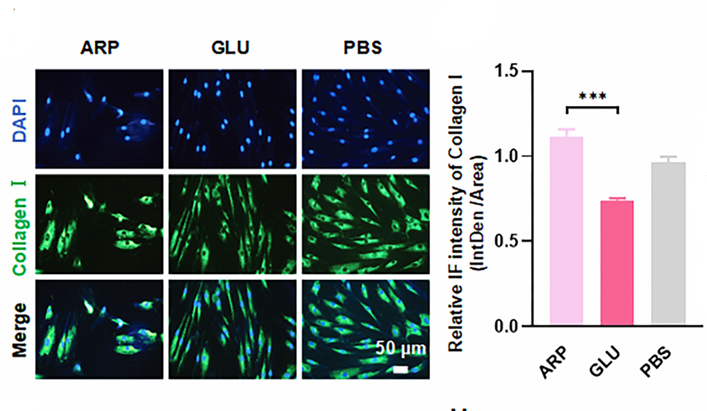
IF analysis revealed a notable increase in collagen I fluorescence intensity in the HFSFs treated with ARP-100 (ARP, 100 μM, 24 h), whereas a reduction in collagen I fluorescence intensity was found in HFSFs treated with glutathione oxidized (GLU, 2.48 mg/mL, 24 h).
ARP-100 purchased from MedChemExpress. Usage Cited in: Invest Ophthalmol Vis Sci. 2025 May 1;66(5):19. [Abstract]
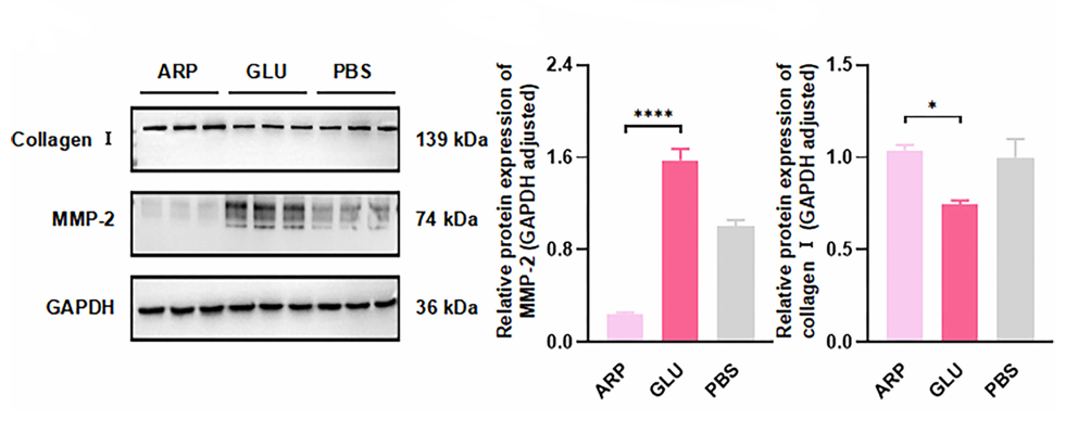
WB analysis of MMP-2 and collagen I expression in HFSFs following treatment with ARP-100 (ARP, 100 μM), glutathione oxidized (GLU, 2.48 mg/mL), or PBS for 24 hours.
ARP-100 purchased from MedChemExpress. Usage Cited in: Invest Ophthalmol Vis Sci. 2025 May 1;66(5):19. [Abstract]
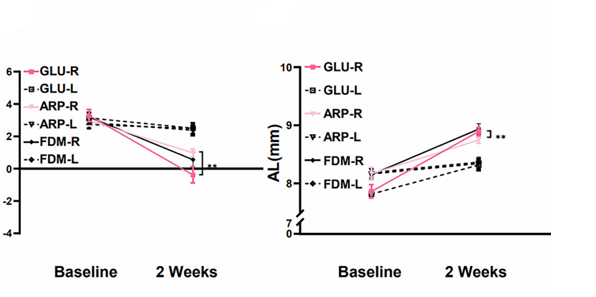
Compared with guinea pigs receiving periocular injections of vehicle (5 mM, 10 μL on days 1, 4, 7, 10, and 13 over two weeks), those treated with glutathione oxidized (Glu, 20 mg/mL, 10 μL on the same schedule) exhibited a 1.76-fold increase in relative myopia, along with a 1.78-fold increase in axial length (AL).


Solvent & Solubility
In Vitro:
DMSO : 100 mg/mL (274.41 mM; Need ultrasonic; Hygroscopic DMSO has a significant impact on the solubility of product, please use newly opened DMSO)
Please refer to the solubility information to select the appropriate solvent. Once prepared, please aliquot and store the solution to prevent product inactivation from repeated freeze-thaw cycles.
Storage method and period of stock solution: -80°C, 6 months; -20°C, 1 month. When stored at -80°C, please use it within 6 months. When stored at -20°C, please use it within 1 month.
Please refer to the solubility information to select the appropriate solvent. Once prepared, please aliquot and store the solution to prevent product inactivation from repeated freeze-thaw cycles.
Storage method and period of stock solution: -80°C, 6 months; -20°C, 1 month. When stored at -80°C, please use it within 6 months. When stored at -20°C, please use it within 1 month.
Concentration (start) × Volume (start) = Concentration (final) × Volume (final)
In Vivo:
Select the appropriate dissolution method based on your experimental animal and administration route.
- For the following dissolution methods, please ensure to first prepare a clear stock solution using an In Vitro approach and then sequentially add co-solvents:
- To ensure reliable experimental results, the clarified stock solution can be appropriately stored based on storage conditions. As for the working solution for In Vivo experiments, it is recommended to prepare freshly and use it on the same day.
- The percentages shown for the solvents indicate their volumetric ratio in the final prepared solution. If precipitation or phase separation occurs during preparation, heat and/or sonication can be used to aid dissolution.
Add each solvent one by one: 10% DMSO 40% PEG300 5% Tween-80 45% Saline
Solubility: ≥ 2.5 mg/mL (6.86 mM); Clear solution
This protocol yields a clear solution of ≥ 2.5 mg/mL (saturation unknown).
Taking 1 mL working solution as an example, add 100 μL DMSO stock solution (25.0 mg/mL) to 400 μL PEG300, and mix evenly; then add 50 μL Tween-80 and mix evenly; then add 450 μL Saline to adjust the volume to 1 mL.
Preparation of Saline: Dissolve 0.9 g sodium chloride in ddH₂O and dilute to 100 mL to obtain a clear Saline solution.
Add each solvent one by one: 10% DMSO 90% (20% SBE-β-CD in Saline)
Solubility: 2.5 mg/mL (6.86 mM); Suspended solution; Need ultrasonic
This protocol yields a suspended solution of 2.5 mg/mL. Suspended solution can be used for oral and intraperitoneal injection.
Taking 1 mL working solution as an example, add 100 μL DMSO stock solution (25.0 mg/mL) to 900 μL 20% SBE-β-CD in Saline, and mix evenly.
Preparation of 20% SBE-β-CD in Saline (4°C, storage for one week): 2 g SBE-β-CD powder is dissolved in 10 mL Saline, completely dissolve until clear.
In Vivo Dissolution Calculator
Please enter the basic information of animal experiments:
-
-
-
-
Recommended: Prepare an additional quantity of animals to account for potential losses during experiments.
Please enter your animal formula composition:
-
%DMSO +
Recommended: Keep the proportion of DMSO in working solution below 2% if your animal is weak.
-
%+
-
+%Tween-80 + +
-
%Saline +
The co-solvents required include: DMSO, . All of co-solvents are available by MedChemExpress (MCE). , Tween 80. All of co-solvents are available by MedChemExpress (MCE).
Working solution concentration: 0.22 mg/mL
Method for preparing stock solution: mg drug dissolved in μL DMSO. Stock solution concentration: mg/mL.
1. Take μL DMSO stock solution;
2. Add μL .
μL , mix evenly;
3. Then add μL Tween 80, mix evenly;
4. Then add μL
Please ensure that the stock solution in the first step is dissolved to a clear state, and add co-solvents in sequence. You can use ultrasonic heating (ultrasonic cleaner, recommended frequency 20-40 kHz), vortexing, etc. to assist dissolution.


Purity & Documentation
-
Data Sheet (276 KB)
-
SDS (604 KB)
- English - EN (604 KB)
- Français - FR (604 KB)
- Deutsch - DE (604 KB)
- Norwegian - NO (604 KB)
- Español - ES (604 KB)
- Swedish - SV (604 KB)
- Italian - IT (604 KB)
- Korean - KR (604 KB)
- Portuguese - PT (604 KB)
-
Handling Instructions (2659 KB)
References
[1]. Rossello A, et al. New N-arylsulfonyl-N-alkoxyaminoacetohydroxamic acids as selective inhibitors of gelatinase A (MMP-2). Bioorg Med Chem. 2004 May 1;12(9):2441-50. [Content Brief]
[2]. Tuccinardi T, et al. Amber force field implementation, molecular modelling study, synthesis and MMP-1/MMP-2 inhibition profile of (R)- and (S)-N-hydroxy-2-(N-isopropoxybiphenyl-4-ylsulfonamido)-3-methylbutanamides. Bioorg Med Chem. 2006 Jun 15;14(12):4260-76. [Content Brief]


Complete Stock Solution Preparation Table
Please refer to the solubility information to select the appropriate solvent. Once prepared, please aliquot and store the solution to prevent product inactivation from repeated freeze-thaw cycles.
Storage method and period of stock solution: -80°C, 6 months; -20°C, 1 month. When stored at -80°C, please use it within 6 months. When stored at -20°C, please use it within 1 month.
| Optional Solvent | Concentration Solvent Mass | 1 mg | 5 mg | 10 mg | 25 mg |
|---|---|---|---|---|---|
| DMSO | 1 mM | 2.7441 mL | 13.7204 mL | 27.4409 mL | 68.6022 mL |
| 5 mM | 0.5488 mL | 2.7441 mL | 5.4882 mL | 13.7204 mL | |
| 10 mM | 0.2744 mL | 1.3720 mL | 2.7441 mL | 6.8602 mL | |
| 15 mM | 0.1829 mL | 0.9147 mL | 1.8294 mL | 4.5735 mL | |
| 20 mM | 0.1372 mL | 0.6860 mL | 1.3720 mL | 3.4301 mL | |
| 25 mM | 0.1098 mL | 0.5488 mL | 1.0976 mL | 2.7441 mL | |
| 30 mM | 0.0915 mL | 0.4573 mL | 0.9147 mL | 2.2867 mL | |
| 40 mM | 0.0686 mL | 0.3430 mL | 0.6860 mL | 1.7151 mL | |
| 50 mM | 0.0549 mL | 0.2744 mL | 0.5488 mL | 1.3720 mL | |
| 60 mM | 0.0457 mL | 0.2287 mL | 0.4573 mL | 1.1434 mL | |
| 80 mM | 0.0343 mL | 0.1715 mL | 0.3430 mL | 0.8575 mL | |
| 100 mM | 0.0274 mL | 0.1372 mL | 0.2744 mL | 0.6860 mL |


ARP-100 Related Classifications
HY-103444 Related Classifications
 Powered by Bioz