



Calcimycin
Based on 27 publication(s) in Google Scholar
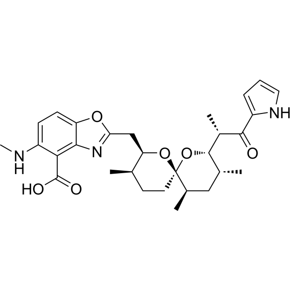
Calcimycin (A-23187) is an antibiotic and a unique divalent cation ionophore (like calcium and magnesium). Calcimycin induces Ca2+-dependent cell death by increasing intracellular calcium concentration. Calcimycin inhibits the growth of Gram-positive bacteria and some fungi. Calcimycin also inhibits the activity of ATPase and uncouples oxidative phosphorylation (OXPHOS) of mammalian cells. Calcimycin induces apoptosis and autophagy.
Para uso exclusivo en investigación. No vendemos a pacientes.
- Pureza : 99.10%
- No. CAS: 52665-69-7
- Fòrmula: C29H37N3O6
- Peso molecular:523.62
-
Almacenamiento:
4°C, sealed storage, away from moisture
* In solvent : -80°C, 1 year; -20°C, 6 months (sealed storage, away from moisture)

To place orders, for customer services and technical support, please contact: MedChemExpress USA
Tel: 609-228-6898 E-mail: [email protected] [email protected]


-
 Actividad biológica
-
Chemical Information
-
Solvente y solubilidad
- Protocolo
- Pureza y Documentación
- Referencias
-
 Help & FAQs


-
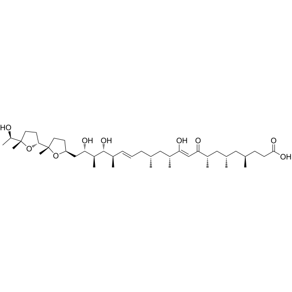
HY-13434
IonomycinCalcium Ionophore -

HY-18739
Phorbol 12-myristate 13-acetatePKC Activator -
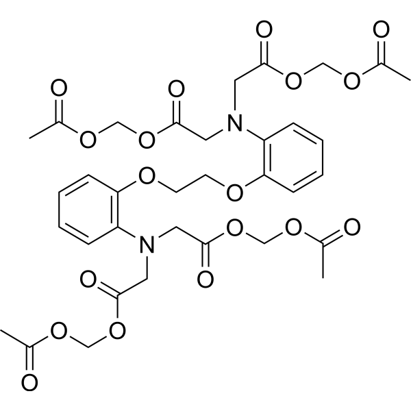
HY-100545
BAPTA-AMCa2+ Chelator -
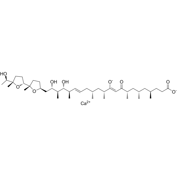
HY-13434A
Ionomycin calciumCalcium Ionophore -
HY-13433
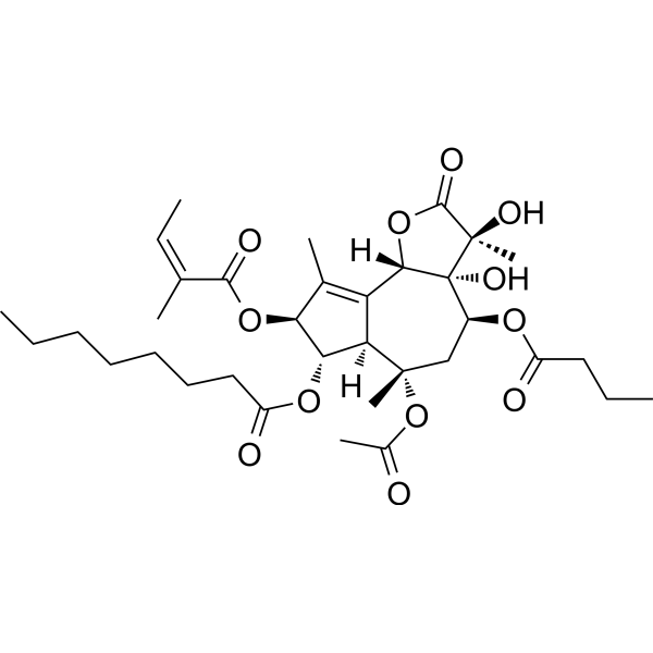
ThapsigarginER Stress Inducer -
HY-109590
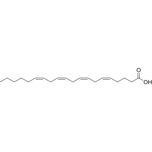
Arachidonic acidPolyunsaturated Fatty Acid -
HY-16592
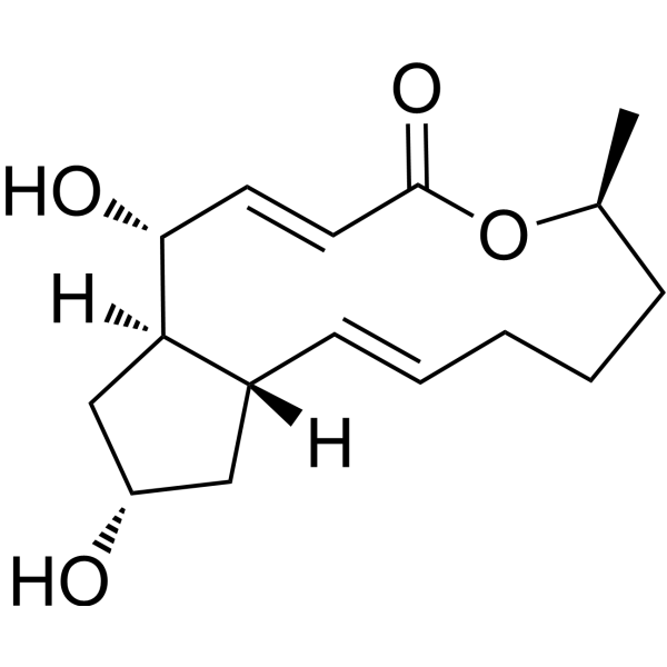
Brefeldin AProtein Transport Inhibitor -
HY-100965
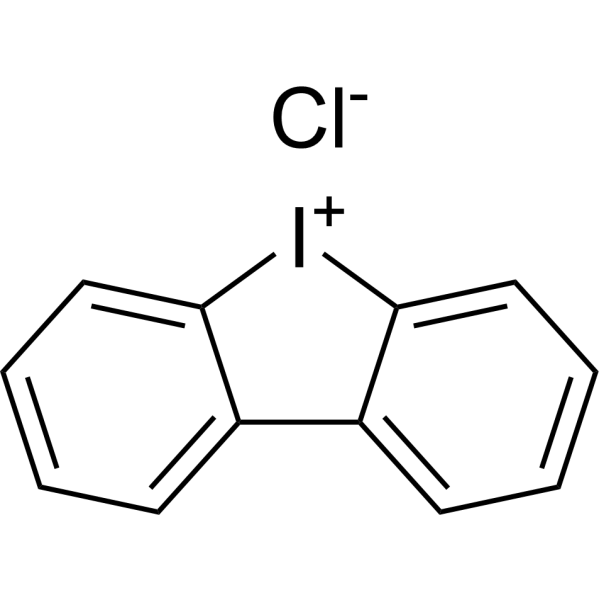
Diphenyleneiodonium chlorideNOX Inhibitor -
HY-13966
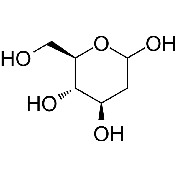
2-Deoxy-D-glucoseGlycolysis Inhibitor -
HY-D0861
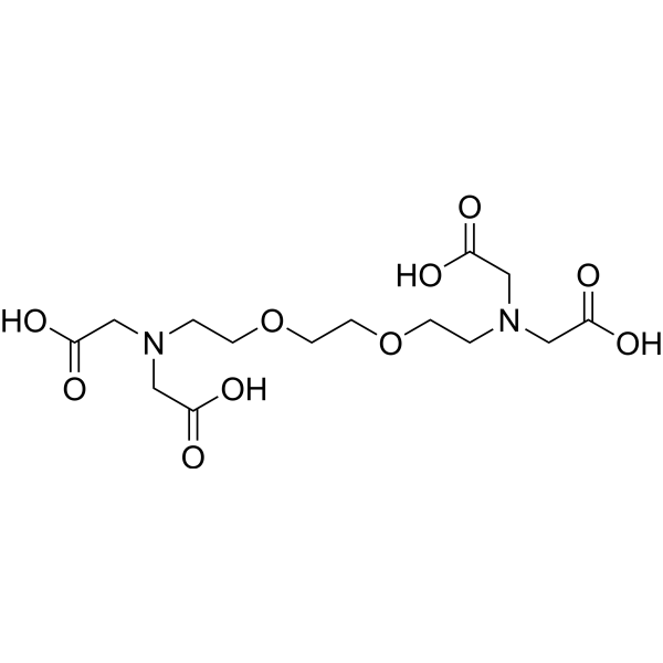
EGTACalcium Ion Chelator -

HY-15371
ForskolinAdenylate Cyclase Activator -
HY-B0627
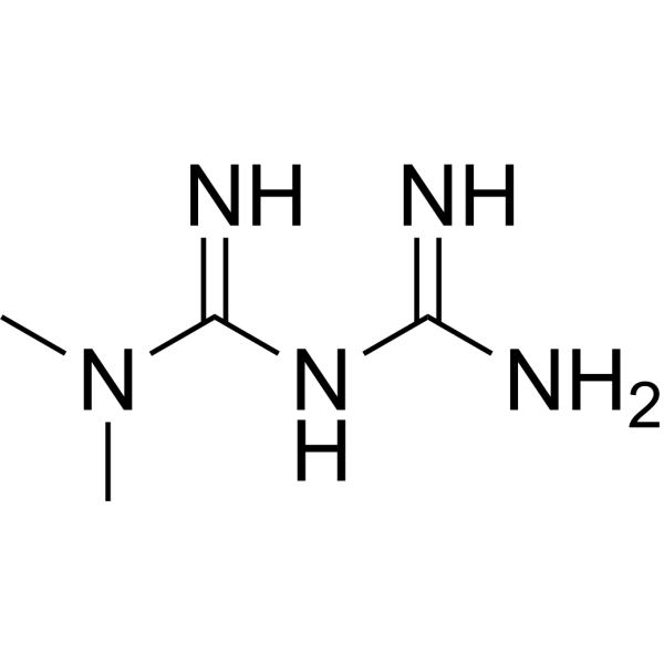
MetforminAntihyperglycemic Agent
Publications Citing Use of MedChemExpress (MCE) Calcimycin
More- Cancer Commun (Lond). 2023 Jan;43(1):123-149. [Abstract]
- Adv Sci (Weinh). 2025 Aug 20:e11873. [Abstract]
- Brain. 2024 Nov 4;147(11):3764-3779. [Abstract]
- Mol Ther. 2022 Dec 7;30(12):3694-3713. [Abstract]
- Phytomedicine. 2024 Apr:126:155200. [Abstract]
- Proc Natl Acad Sci U S A. 2025 Sep 2;122(35):e2513155122. [Abstract]
- Int J Biol Macromol. 2025 Jun;311(Pt 3):144068. [Abstract]
- Extracell Vesicles Circ Nucl Acids. 2026 Jul 31;7(3):1228-48.
- Antioxidants (Basel). 2022 Jul 26;11(8):1455. [Abstract]
- EMBO Mol Med. 2021 Dec 7;13(12):e14072. [Abstract]
- ACS Appl Mater Interfaces. 2026 Apr 22;18(15):21604-21621. [Abstract]
- J Med Chem. 2025 Oct 23;68(20):21520-21533. [Abstract]
- Virulence. 2025 Dec;16(1):2490208. [Abstract]
- Ecotoxicol Environ Saf. 2025 Jul 15:300:118435. [Abstract]
- Life Sci. 2022 Nov 1:308:120921. [Abstract]
- Life Metab. 2025 Jul 16;4(6):loaf027. [Abstract]
- EMBO Rep. 2022 Feb 3;23(3):e53373. [Abstract]
- Mbio. 2022 Apr 21;e0069022. [Abstract]
- PLoS Pathog. 2026 Mar 17;22(3):e1014062. [Abstract]
- BMC Cancer. 2025 May 28;25(1):956. [Abstract]
- J Proteome Res. 2026 Feb 25;25(3):1584-1600.
- Exp Cell Res. 2026 Aug 1;461(1):115103. [Abstract]
- Thromb J. 2025 May 7;23(1):44. [Abstract]
- Res Sq. 2026 Feb 12.
- Cell Biomater. 2026 Jan 12.
- bioRxiv. 2025 May 05.
- Chemosphere. 2024 Jan:346:140532. [Abstract]
 Customer Validation & Images

-
Cell Proliferation/Viability Assay
-
IF
-
WB
-
IP
-
Cell Proliferation/Viability Assay
Ver todos los productos específicos de isoformas Antibiotic
More

Actividad biológica
Descripciòn
In Vitro
In Vivo
MedChemExpress (MCE) has not independently confirmed the accuracy of these methods. They are for reference only.
-
Animal Model:Mice (ICR, 25-30 g)[5]
-
Dosage:2.5 or 7.5 nM
-
Administration:Intrapleurally
-
Result:Two hours after 2.5 nM, or three hours after 7.5 nM, challenge the protein levels in the pleural cavity were equivalent to about a half of their corresponding peak values.
- Western Blot
- Mammalian live/dead viability and cytotoxicity staining
- Annexin V plus membrane-impermeant dye apoptosis staining
- Ca2+ Staining Technique
- Gram Staining of Tissue Sections
- Kinase activity and phosphorylation assays
- Detection of Apoptosis
- TUNEL staining for apoptotic DNA fragmentation
- Cell Cytotoxicity Assay
- Autophagy
- Apoptosis Solutions
- Calcium Spark Assay
- Protocol for Kinase activity and phosphorylation assays
- Lysosome and acidic-vesicle live-cell staining
- Macroautophagy Solutions
Ensayo clínico
| NCT Number | Sponsor | Condition | Start Date |
Phase

|
|---|---|---|---|---|
| NCT01329991 | Plexxikon| | 2011-05 | PHASE1 |


Chemical Information
-
No. CAS 52665-69-7
-
Appearance Solid
-
Peso molecular 523.62
-
Fòrmula C29H37N3O6
-
Color White to off-white
-
SMILES
O=C(C1=C2N=C(C[C@H]3O[C@]4(O[C@H]([C@H](C)C(C5=CC=CN5)=O)[C@H](C)C[C@H]4C)CC[C@H]3C)OC2=CC=C1NC)O
-
Synonyms
A-23187; Antibiotic A-23187
-
Structure Classification
-
Initial Source
Streptomyces chartreusensis
-
Envío
Room temperature in continental US; may vary elsewhere.
-
Almacenamiento
4°C, sealed storage, away from moisture
* In solvent : -80°C, 1 year; -20°C, 6 months (sealed storage, away from moisture)


Publications (27)
-
Journal Impact Factor
-
Most Recent
-
Cancer Commun (Lond)
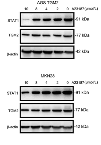
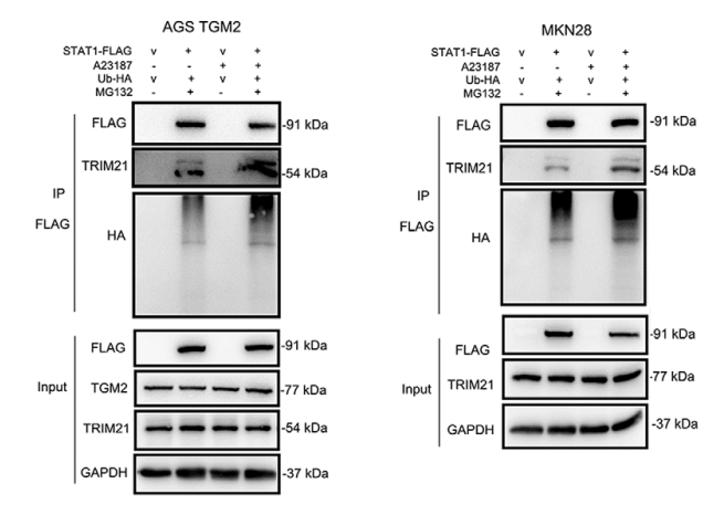
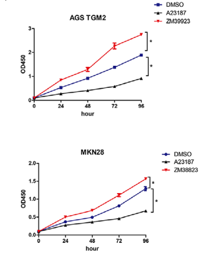
Cytosolic TGM2 promotes malignant progression in gastric cancer by suppressing the TRIM21-mediated ubiquitination/degradation of STAT1 in a GTP binding-dependent modality. [Abstract]2023 Jan;43(1):123-149. PMID: 36353796
Calcimycin purchased from MedChemExpress. Usage Cited in: Cancer Commun (Lond). 2023 Jan;43(1):123-149. [Abstract]
Cells were treated with different concentrations of Calcimycin (A23187: 2 µmol/L, 4 µmol/L, 8 µmol/L, 10 µmol/L; 24 h), and the effect of A23187 on STAT1 expression was assessed by Western blotting.
Calcimycin purchased from MedChemExpress. Usage Cited in: Cancer Commun (Lond). 2023 Jan;43(1):123-149. [Abstract]
Cells were treated with A23187 (4 µmol/L; 24 h) to examine the effect of A21187 on the binding ability of TRIM21 and STAT1 by Co‐IP and western blotting, and the result showed that A23187 reversed the dissociation of TRIM21 and STAT1 in the context of TGM2 and increased ubiquitination of STAT1.
Calcimycin purchased from MedChemExpress. Usage Cited in: Cancer Commun (Lond). 2023 Jan;43(1):123-149. [Abstract]
The effect of A23187 (4 µmol/L; 0-96 h) and ZM39923 (20 nmol/L) on GC cell proliferation was evaluated by CCK8 assay. The result showed that ZM39923 promoted cell proliferation while A23187 had an inhibitory role in GC cell proliferation.
-
Adv Sci (Weinh)
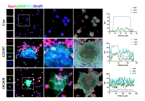
Succinate Dehydrogenase Subunit A (SDHA) Mediated Microglia Extracellular Traps Formation Participating in Cerebral Ischemic Reperfusion Injury. [Abstract]2025 Aug 20:e11873. PMID: 40832885
Calcimycin purchased from MedChemExpress. Usage Cited in: Adv Sci (Weinh). 2025 Aug 20:e11873. [Abstract]
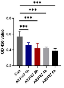
A23187 (5 µM; 1–6 h) induced apoptosis in microglia in vitro. Microglia viability was assessed using the CCK-8 assay.
Calcimycin purchased from MedChemExpress. Usage Cited in: Adv Sci (Weinh). 2025 Aug 20:e11873. [Abstract]
A23187 (5 µM; 4 h). Representative images of microglia stained with Iba1, citH3, and MPO triple staining, and colocalization analysis of MPO, citH3, and DNA in the three cell groups.
-
Brain
Hippocampal excitation-inhibition balance underlies the 5-HT2C receptor in modulating depressive behaviours. [Abstract]2024 Nov 4;147(11):3764-3779. PMID: 38701344 -
Mol Ther
mmu-lncRNA 121686/hsa-lncRNA 520657 induced by METTL3 drive the progression of AKI by targeting miR-328-5p/HtrA3 signaling axis. [Abstract]2022 Dec 7;30(12):3694-3713. PMID: 35869629 -
Phytomedicine
Sinapine targeting PLCβ3 EF hands disrupts Gαq-PLCβ3 interaction and ameliorates cardiovascular diseases. [Abstract]2024 Apr:126:155200. PMID: 38387273 -
Proc Natl Acad Sci U S A
2025 Sep 2;122(35):e2513155122. PMID: 40880539 -
Int J Biol Macromol
FKBP5 inhibitor suppresses platelet activation and thrombosis by inhibiting IKBKE/PI3K/Rap1 pathway. [Abstract]2025 Jun;311(Pt 3):144068. PMID: 40345291 -
-
Antioxidants (Basel)
Muscone and (+)-Borneol Cooperatively Strengthen CREB Induction of Claudin 5 in IL-1 β-Induced Endothelium Injury. [Abstract]2022 Jul 26;11(8):1455. PMID: 35892657 -
EMBO Mol Med
Conformational change of adenine nucleotide translocase-1 mediates cisplatin resistance induced by EBV-LMP1. [Abstract]2021 Dec 7;13(12):e14072. PMID: 34755470 -
ACS Appl Mater Interfaces
Disrupting Neutrophil Extracellular Traps with Targeted Cerium Oxide Nanoparticles Ameliorates Diabetic Periodontitis. [Abstract]2026 Apr 22;18(15):21604-21621. PMID: 41972905 -
J Med Chem
2025 Oct 23;68(20):21520-21533. PMID: 41041960 -
Virulence
2025 Dec;16(1):2490208. PMID: 40202859 -
Ecotoxicol Environ Saf
HFPO homologues, the novel alternatives to PFOA, impair male reproduction in mice through inducing mitochondrial dysfunction. [Abstract]2025 Jul 15:300:118435. PMID: 40460775 -
Life Sci
MiR-6918-5p prevents renal tubular cell apoptosis by targeting MBD2 in ischemia/reperfusion-induced AKI. [Abstract]2022 Nov 1:308:120921. PMID: 36057400 -
Life Metab
Energy stress-induced PKCζ S-glutathionylation is essential for LKB1 cytoplasmic translocation and AMPK activation. [Abstract]2025 Jul 16;4(6):loaf027. PMID: 41070197 -
EMBO Rep
Amino acid starvation-induced LDLR trafficking accelerates lipoprotein endocytosis and LDL clearance. [Abstract]2022 Feb 3;23(3):e53373. PMID: 34994492 -
Mbio
Calmodulin Binding Activates Chromobacterium CopC Effector to ADP-Riboxanate Host Apoptotic Caspases. [Abstract]2022 Apr 21;e0069022. PMID: 35446120 -
PLoS Pathog
LL-37 selectively targets Plasmodium-infected erythrocytes and exhibits antimalarial activity. [Abstract]2026 Mar 17;22(3):e1014062. PMID: 41843625 -
BMC Cancer
TRPM2 channels mediate ROS-induced actin remodeling and cell migration of prostate cancer cells. [Abstract]2025 May 28;25(1):956. PMID: 40437388 -
-
Exp Cell Res
2026 Aug 1;461(1):115103. PMID: 42276196 -
Thromb J
Endoplasmic reticulum protein 29 negatively regulates platelet functions and thrombosis in mice. [Abstract]2025 May 7;23(1):44. PMID: 40336049 -
-
-
-
Chemosphere
PCB126 impairs human sperm functions by affecting post-translational modifications and mitochondrial functions. [Abstract]2024 Jan:346:140532. PMID: 37918541


Solvente y solubilidad
In Vitro:
DMSO : 33.33 mg/mL (63.65 mM; Need ultrasonic; Hygroscopic DMSO has a significant impact on the solubility of product, please use newly opened DMSO)
Please refer to the solubility information to select the appropriate solvent. Once prepared, please aliquot and store the solution to prevent product inactivation from repeated freeze-thaw cycles.
Storage method and period of stock solution: -80°C, 1 year; -20°C, 6 months (sealed storage, away from moisture). When stored at -80°C, please use it within 1 year. When stored at -20°C, please use it within 6 months.
Please refer to the solubility information to select the appropriate solvent. Once prepared, please aliquot and store the solution to prevent product inactivation from repeated freeze-thaw cycles.
Storage method and period of stock solution: -80°C, 1 year; -20°C, 6 months (sealed storage, away from moisture). When stored at -80°C, please use it within 1 year. When stored at -20°C, please use it within 6 months.
Concentration (start) × Volume (start) = Concentration (final) × Volume (final)
In Vivo:
Select the appropriate dissolution method based on your experimental animal and administration route.
- For the following dissolution methods, please ensure to first prepare a clear stock solution using an In Vitro approach and then sequentially add co-solvents:
- To ensure reliable experimental results, the clarified stock solution can be appropriately stored based on storage conditions. As for the working solution for In Vivo experiments, it is recommended to prepare freshly and use it on the same day.
- The percentages shown for the solvents indicate their volumetric ratio in the final prepared solution. If precipitation or phase separation occurs during preparation, heat and/or sonication can be used to aid dissolution.
Add each solvent one by one: 10% DMSO 40% PEG300 5% Tween-80 45% Saline
Solubility: ≥ 2.5 mg/mL (4.77 mM); Clear solution
This protocol yields a clear solution of ≥ 2.5 mg/mL (saturation unknown).
Taking 1 mL working solution as an example, add 100 μL DMSO stock solution (25.0 mg/mL) to 400 μL PEG300, and mix evenly; then add 50 μL Tween-80 and mix evenly; then add 450 μL Saline to adjust the volume to 1 mL.
Preparation of Saline: Dissolve 0.9 g sodium chloride in ddH₂O and dilute to 100 mL to obtain a clear Saline solution.
In Vivo Dissolution Calculator
Please enter the basic information of animal experiments:
-
-
-
-
Recommended: Prepare an additional quantity of animals to account for potential losses during experiments.
Please enter your animal formula composition:
-
%DMSO +
Recommended: Keep the proportion of DMSO in working solution below 2% if your animal is weak.
-
%+
-
+%Tween-80 + +
-
%Saline +
The co-solvents required include: DMSO, . All of co-solvents are available by MedChemExpress (MCE). , Tween 80. All of co-solvents are available by MedChemExpress (MCE).
Working solution concentration: 0.22 mg/mL
Method for preparing stock solution: mg drug dissolved in μL DMSO. Stock solution concentration: mg/mL. * In solvent : -80°C, 1 year; -20°C, 6 months (sealed storage, away from moisture)
1. Take μL DMSO stock solution;
2. Add μL .
μL , mix evenly;
3. Then add μL Tween 80, mix evenly;
4. Then add μL
Please ensure that the stock solution in the first step is dissolved to a clear state, and add co-solvents in sequence. You can use ultrasonic heating (ultrasonic cleaner, recommended frequency 20-40 kHz), vortexing, etc. to assist dissolution.


Pureza y Documentación
-
Ficha de datos (277 KB)
-
SDS (396 KB)
- English - EN (396 KB)
- Français - FR (396 KB)
- Deutsch - DE (396 KB)
- Norwegian - NO (396 KB)
- Español - ES (396 KB)
- Swedish - SV (396 KB)
- Italian - IT (396 KB)
- Korean - KR (396 KB)
- Portuguese - PT (396 KB)
-
Instrucciones de manejo (2659 KB)
Referencias
[1]. Wu Q, et al. Characterization of the biosynthesis gene cluster for the pyrrole polyether antibiotic calcimycin(A23187) in Streptomyces chartreusis NRRL 3882. Antimicrob Agents Chemother. 2011 Mar;55(3):974-82. [Content Brief]
[2]. Elliott JI, et al. IKCa1 activity is required for cell shrinkage, phosphatidylserine translocation and death in Tlymphocyte apoptosis. EMBO Rep. 2003 Feb;4(2):189-94. [Content Brief]
[3]. Engedal N, et al. Modulation of intracellular calcium homeostasis blocks autophagosome formation. Autophagy. 2013 Oct;9(10):1475-90. [Content Brief]
[4]. Mawatwal S, et al. Calcimycin mediates mycobacterial killing by inducing intracellular calcium-regulated autophagy in a P2RX7 dependent manner. Biochim Biophys Acta Gen Subj. 2017 Dec;1861(12):3190-3200. [Content Brief]
[5]. Wang JP, et al. Effect of norathyriol, isolated from Tripterospermum lanceolatum, on A23187-induced pleurisy and analgesia in mice. Naunyn Schmiedebergs Arch Pharmacol. 1994 Jul;350(1):90-5. [Content Brief]


Complete Stock Solution Preparation Table
Please refer to the solubility information to select the appropriate solvent. Once prepared, please aliquot and store the solution to prevent product inactivation from repeated freeze-thaw cycles.
Storage method and period of stock solution: -80°C, 1 year; -20°C, 6 months (sealed storage, away from moisture). When stored at -80°C, please use it within 1 year. When stored at -20°C, please use it within 6 months.
| Optional Solvent | Concentration Solvent Mass | 1 mg | 5 mg | 10 mg | 25 mg |
|---|---|---|---|---|---|
| DMSO | 1 mM | 1.9098 mL | 9.5489 mL | 19.0978 mL | 47.7445 mL |
| 5 mM | 0.3820 mL | 1.9098 mL | 3.8196 mL | 9.5489 mL | |
| 10 mM | 0.1910 mL | 0.9549 mL | 1.9098 mL | 4.7745 mL | |
| 15 mM | 0.1273 mL | 0.6366 mL | 1.2732 mL | 3.1830 mL | |
| 20 mM | 0.0955 mL | 0.4774 mL | 0.9549 mL | 2.3872 mL | |
| 25 mM | 0.0764 mL | 0.3820 mL | 0.7639 mL | 1.9098 mL | |
| 30 mM | 0.0637 mL | 0.3183 mL | 0.6366 mL | 1.5915 mL | |
| 40 mM | 0.0477 mL | 0.2387 mL | 0.4774 mL | 1.1936 mL | |
| 50 mM | 0.0382 mL | 0.1910 mL | 0.3820 mL | 0.9549 mL | |
| 60 mM | 0.0318 mL | 0.1591 mL | 0.3183 mL | 0.7957 mL |


Calcimycin Related Classifications
HY-N6687 Related Classifications
 Powered by Bioz