



Ossirene
Based on 2 publication(s) in Google Scholar
Ossirene (AS101), an immunomodulatory tellurium compound, is a potent IL-1β inhibitor. Ossirene abolishes phosphorylation of STAT3 by inhibiting IL-10. Ossirene potently inhibits Caspase-1 and is used for the autoimmune diseases and certain malignancies.
Para uso exclusivo en investigación. No vendemos a pacientes.
- Pureza : 98.0%
- No. CAS: 106566-58-9
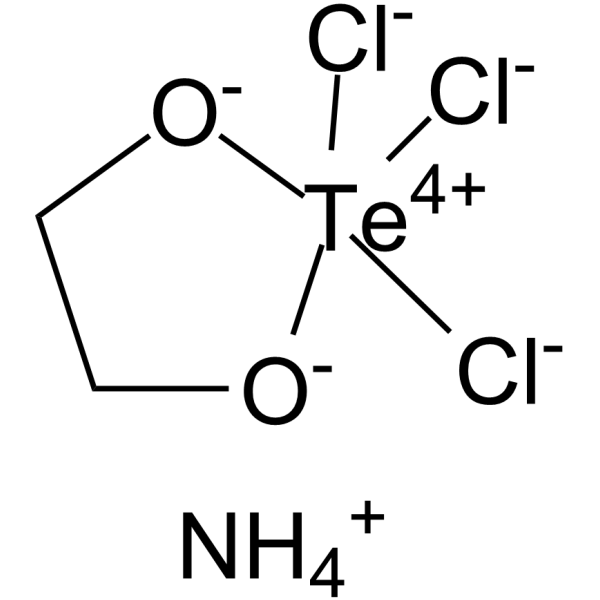
- Fòrmula: C2H8Cl3NO2Te
- Peso molecular:312.05
-
Almacenamiento:
-20°C, stored under nitrogen
* In solvent : -80°C, 6 months; -20°C, 1 month (stored under nitrogen)

To place orders, for customer services and technical support, please contact: MedChemExpress USA
Tel: 609-228-6898 E-mail: [email protected] [email protected]


-
 Actividad biológica
-
Chemical Information
-
Solvente y solubilidad
- Protocolo
- Pureza y Documentación
- Referencias
-
 Help & FAQs


-
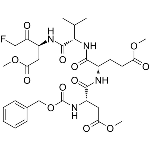
HY-12466
Z-DEVD-FMKCaspase-3 Inhibitor -
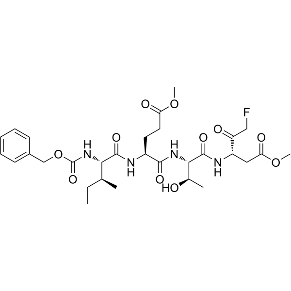
HY-101297
Z-IETD-FMKCaspase-8 Inhibitor -

HY-P70751
IL-10 Protein, Human (HEK293) -
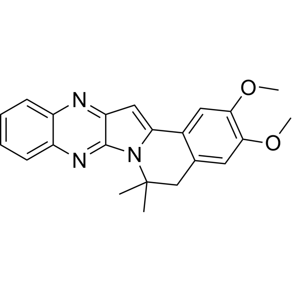
HY-19969
YM-90709IL-5 Receptor Antagonist -
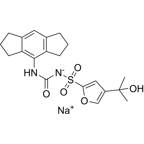
HY-12815A
MCC950 sodiumNLRP3 Inhibitor -
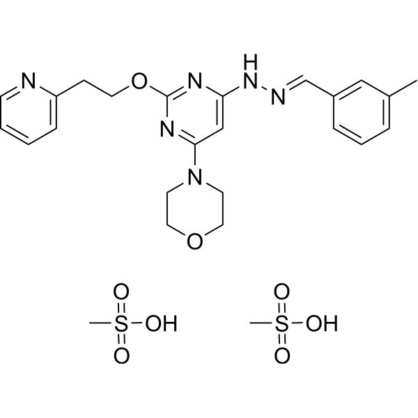
HY-14644A
Apilimod mesylateIL-12/IL-23 Inhibitor -
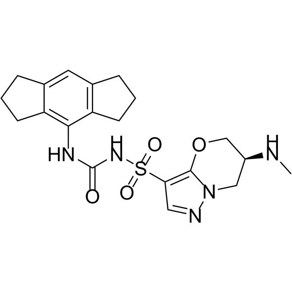
HY-148258
GDC-2394NLRP3 Inhibitor -
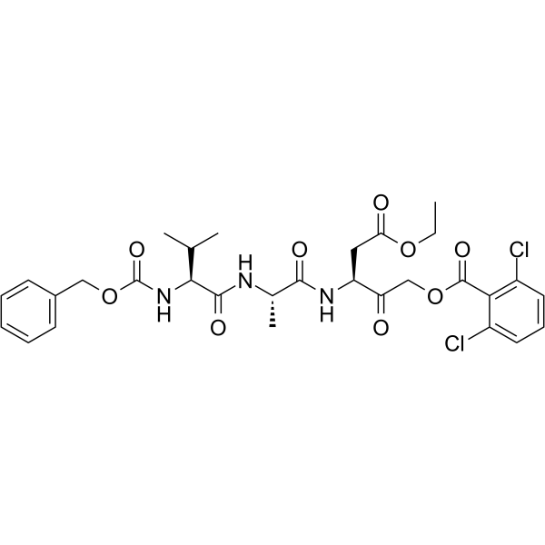
HY-141622
SDZ 224-015IL-1β Converting Enzyme Inhibitor -

HY-12815AR
MCC950 sodium (Standard)NLRP3 Inhibitor
Publications Citing Use of MedChemExpress (MCE) Ossirene
More Customer Validation & Images

-
RT-PCR

-
Histological Imaging/Staining

-
IHC
Ver todos los productos específicos de isoformas Caspase
More

Actividad biológica
Descripciòn
|
IL-1β |
IL-6 |
IL-8 |
IL-10 |
Caspase-1 |
In Vitro
Ossirene (AS101; 1 μg/mL; for 24 hours) almost completely abrogates expression of pStat3. Ossirene may reduce expression of Bcl-2 after inhibition of Stat3 activation via IL-10 inhibition[2].
AS101 (0.5, 5 mg/mL; 24 hours) inhibits IL-1β-induced mRNA expression of inflammatory mediators in the RPE in a dose-dependent manner. AS101 inhibits IL-1β-induced mRNA expression and protein production of IL-6 and IL-8 in RPE cells. AS101 (5 mg/mL; 1 hour) inhibits the phosphorylation of the p65 component of the NFκB complex activated by IL-1β[1].
Ossirene (0.1, 0.5, 1, 2.5 μg/mL) significantly decreases B16 melanoma, stomach adenocarcinoma, and human glioblastoma multiforme (GBM) cells proliferation[2].
AS101 (0.5 μg/mL; for 24 hours) sensitizes GBM tumor cells to paclitaxel in an IL-10-dependent manner[2].
MedChemExpress (MCE) has not independently confirmed the accuracy of these methods. They are for reference only. Further protocols information, click here.
-
Cell Line:B16 melanoma cells
-
Concentration:1 μg/mL
-
Incubation Time:For 24 hours
-
Result:Almost completely abrogated expression of pStat3.
-
Cell Line:ARPE19 cells
-
Concentration:0.5, 5 mg/mL
-
Incubation Time:24 hours
-
Result:Inhibited IL-1β-induced mRNA expression of inflammatory mediators in the RPE in a dose-dependent manner.
In Vivo
MedChemExpress (MCE) has not independently confirmed the accuracy of these methods. They are for reference only.
-
Animal Model:SCID mice with GBM cells[2]
-
Dosage:0.5 mg/kg
-
Administration:IP; daily; 25 days
-
Result:Significantly increased survival of GBM tumor-bearing mice.
Ensayo clínico
| NCT Number | Sponsor | Condition | Start Date |
Phase

|
|---|---|---|---|---|
| NCT01329991 | Plexxikon| | 2011-05 | PHASE1 |


Chemical Information
-
No. CAS 106566-58-9
-
Appearance Solid
-
Peso molecular 312.05
-
Fòrmula C2H8Cl3NO2Te
-
Color White to off-white
-
SMILES
[Cl-][Te+4]1([Cl-])([Cl-])[O-]CC[O-]1.[NH4+]
-
Synonyms
AS101
-
Envío
Room temperature in continental US; may vary elsewhere.
-
Almacenamiento
-20°C, stored under nitrogen
* In solvent : -80°C, 6 months; -20°C, 1 month (stored under nitrogen)


Publications (2)
-
Journal Impact Factor
-
Most Recent
-
Cell Death Dis
Targeting miR-18a sensitizes chondrocytes to anticytokine therapy to prevent osteoarthritis progression. [Abstract]2020 Nov 3;11(11):947. PMID: 33144571
Ossirene purchased from MedChemExpress. Usage Cited in: Cell Death Dis. 2020 Nov 3;11(11):947. [Abstract]
qRT-PCR shows the effect of miR-18a ectopic expression on chondrocyte hypertrophy in cells treated with either AS101 or JSH23.
Ossirene purchased from MedChemExpress. Usage Cited in: Cell Death Dis. 2020 Nov 3;11(11):947. [Abstract]
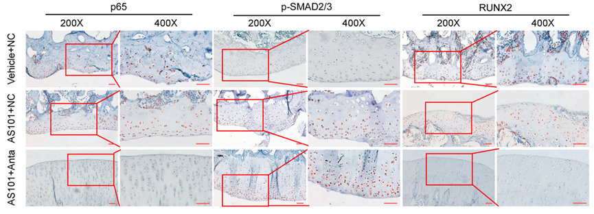
AS101 (1 mg/kg) was intraperitoneal injected, and 10 μL antagomir of miR-18a (250 nM) were intra-articular injected in rat model of knee OA every 3 days, followed by dissociation of knee joint after 4 weeks. HE staining of p65, p-SMAD2/3, and RUNX2 were conducted.
Ossirene purchased from MedChemExpress. Usage Cited in: Cell Death Dis. 2020 Nov 3;11(11):947. [Abstract]
AS101 (1 mg/kg) was intraperitoneal injected, and 10 μL antagomir of miR-18a (250 nM) were intra-articular injected in rat model of knee OA every 3 days, followed by dissociation of knee joint after 4 weeks. IHC of p65, p-SMAD2/3, and RUNX2 were conducted.
-
Inflamm Res
Alpha-galactosylceramide pre-treatment attenuates clinical symptoms of LPS-induced acute neuroinflammation by converting pathogenic iNKT cells to anti-inflammatory iNKT10 cells in the brain. [Abstract]2024 Sep;73(9):1511-1527. PMID: 39028491


Solvente y solubilidad
In Vitro:
DMSO : 12.5 mg/mL (40.06 mM; ultrasonic and warming and heat to 60°C; Hygroscopic DMSO has a significant impact on the solubility of product, please use newly opened DMSO)
Please refer to the solubility information to select the appropriate solvent. Once prepared, please aliquot and store the solution to prevent product inactivation from repeated freeze-thaw cycles.
Storage method and period of stock solution: -80°C, 6 months; -20°C, 1 month (stored under nitrogen). When stored at -80°C, please use it within 6 months. When stored at -20°C, please use it within 1 month.
Please refer to the solubility information to select the appropriate solvent. Once prepared, please aliquot and store the solution to prevent product inactivation from repeated freeze-thaw cycles.
Storage method and period of stock solution: -80°C, 6 months; -20°C, 1 month (stored under nitrogen). When stored at -80°C, please use it within 6 months. When stored at -20°C, please use it within 1 month.
Concentration (start) × Volume (start) = Concentration (final) × Volume (final)

Protocolo
-
Kinase activity and phosphorylation assays
Kinase activity assays measure the ability of kinases to transfer phosphate groups from ATP to specific substrates, while phosphorylation assays detect the presence and levels of phosphorylated proteins. Common methods include radiolabeled ATP incorporation (e. g. ,), ADP release detection via bioluminescence (e. g. ,[3]), enzyme-linked immunosorbent assays (ELISA) for phospho-specific epitopes (e. g. ,[6]), and microtiter-based formats for high-throughput screening (e. g. ,[8]). The ADP-Glo assay quantifies kinase activity by measuring ADP produced during phosphorylation using a luciferase-based system. Radiometric assays involve autoradiography or scintillation counting after incorporation of 32P-labeled ATP into substrate proteins. ELISA-based approaches rely on phospho-specific antibodies to detect activated kinases in cell lysates or purified samples.
-
Western Blot
Western blotting (WB) is a commonly used experimental method in molecular biology, biochemistry, and immunogenetics for identifying and quantifying target proteins. It combines gel electrophoresis with immunoassay, enabling researchers to analyze protein expression, post-translational modifications, and molecular weight.
-
Apoptosis Solutions
Apoptosis is a regulated, generally non-lytic cell-death pathway that removes unwanted, damaged, infected, or abnormal cells through coordinated morphological changes, caspase activation, DNA fragmentation, and membrane remodeling. The intrinsic apoptosis pathway is controlled mainly by mitochondrial outer membrane permeabilization, BCL-2 family proteins, cytochrome c release, apoptosome formation, caspase-9 activation, and downstream executioner caspase-3/7 activation. The extrinsic apoptosis pathway is initiated by death receptors such as Fas, TNFR, and TRAIL receptors, which recruit adaptor proteins and activate caspase-8 before engaging executioner caspases or mitochondrial amplification through BID cleavage. Apoptosis is linked to many phenotypes, including cancer cell killing, tissue homeostasis, immune regulation, neurodegeneration, infection response, and treatment-induced cytotoxicity; unresolved questions include how apoptosis interacts with necroptosis, pyroptosis, ferroptos
-
Protocol for Kinase activity and phosphorylation assays
Kinase activity assays measure transfer of phosphate from ATP to a protein or peptide substrate, generating phosphorylated substrate, ADP, or incorporated radiolabeled phosphate as the readout; phosphorylation assays measure site-specific phosphorylation in cells or tissues as a proxy for kinase-pathway activation, inhibition, or substrate regulation. Phosphorylation can be detected by phospho-specific Western blot, immunoprecipitation kinase assay, phospho-immunofluorescence, phospho-flow cytometry, luminescent ADP detection, radiolabeled ATP incorporation, or reporter-based pathway assays, and these readouts can be applied to cancer cells, primary neurons, mouse tumors, organoids, inflammatory macrophages, ferroptosis studies, and mitophagy studies when the kinase target is biologically relevant.
-
Pyroptosis Solutions
Pyroptosis is a lytic inflammatory cell-death pathway executed by gasdermin pores, most classically through inflammasome-mediated activation of caspase-1, cleavage of gasdermin D, membrane pore formation, LDH release, and secretion of IL-1β and IL-18. The canonical pathway is commonly modeled by priming cells with an inflammatory signal such as LPS to induce pro-IL-1β and inflammasome components, followed by an activation signal such as ATP or nigericin to activate NLRP3, ASC speck formation, caspase-1 cleavage, GSDMD cleavage, cytokine release, and pyroptotic membrane rupture. The non-canonical pathway is triggered when cytosolic LPS activates mouse caspase-11 or human caspase-4/5, leading to GSDMD cleavage and pyroptosis, and this can secondarily activate NLRP3-dependent IL-1β release. Pyroptosis is linked to inflammatory injury, infection, cancer, liver disease, ocular disease, placental inflammation, and other disease phenotypes, but unresolved questions include which gasdermin fam


Pureza y Documentación
-
Ficha de datos (284 KB)
-
SDS (394 KB)
- English - EN (394 KB)
- Français - FR (394 KB)
- Deutsch - DE (394 KB)
- Norwegian - NO (394 KB)
- Español - ES (394 KB)
- Swedish - SV (394 KB)
- Italian - IT (394 KB)
- Korean - KR (394 KB)
- Portuguese - PT (394 KB)
-
Instrucciones de manejo (2659 KB)
Referencias
[1]. Diamond Ling, et al. The Tellurium Redox Immunomodulating Compound AS101 Inhibits IL-1β-activated Inflammation in the Human Retinal Pigment Epithelium. Br J Ophthalmol. 2013 Jul;97(7):934-8. [Content Brief]
[2]. Sredni B, et al. Ammonium trichloro(dioxoethylene-o,o')tellurate (AS101) sensitizes tumors to chemotherapy by inhibiting the tumor interleukin 10 autocrine loop. Cancer Res. 2004 Mar 1;64(5):1843-52. [Content Brief]
[3]. Yona Kalechman, et al. Inhibition of interleukin-10 by the Immunomodulator AS101 Reduces Mesangial Cell Proliferation in Experimental Mesangioproliferative Glomerulonephritis: Association With Dephosphorylation of STAT3. J Biol Chem. 2004 Jun 4;279(23):24724-32. [Content Brief]
[4]. Yafit Hachmo, et al. The Small Tellurium Compound AS101 Ameliorates Rat Crescentic Glomerulonephritis: Association With Inhibition of Macrophage Caspase-1 Activity via Very Late Antigen-4 Inactivation. Front Immunol. 2017 Mar 7;8:240. [Content Brief]


Complete Stock Solution Preparation Table
Please refer to the solubility information to select the appropriate solvent. Once prepared, please aliquot and store the solution to prevent product inactivation from repeated freeze-thaw cycles.
Storage method and period of stock solution: -80°C, 6 months; -20°C, 1 month (stored under nitrogen). When stored at -80°C, please use it within 6 months. When stored at -20°C, please use it within 1 month.
| Optional Solvent | Concentration Solvent Mass | 1 mg | 5 mg | 10 mg | 25 mg |
|---|---|---|---|---|---|
| DMSO | 1 mM | 3.2046 mL | 16.0231 mL | 32.0461 mL | 80.1154 mL |
| 5 mM | 0.6409 mL | 3.2046 mL | 6.4092 mL | 16.0231 mL | |
| 10 mM | 0.3205 mL | 1.6023 mL | 3.2046 mL | 8.0115 mL | |
| 15 mM | 0.2136 mL | 1.0682 mL | 2.1364 mL | 5.3410 mL | |
| 20 mM | 0.1602 mL | 0.8012 mL | 1.6023 mL | 4.0058 mL | |
| 25 mM | 0.1282 mL | 0.6409 mL | 1.2818 mL | 3.2046 mL | |
| 30 mM | 0.1068 mL | 0.5341 mL | 1.0682 mL | 2.6705 mL | |
| 40 mM | 0.0801 mL | 0.4006 mL | 0.8012 mL | 2.0029 mL |


Ossirene Related Classifications
HY-101019 Related Classifications
 Powered by Bioz