



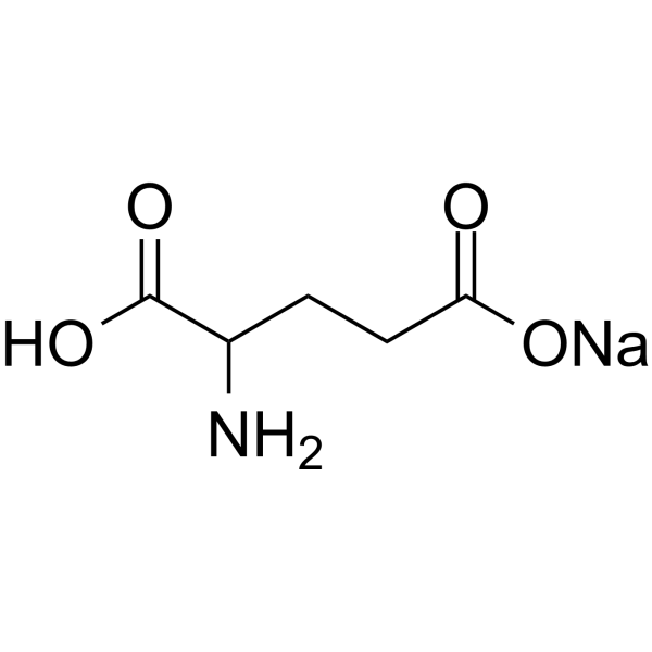
Glutamic acid sodium salt
Based on 1 publication(s) in Google Scholar
Glutamic acid sodium salt (Monosodium glutamate) is an orally active food flavor enhancer. Glutamic acid sodium salt causes ROS generation, mitochondrial dysfunction, and Apoptosis. Glutamic acid sodium salt upregulates CHOP, Grp78, and Bcl-2. Glutamic acid sodium salt impairs cognition, induces depressive-like behavior, induces hyperalgesia, and induces obesity and insulin resistance. Glutamic acid sodium salt can be used to study neurotoxicity (e.g., brain damage, cognitive impairment), metabolic disorders (e.g., obesity, insulin resistance), hepatotoxicity, and renal toxicity, as well as pain-related disorders.
For research use only. We do not sell to patients.
- Purity : 99.92%
- CAS No.: 32221-81-1
- Formula: C5H8NNaO4
- Molecular Weight:169.11
-
Storage:
4°C, sealed storage, away from moisture and light
* In solvent : -80°C, 6 months; -20°C, 1 month (sealed storage, away from moisture and light)

To place orders, for customer services and technical support, please contact: MedChemExpress USA
Tel: 609-228-6898 E-mail: [email protected] [email protected]


-
 Biological Activity
-
Chemical Information
-
Solvent & Solubility
- Protocols
- Purity & Documentation
- References
-
 Help & FAQs


-
HY-N0390
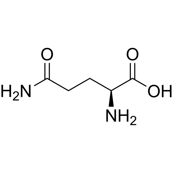
L-GlutamineEndogenous Metabolite -
HY-N0666C
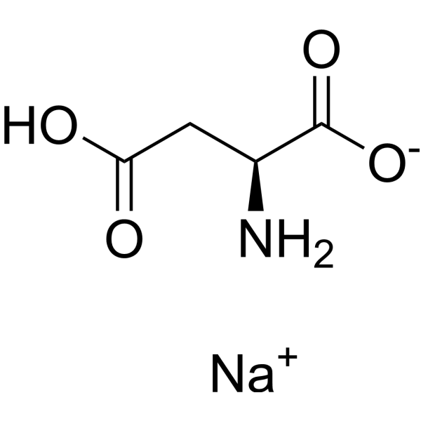
L-Aspartic aicd sodiumNMDA Receptor Agonist -
HY-13417
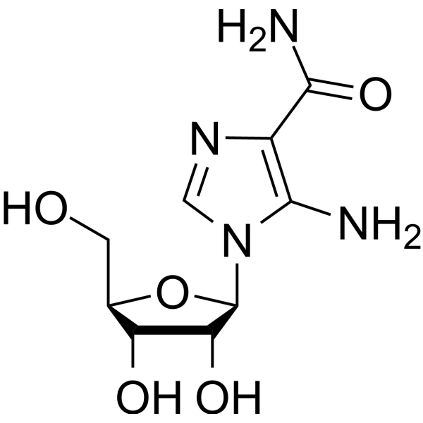
AICARAMPK Activator -
HY-W015410
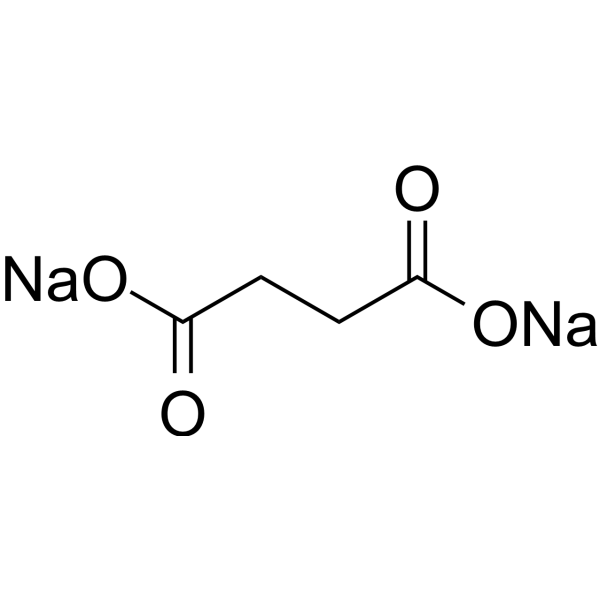
Disodium succinateFood Additive, Platform chemical -
HY-W017443
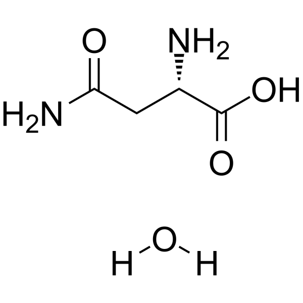
L-Asparagine monohydrateL-Asparaginase Substrate -

HY-W250303A
Malic acid disodium hydrateBiochemical Assay Reagent
Publications Citing Use of MedChemExpress (MCE) Glutamic acid sodium salt
More

Biological Activity
Description
|
Grp78 |
HSPA5 |
Bcl-2 |
In Vitro
Monosodium glutamate (20 mM; 12-24 h) induces apoptotic cell death in C6 astrocytic cells by promoting reactive oxygen species generation, glutathione depletion, mitochondrial dysfunction, and endoplasmic reticulum stress[1].
MedChemExpress (MCE) has not independently confirmed the accuracy of these methods. They are for reference only. Further protocols information, click here.
-
Cell Line:C6 astrocytic cells
-
Concentration:20 mM
-
Incubation Time:12 h
-
Result:Up-regulated protein levels of CHOP, Grp78, and Bcl-2.
In Vivo
Monosodium glutamate (4 mg/g b.w.; i.p.) induces liver and kidney damage in Wistar rats[3].
Monosodium glutamate (8 mg/kg; p.o. via drinking water; 1 month) impairs cognitive behavior in male Swiss albino mice, and its combination with Aspartame (HY-B0361) exacerbates neurotransmitter (dopamine, serotonin) reduction and oxidative stress in the forebrain[4].
Monosodium glutamate (100-500 mg/kg; p.o.; daily for 21 days) induces depressive-like behavior, brain oxidative stress, and elevated liver enzymes in male Swiss mice[5].
Monosodium glutamate (300 mg/kg; p.o.; 21 days) reduces pain threshold and increases brain nitric oxide levels in male NMRI mice, inducing hyperalgesia[6].
Monosodium glutamate (4 g/kg b.w.; s.c.; once daily for 7 consecutive days) induces obesity and insulin resistance in female ICR mice[7].
Monosodium glutamate (0.5%-5% in diet or oral gavage) does not affect pulmonary inflammation, Th2 cytokine production, IgE levels, or airway hyperresponsiveness in Ovalbumin-induced asthmatic BALB/c mice[8].
MedChemExpress (MCE) has not independently confirmed the accuracy of these methods. They are for reference only.
-
Animal Model:Male Wistar rats (225-250 g)[3]
-
Dosage:4 mg/g b.w.
-
Administration:Intraperitoneal injection
-
Result:Caused turbid swelling, vacuolar degeneration and necrosis in the liver, glomerular compaction and tubular destruction in the kidneys.
Increased serum ALAT, ASAT and lipid peroxidation products (MDA, 4-HDA).
Clinical Trial
| NCT Number | Sponsor | Condition | Start Date |
Phase

|
|---|---|---|---|---|
| NCT01329991 | Plexxikon| | 2011-05 | PHASE1 |


Chemical Information
-
CAS No. 32221-81-1
-
Appearance Solid
-
Molecular Weight 169.11
-
Formula C5H8NNaO4
-
Color White to off-white
-
SMILES
O=C(CCC(C(O)=O)N)O[Na]
-
Synonyms
Monosodium glutamate
-
Shipping
Room temperature in continental US; may vary elsewhere.
-
Storage
4°C, sealed storage, away from moisture and light
* In solvent : -80°C, 6 months; -20°C, 1 month (sealed storage, away from moisture and light)


Publications (1)
-
Journal Impact Factor
-
Most Recent
-
Microorganisms
Monosodium Glutamate Inhibits Pseudomonas aeruginosa-Induced Acute Lung Injury by Targeting the Type III Secretion Systems and Modulating Host Immunity. [Abstract]2026 Mar 23;14(3):725. PMID: 41900484


Solvent & Solubility
In Vitro:
H2O : 250 mg/mL (1478.33 mM; Need ultrasonic)
Please refer to the solubility information to select the appropriate solvent. Once prepared, please aliquot and store the solution to prevent product inactivation from repeated freeze-thaw cycles.
Storage method and period of stock solution: -80°C, 6 months; -20°C, 1 month (sealed storage, away from moisture and light). When stored at -80°C, please use it within 6 months. When stored at -20°C, please use it within 1 month.
* Note: If you choose water as the stock solution, please dilute it to the working solution, then filter and sterilize it with a 0.22 μm filter before use.
Please refer to the solubility information to select the appropriate solvent. Once prepared, please aliquot and store the solution to prevent product inactivation from repeated freeze-thaw cycles.
Storage method and period of stock solution: -80°C, 6 months; -20°C, 1 month (sealed storage, away from moisture and light). When stored at -80°C, please use it within 6 months. When stored at -20°C, please use it within 1 month.
* Note: If you choose water as the stock solution, please dilute it to the working solution, then filter and sterilize it with a 0.22 μm filter before use.
Concentration (start) × Volume (start) = Concentration (final) × Volume (final)

Protocols
-
Neurotoxicity Study
This protocol assesses in vitro neurotoxicity by combining neuronal viability, mitochondrial/metabolic activity, neurite outgrowth, and optional neuronal network function readouts. Calcein-AM or resazurin/PrestoBlue readouts estimate viable or metabolically active cells; βIII-tubulin immunofluorescence detects neuronal morphology and neurite networks; TMRE detects mitochondrial membrane potential; and MEA recordings detect functional changes in neuronal network activity.
-
Nephrotoxicity Study
This protocol assesses nephrotoxicity by combining functional kidney injury readouts, urinary/tissue injury biomarkers, and renal histopathology. Serum creatinine and BUN reflect impaired kidney function, while KIM-1, NGAL, clusterin, osteopontin, IL-18, cystatin C, nephrin, Oat5, urinary protein, glucose, and alkaline phosphatase have been used to detect tubular injury in cisplatin-, gentamicin-, and acetaminophen-induced nephrotoxicity models.
-
Annexin V plus membrane-impermeant dye apoptosis staining
Annexin V-based apoptosis assays rely on the detection of phosphatidylserine (PS) externalization from the inner leaflet of the plasma membrane to the outer leaflet, an early biochemical hallmark of apoptosis. Fluorescently labeled Annexin V binds PS in a calcium-dependent manner, enabling identification of early apoptotic cells by flow cytometry or fluorescence microscopy. When combined with a membrane-impermeant DNA-binding dye (e. g. , propidium iodide), this approach allows discrimination between viable (Annexin V−/dye−), early apoptotic (Annexin V+/dye−), and late apoptotic or necrotic (Annexin V+/dye+) cell populations by assessing membrane integrity and PS exposure.
-
ROS/oxidative-stress fluorescent staining
ROS/oxidative-stress fluorescent staining uses cell-permeant fluorogenic probes that become fluorescent after oxidation inside cells or tissues; commonly used examples include DCFH-DA/DCFDA for broad cellular oxidant detection, DHE for superoxide-related signal detection, MitoSOX for mitochondrial superoxide-related signal detection, and CellROX probes for oxidative-stress-associated fluorescence readouts. The assay detects probe oxidation rather than a single ROS species unless the probe and analysis method have been chemically validated for that species. DCFH-DA enters cells, is deacetylated by intracellular esterases to DCFH, and produces fluorescent DCF after oxidation, so the readout is used as an operational measure of total cellular oxidative stress rather than a species-specific ROS measurement. DHE and MitoSOX can report superoxide-related oxidation, but red fluorescence alone can include non-specific ethidium-like oxidation products; HPLC or optimized spectral approaches are
-
Protocol for Tail Suspension Test (TST)
The Tail Suspension Test is a mouse behavioral assay in which an animal is suspended by the tail in an inescapable position, causing alternating escape-directed activity and immobility; the main readout is immobility time, and antidepressant-like treatments generally reduce immobility compared with vehicle controls. The assay detects behavioral response to acute inescapable stress rather than a molecular event; immobility is interpreted as passive stress-coping behavior, while reduced immobility is used as a predictive screen for antidepressant-like activity, with important limitations related to strain, locomotor activity, and tail-climbing behavior.
-
Research Protocol for Metabolic Diseases
AMP-activated protein kinase, AMPK, is a conserved cellular energy sensor that responds to reduced cellular energy status and coordinates metabolism by increasing ATP-generating catabolic pathways while suppressing ATP-consuming anabolic processes. In metabolic disease research, the AMPK pathway is experimentally relevant because it regulates hepatic lipid synthesis, fatty acid oxidation, glucose production, skeletal-muscle glucose disposal, mTORC1-linked biosynthesis, autophagy, mitochondrial homeostasis, and whole-body energy balance. The central pathway logic is that energy stress, metformin, exercise-like stimulation, or direct AMPK activators increase AMPKα Thr172 phosphorylation and downstream substrate phosphorylation, including ACC and RAPTOR. Phosphorylation of ACC suppresses lipogenesis and supports fatty acid oxidation, whereas phosphorylation of RAPTOR suppresses mTORC1 signaling and links cellular energy status to growth and protein synthesis control. The pathway is linked
-
Alzheimer’s Disease Modeling
Alzheimer’s Disease (AD) is a neurodegenerative disorder characterized by a progressive decline in cognitive functions and loss of specific types of neurons and synapses. Alzheimer's symptoms can be simulated in mice by injecting drugs (such as Aβ) or genetically modified.
-
Apoptosis
Apoptosis, also called programmed cell death, is generally characterized by distinct morphological characteristics.
-
TUNEL staining for apoptotic DNA fragmentation
TUNEL staining detects DNA strand breaks by using terminal deoxynucleotidyl transferase to add labeled nucleotides to exposed 3′-OH DNA termini, generating either microscopic staining in fixed cells or tissue sections, or fluorescence/cytometric signal in cell suspensions. TUNEL positivity reflects DNA fragmentation but should not be interpreted alone as definitive apoptosis, because TUNEL can also label necrotic, autolytic, mechanically damaged, or DNA-repair-associated DNA breaks.
-
Research Protocol for Endocrine Diseases
Endocrine diseases often arise from disrupted hormone production, hormone signaling, or target-tissue responsiveness; for diabetes-focused endocrine disease models, insulin signaling regulates glucose uptake, hepatic glucose output, lipid metabolism, and β-cell compensation. Type 2 diabetes develops through interacting defects in insulin resistance, β-cell dysfunction, adipose inflammation, hepatic glucose overproduction, altered incretin signaling, and ectopic lipid metabolism. A major unresolved question is whether endocrine dysfunction is driven primarily by target-tissue insulin resistance, intrinsic β-cell failure, immune/inflammatory stress, or combined multi-organ failure that differs by disease stage.
-
Mitochondrial membrane-potential and mitochondrial mass staining
Mitochondrial membrane potential staining measures the electrochemical polarization across the mitochondrial inner membrane in live cells using lipophilic cationic fluorescent probes; early rhodamine-based work showed that selective mitochondrial dye accumulation is lost when the mitochondrial transmembrane potential is dissipated. JC-1 reports mitochondrial polarization by shifting from green monomer fluorescence to red J-aggregate fluorescence as dye concentration increases within energized mitochondria; therefore, the red/green fluorescence ratio is used as a relative readout of mitochondrial membrane potential. TMRE or TMRM staining provides a single-channel relative readout because these cationic rhodamine esters accumulate in polarized mitochondria, and lower fluorescence indicates reduced mitochondrial polarization when acquisition and dye-loading conditions are controlled. Mitochondrial mass staining is commonly performed with MitoTracker Green FM or related MitoTracker dyes as
-
Apoptosis Solutions
Apoptosis is a regulated, generally non-lytic cell-death pathway that removes unwanted, damaged, infected, or abnormal cells through coordinated morphological changes, caspase activation, DNA fragmentation, and membrane remodeling. The intrinsic apoptosis pathway is controlled mainly by mitochondrial outer membrane permeabilization, BCL-2 family proteins, cytochrome c release, apoptosome formation, caspase-9 activation, and downstream executioner caspase-3/7 activation. The extrinsic apoptosis pathway is initiated by death receptors such as Fas, TNFR, and TRAIL receptors, which recruit adaptor proteins and activate caspase-8 before engaging executioner caspases or mitochondrial amplification through BID cleavage. Apoptosis is linked to many phenotypes, including cancer cell killing, tissue homeostasis, immune regulation, neurodegeneration, infection response, and treatment-induced cytotoxicity; unresolved questions include how apoptosis interacts with necroptosis, pyroptosis, ferroptos
-
Hepatotoxicity Study
This protocol evaluates hepatotoxicity using complementary in vivo mouse APAP acute liver injury and in vitro hepatocyte-based cytotoxicity readouts. In vivo APAP injury is assessed by serum ALT/AST, liver histology, hepatic glutathione, protein adducts, necrosis, inflammation, and regeneration-related endpoints. In vitro hepatotoxicity is assessed by loss of viability, leakage of ALT/AST/LDH, oxidative-stress markers, mitochondrial function, nuclear morphology, intracellular calcium, and high-content imaging endpoints.
-
Protocol for Forced Swim Test (FST)
The Forced Swim Test is a rodent behavioral assay in which a mouse or rat is placed in an inescapable cylinder of water, and the main readout is the time spent immobile versus active escape-related behaviors such as swimming or climbing. Reduced immobility after treatment has historically been interpreted as antidepressant-like activity, but the assay should be interpreted as a behavioral response to acute inescapable stress rather than a complete model of human depression.
-
Protocol for Sucrose Preference Test (SPT)
The Sucrose Preference Test is a rodent two-bottle choice assay used to estimate reward-related behavior by measuring preference for a sweet sucrose solution over water. Reduced sucrose preference is commonly interpreted as an anhedonia-like phenotype in stress-based depression models, but it can also be affected by thirst, hunger, body weight, learning, motivation, and general fluid intake.


Purity & Documentation
-
Data Sheet (276 KB)
-
SDS (393 KB)
- English - EN (393 KB)
- Français - FR (393 KB)
- Deutsch - DE (393 KB)
- Norwegian - NO (393 KB)
- Español - ES (393 KB)
- Swedish - SV (393 KB)
- Italian - IT (393 KB)
- Korean - KR (393 KB)
- Portuguese - PT (393 KB)
-
Handling Instructions (2659 KB)
References
[1]. Park E, et al. Protective effects of N-acetylcysteine against monosodium glutamate-induced astrocytic cell death. Food Chem Toxicol. 2014 May;67:1-9. [Content Brief]
[2]. Rycerz K, et al. Effects of monosodium glutamate treatment on calretinin-immunoreactive neurons in hippocampus of postnatal rats. Folia Histochem Cytobiol. 2014;52(4):281-8. [Content Brief]
[3]. Ortiz GG, et al. Monosodium glutamate-induced damage in liver and kidney: a morphological and biochemical approach. Biomed Pharmacother. 2006 Feb;60(2):86-91. [Content Brief]
[4]. Abu-Taweel GM, et al. Cognitive and biochemical effects of monosodium glutamate and aspartame, administered individually and in combination in male albino mice. Neurotoxicol Teratol. 2014 Mar-Apr;42:60-7. [Content Brief]
[6]. Zanfirescu A, et al. Chronic Monosodium Glutamate Administration Induced Hyperalgesia in Mice. Nutrients. 2017 Dec 21;10(1):1 [Content Brief]
[7]. Zhang N , et al. Atorvastatin improves insulin sensitivity in mice with obesity induced by monosodium glutamate. Acta Pharmacol Sin. 2010 Jan;31(1):35-42. [Content Brief]
[8]. Yoneda J, et al. Effects of oral monosodium glutamate in mouse models of asthma. Food Chem Toxicol. 2011 Jan;49(1):299-304. [Content Brief]


Complete Stock Solution Preparation Table
Please refer to the solubility information to select the appropriate solvent. Once prepared, please aliquot and store the solution to prevent product inactivation from repeated freeze-thaw cycles.
Storage method and period of stock solution: -80°C, 6 months; -20°C, 1 month (sealed storage, away from moisture and light). When stored at -80°C, please use it within 6 months. When stored at -20°C, please use it within 1 month.
| Optional Solvent | Concentration Solvent Mass | 1 mg | 5 mg | 10 mg | 25 mg |
|---|---|---|---|---|---|
| H2O | 1 mM | 5.9133 mL | 29.5666 mL | 59.1331 mL | 147.8328 mL |
| 5 mM | 1.1827 mL | 5.9133 mL | 11.8266 mL | 29.5666 mL | |
| 10 mM | 0.5913 mL | 2.9567 mL | 5.9133 mL | 14.7833 mL | |
| 15 mM | 0.3942 mL | 1.9711 mL | 3.9422 mL | 9.8555 mL | |
| 20 mM | 0.2957 mL | 1.4783 mL | 2.9567 mL | 7.3916 mL | |
| 25 mM | 0.2365 mL | 1.1827 mL | 2.3653 mL | 5.9133 mL | |
| 30 mM | 0.1971 mL | 0.9856 mL | 1.9711 mL | 4.9278 mL | |
| 40 mM | 0.1478 mL | 0.7392 mL | 1.4783 mL | 3.6958 mL | |
| 50 mM | 0.1183 mL | 0.5913 mL | 1.1827 mL | 2.9567 mL | |
| 60 mM | 0.0986 mL | 0.4928 mL | 0.9856 mL | 2.4639 mL | |
| 80 mM | 0.0739 mL | 0.3696 mL | 0.7392 mL | 1.8479 mL | |
| 100 mM | 0.0591 mL | 0.2957 mL | 0.5913 mL | 1.4783 mL |
* Note: If you choose water as the stock solution, please dilute it to the working solution, then filter and sterilize it with a 0.22 μm filter before use.


Glutamic acid sodium salt Related Classifications
HY-W250122 Related Classifications
 Powered by Bioz