



HDAC-IN-71
Based on 1 publication(s) in Google Scholar
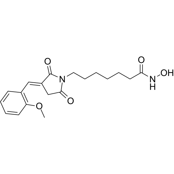
HDAC-IN-71 (Compound 17q) is a potent HDAC inhibitor with IC50 values of 12.6, 14.1, 20, 3, and 72 nM for HDAC1, HDAC2, HDAC3, HDAC6, and HDAC10, respectively. HDAC-IN-71 induces apoptosis and can be used in cancer research.
Nur für Forschungszwecke. Wir verkaufen nicht an Patienten.
- CAS. Nr.: 2995354-52-2
- Formel: C19H24N2O5
- Molecular Weight:360.40
-
Speicherung:
Please store the product under the recommended conditions in the Certificate of Analysis.

To place orders, for customer services and technical support, please contact: MedChemExpress USA
Tel: 609-228-6898 E-mail: [email protected] [email protected]


-
 Biologische Aktivität
-
Chemical Information
- Protokoll
- Reinheit & Dokumentation
- Verweise
-
 Help & FAQs


Publications Citing Use of MedChemExpress (MCE) HDAC-IN-71
More

Biologische Aktivität
Beschreibung
IC50 & Target
|
HDAC1 12.6 nM (IC50) |
HDAC2 14.1 nM (IC50) |
HDAC3 20 nM (IC50) |
HDAC6 3 nM (IC50) |
HDAC10 72 nM (IC50) |
Cellular Effect
|
Cell Line

|
Type | Value | Description | References |
|---|---|---|---|---|
| A549 | IC50 |
1.39 μM
Compound: 17q
|
Antiproliferative activity against human A549 cells incubated for 96 hrs by MTT assay
Antiproliferative activity against human A549 cells incubated for 96 hrs by MTT assay
|
[PMID: 38503167] |
| DU-145 | IC50 |
0.16 μM
Compound: 17q
|
Antiproliferative activity against human DU-145 cells incubated for 96 hrs by MTT assay
Antiproliferative activity against human DU-145 cells incubated for 96 hrs by MTT assay
|
[PMID: 38503167] |
| HCT-116 | IC50 |
0.47 μM
Compound: 17q
|
Antiproliferative activity against human HCT-116 cells incubated for 96 hrs by MTT assay
Antiproliferative activity against human HCT-116 cells incubated for 96 hrs by MTT assay
|
[PMID: 38503167] |
| HT-29 | IC50 |
1.05 μM
Compound: 17q
|
Antiproliferative activity against human HT-29 cells incubated for 96 hrs by MTT assay
Antiproliferative activity against human HT-29 cells incubated for 96 hrs by MTT assay
|
[PMID: 38503167] |
| LNCaP | IC50 |
0.63 μM
Compound: 17q
|
Antiproliferative activity against human LNCaP cells incubated for 96 hrs by MTT assay
Antiproliferative activity against human LNCaP cells incubated for 96 hrs by MTT assay
|
[PMID: 38503167] |
| MCF7 | IC50 |
0.35 μM
Compound: 17q
|
Antiproliferative activity against human MCF7 cells incubated for 96 hrs by MTT assay
Antiproliferative activity against human MCF7 cells incubated for 96 hrs by MTT assay
|
[PMID: 38503167] |
| MDA-MB-231 | IC50 |
0.31 μM
Compound: 17q
|
Antiproliferative activity against human MDA-MB-231 cells incubated for 96 hrs by MTT assay
Antiproliferative activity against human MDA-MB-231 cells incubated for 96 hrs by MTT assay
|
[PMID: 38503167] |
| NCI-H1650 | IC50 |
3.56 μM
Compound: 17q
|
Antiproliferative activity against human NCI-H1650 cells incubated for 96 hrs by MTT assay
Antiproliferative activity against human NCI-H1650 cells incubated for 96 hrs by MTT assay
|
[PMID: 38503167] |
| NCI-H446 | IC50 |
0.65 μM
Compound: 17q
|
Antiproliferative activity against human NCI-H446 cells incubated for 96 hrs by MTT assay
Antiproliferative activity against human NCI-H446 cells incubated for 96 hrs by MTT assay
|
[PMID: 38503167] |
| PC-3 | IC50 |
0.72 μM
Compound: 17q
|
Antiproliferative activity against human PC-3 cells incubated for 96 hrs by MTT assay
Antiproliferative activity against human PC-3 cells incubated for 96 hrs by MTT assay
|
[PMID: 38503167] |
In Vitro
HDAC-IN-71 (0-1.6 μM; 24 h) inhibits colony formation in a dose-dependent manner and completely inhibits colony formation of DU145 cells at a concentration of 3.2 μM[1].
HDAC-IN-71 (0-1.6 μM; 72 h) arrests cells in G2/M phase and induces apoptosis in DU145 cell line in a concentration-dependent manner[1].
MedChemExpress (MCE) has not independently confirmed the accuracy of these methods. They are for reference only.
-
Cell Line:DU145 cells
-
Concentration:0, 0.2, 0.4, 0.8, 1.6 μM
-
Incubation Time:12 h
-
Result:Inhibited cancer cell migration.
-
Cell Line:DU145 cells
-
Concentration:0, 0.2, 0.4, 0.8, 1.6 μM
-
Incubation Time:72 h
-
Result:Arrested cells in G2/M phase in a dose-dependent manner.
-
Cell Line:DU145 cells
-
Concentration:0.8, 1.6, 3.2 μM
-
Incubation Time:24 h
-
Result:Induced histone acetylation and inhibited HDAC6 expression.
In Vivo
Pharmacokinetic analysis[1]
| Parameters | I.V. (2 mg/kg) | P.O. (10 mg/kg) |
| AUC0-∞ (h ng/mL) | 1633.35 | 3799.46 |
| T1/2 (h) | 3.12 | 2.87 |
| CL (L/h/kg) | 1.57 | |
| V (L/kg) | 4.63 | |
| Cmax(ng/mL) | 851.5 | 774.0 |
| F | 46.52% |
MedChemExpress (MCE) has not independently confirmed the accuracy of these methods. They are for reference only.
-
Animal Model:Balb/c nude mice[1]
-
Dosage:100, 200 mg/kg
-
Administration:Oral administration; daily; 40 days
-
Result:Significantly inhibited prostate cancer (PCa) tumor growth.


Chemical Information
-
CAS. Nr. 2995354-52-2
-
Molecular Weight 360.40
-
Formel C19H24N2O5
-
SMILES
COC1=C(C=CC=C1)/C=C2CC(N(C\2=O)CCCCCCC(NO)=O)=O
-
Versand
Room temperature in continental US; may vary elsewhere.
-
Speicherung
Please store the product under the recommended conditions in the Certificate of Analysis.


Publications (1)
-
Journal Impact Factor
-
Most Recent


Reinheit & Dokumentation
Verweise

Calculators
Konzentration (Stammlösung) × Volumen (Stammlösung) = Konzentration (Ziellösung) × Volumen (Ziellösung)


HDAC-IN-71 Related Classifications
HY-163430 Related Classifications
 Powered by Bioz