



Hibifolin

Based on 1 Customer Validation
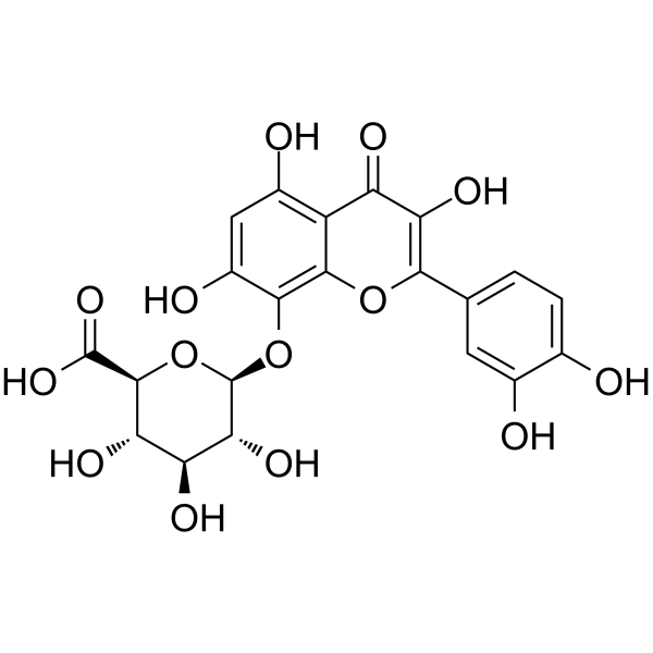
Hibifolin is a flavonol glycoside that can be isolated from Helicteres isora. Hibifolin is an inhibitor of adenosine deaminase (ADA) (Ki = 49.92 μM). Hibifolin protects neurons against β-amyloid-induced neurotoxicity. Hibifolin possesses a potent protective activity against cell death induced by aggregated Aβ. Hibifolin can abolish Aβ-induced caspase-3 and caspase-7 activation. Hibifolin induces Akt phosphorylation in cortical neurons. Hibifolin is also a natural sortase A (SrtA) inhibitor (IC50 = 31.2 μM) through direct binding to SrtA protein. Hibifolin attenuates the pathogenic behavior of Staphylococcus aureus including adhesion, invasion, and biofilm formation. Hibifolin improves the survival of pneumonia induced by Staphylococcus aureus in mouse model and alleviates pathological damage. Hibifolin shows a synergistic antibacterial effect with Cefotaxime (HY-A0088A).
For research use only. We do not sell to patients.
- Purity : 99.70%
- CAS No.: 55366-56-8
- Formula: C21H18O14
- Molecular Weight:494.36
-
Storage:
4°C, protect from light
* In solvent : -80°C, 6 months; -20°C, 1 month (protect from light)

To place orders, for customer services and technical support, please contact: MedChemExpress USA
Tel: 609-228-6898 E-mail: [email protected] [email protected]


-
 Biological Activity
-
Chemical Information
-
Solvent & Solubility
- Protocols
- Purity & Documentation
- References
-
 Help & FAQs


-
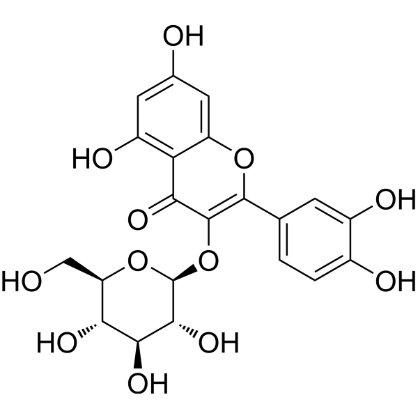
HY-N1445
IsoquercitrinNO Synthase Inhibitor -
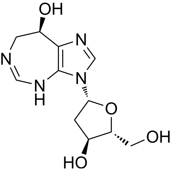
HY-A0006
PentostatinAntibiotic -
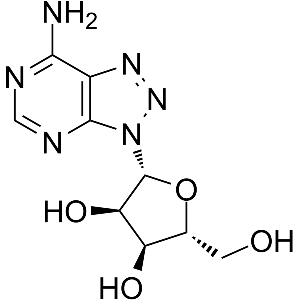
HY-115686
8-AzaadenosineADAR1 Inhibitor -
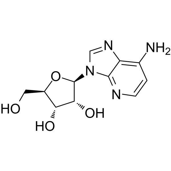
HY-103159
1-DeazaadenosineADA Inhibitor -

HY-N7368R
Hibifolin (Standard)ADA Inhibitor -
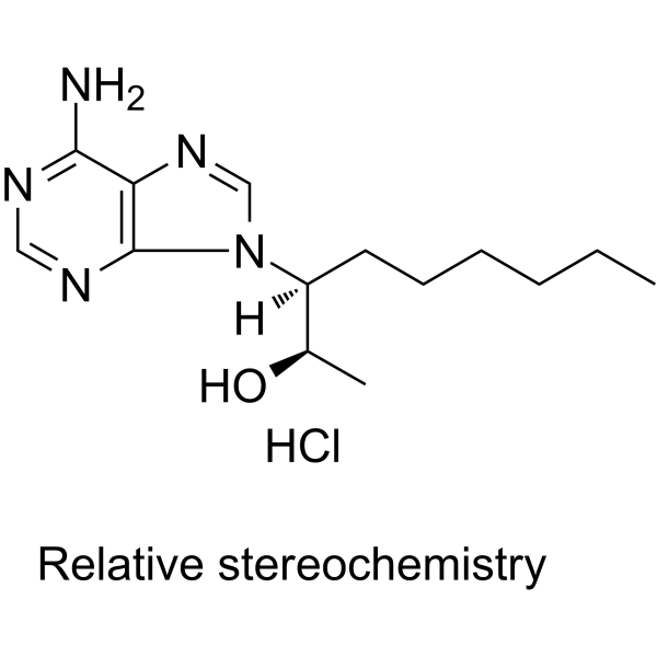
HY-103160A
EHNA hydrochloridePDE2 Inhibitor -
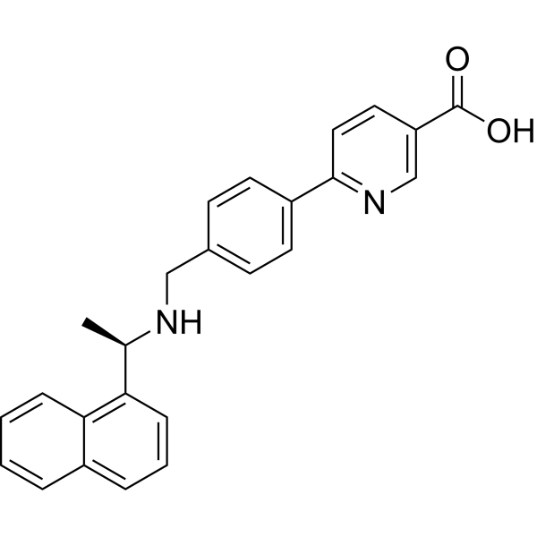
HY-103673
AMPD2 inhibitor 1AMPD2 Inhibitor -

HY-A0006R
Pentostatin (Standard)Antibiotic -
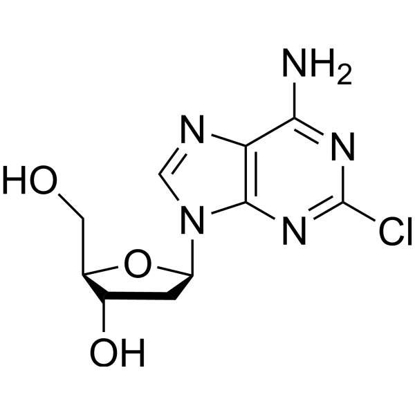
HY-13599
CladribineDNA Synthesis Inhibitor -
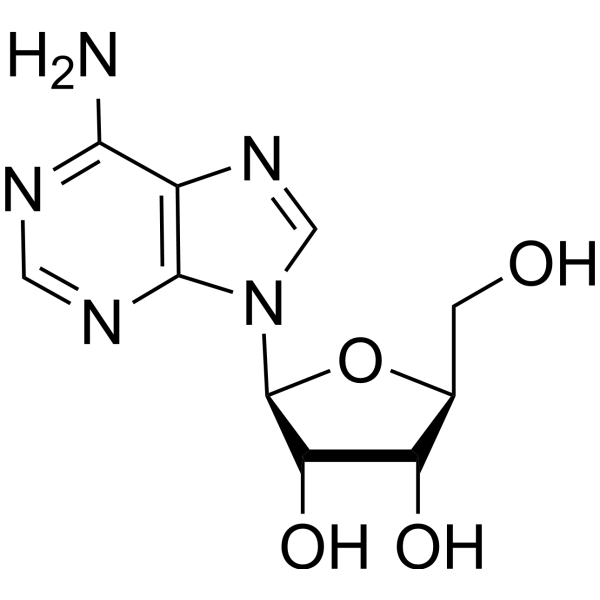
HY-103237
L-AdenosineADA Inhibitor -

HY-103673R
AMPD2 inhibitor 1 (Standard)Reference Standard -

HY-103237R
L-Adenosine (Standard)Reference Standard
All Caspase Isoforms
More

Biological Activity
Description
IC50 & Target
Ki: 49.92 μM (ADA)[1]
In Vitro
Hibifolin (100 μM) shows about 60% inhibition of the ADA (10 μM) enzymatic activity[1].
Hibifolin (0.5-50 μM, 24 h) reduces the release of LDH induced by Aβ in cortical neurons[2].
Hibifolin (50 μM, 0-120 sec) reduces the amount of Aβ-induced Ca2+ by 70% in cortical neurons[2].
Hibifolin (50 μM) induces the phosphorylation of Akt to approximately 2-fold[2].
Hibifolin (0-1000 μg/mL) remarkably inhibits the activity of SrtA with an IC50 of 31.2 μM[3].
Hibifolin (32-256 μg/mL) decreases the adhesion of S. fibrinogen to fribinogen with the concentration of 256 μg/mL and significantly inhibits bacterial invasion of A549 cells[3].
MedChemExpress (MCE) has not independently confirmed the accuracy of these methods. They are for reference only. Further protocols information, click here.
-
Cell Line:Cortical neurons
-
Concentration:0.5, 5, 50 μM
-
Incubation Time:24 h
-
Result:Had protective effect by preventing Aβ-induced cell death dose-dependently.
-
Cell Line:Cortical neurons
-
Concentration:50 μM
-
Incubation Time:24 h
-
Result:Blocked the DNA fragmentation triggered by Aβ.


Chemical Information
-
CAS No. 55366-56-8
-
Appearance Solid
-
Molecular Weight 494.36
-
Formula C21H18O14
-
Color Yellow to orange
-
SMILES
O[C@H]([C@H]([C@@H]([C@@H](C(O)=O)O1)O)O)[C@@H]1OC2=C3C(C(C(O)=C(C4=CC=C(O)C(O)=C4)O3)=O)=C(O)C=C2O
-
Structure Classification
-
Initial Source
-
Shipping
Room temperature in continental US; may vary elsewhere.
-
Storage
4°C, protect from light
* In solvent : -80°C, 6 months; -20°C, 1 month (protect from light)


Solvent & Solubility
In Vitro:
DMSO : 100 mg/mL (202.28 mM; Need ultrasonic; Hygroscopic DMSO has a significant impact on the solubility of product, please use newly opened DMSO)
Please refer to the solubility information to select the appropriate solvent. Once prepared, please aliquot and store the solution to prevent product inactivation from repeated freeze-thaw cycles.
Storage method and period of stock solution: -80°C, 6 months; -20°C, 1 month (protect from light). When stored at -80°C, please use it within 6 months. When stored at -20°C, please use it within 1 month.
Please refer to the solubility information to select the appropriate solvent. Once prepared, please aliquot and store the solution to prevent product inactivation from repeated freeze-thaw cycles.
Storage method and period of stock solution: -80°C, 6 months; -20°C, 1 month (protect from light). When stored at -80°C, please use it within 6 months. When stored at -20°C, please use it within 1 month.
Concentration (start) × Volume (start) = Concentration (final) × Volume (final)
In Vivo:
Select the appropriate dissolution method based on your experimental animal and administration route.
- For the following dissolution methods, please ensure to first prepare a clear stock solution using an In Vitro approach and then sequentially add co-solvents:
- To ensure reliable experimental results, the clarified stock solution can be appropriately stored based on storage conditions. As for the working solution for In Vivo experiments, it is recommended to prepare freshly and use it on the same day.
- The percentages shown for the solvents indicate their volumetric ratio in the final prepared solution. If precipitation or phase separation occurs during preparation, heat and/or sonication can be used to aid dissolution.
Add each solvent one by one: 10% DMSO 90% (20% SBE-β-CD in Saline)
Solubility: ≥ 2.5 mg/mL (5.06 mM); Clear solution
This protocol yields a clear solution of ≥ 2.5 mg/mL (saturation unknown).
Taking 1 mL working solution as an example, add 100 μL DMSO stock solution (25.0 mg/mL) to 900 μL 20% SBE-β-CD in Saline, and mix evenly.
Preparation of 20% SBE-β-CD in Saline (4°C, storage for one week): 2 g SBE-β-CD powder is dissolved in 10 mL Saline, completely dissolve until clear.
In Vivo Dissolution Calculator
Please enter the basic information of animal experiments:
-
-
-
-
Recommended: Prepare an additional quantity of animals to account for potential losses during experiments.
Please enter your animal formula composition:
-
%DMSO +
Recommended: Keep the proportion of DMSO in working solution below 2% if your animal is weak.
-
%+
-
+%Tween-80 + +
-
%Saline +
The co-solvents required include: DMSO, . All of co-solvents are available by MedChemExpress (MCE). , Tween 80. All of co-solvents are available by MedChemExpress (MCE).
Working solution concentration: 0.22 mg/mL
Method for preparing stock solution: mg drug dissolved in μL DMSO. Stock solution concentration: mg/mL. * In solvent : -80°C, 6 months; -20°C, 1 month (protect from light)
1. Take μL DMSO stock solution;
2. Add μL .
μL , mix evenly;
3. Then add μL Tween 80, mix evenly;
4. Then add μL
Please ensure that the stock solution in the first step is dissolved to a clear state, and add co-solvents in sequence. You can use ultrasonic heating (ultrasonic cleaner, recommended frequency 20-40 kHz), vortexing, etc. to assist dissolution.

Protocols
-
Neurotoxicity Study
This protocol assesses in vitro neurotoxicity by combining neuronal viability, mitochondrial/metabolic activity, neurite outgrowth, and optional neuronal network function readouts. Calcein-AM or resazurin/PrestoBlue readouts estimate viable or metabolically active cells; βIII-tubulin immunofluorescence detects neuronal morphology and neurite networks; TMRE detects mitochondrial membrane potential; and MEA recordings detect functional changes in neuronal network activity.
-
Western Blot
Western blotting (WB) is a commonly used experimental method in molecular biology, biochemistry, and immunogenetics for identifying and quantifying target proteins. It combines gel electrophoresis with immunoassay, enabling researchers to analyze protein expression, post-translational modifications, and molecular weight.
-
Research Protocol for Infectious Diseases
Infectious-disease experiments test how pathogens interact with host barriers, innate immune receptors, inflammatory signaling, pathogen replication, and tissue injury; pattern-recognition receptors such as TLRs, RIG-I-like receptors, NOD-like receptors, and inflammasomes detect microbial molecules and activate NF-κB, interferon, and cytokine responses. The central hypothesis is that infection severity reflects the balance between pathogen burden and host response: protective inflammation restricts pathogen growth, whereas excessive or mislocalized inflammation contributes to tissue damage and disease phenotype. Unresolved questions include which host pathways are protective versus pathogenic, why some infection models fail to translate to human disease, and which combined readouts best predict clinically relevant infection outcomes.
-
Mammalian live/dead viability and cytotoxicity staining
Live/dead viability and cytotoxicity staining assays are based on the simultaneous detection of intracellular esterase activity in metabolically active (viable) cells and membrane integrity loss in non-viable cells. In commonly used dual-staining approaches, membrane-permeant fluorogenic substrates are converted by intracellular esterases into fluorescent products in live cells, while impermeant DNA-binding dyes selectively enter cells with compromised plasma membranes and label nucleic acids in dead or dying cells, enabling discrimination between viable and non-viable populations by fluorescence microscopy or flow cytometry.
-
Primary Embryonic Cortical Neuron Culture
Primary embryonic cortical neuron culture isolates cortical tissue from prenatal rodents, dissociates it into single cells, and maintains neurons in vitro so that neurite extension, neuronal marker expression, synapse formation, survival, and treatment responses can be examined outside the intact brain.
-
Amyloid: Congo Red Amyloid Staining
Congo red amyloid staining is a histochemical method used to detect extracellular amyloid deposits in tissue sections based on the affinity of Congo red dye for β-pleated sheet-rich protein aggregates. When bound to amyloid, Congo red produces characteristic apple-green birefringence under polarized light microscopy, which is widely regarded as a diagnostic feature of amyloid deposition in histopathology. The diagnostic principle relies on the combination of dye binding (congophilia) and optical anisotropy under polarized illumination, which distinguishes amyloid from most non-amyloid eosinophilic extracellular deposits in routine histological evaluation. Amyloid identification by Congo red staining remains a cornerstone in diagnostic pathology despite the availability of adjunct methods such as immunohistochemistry and mass spectrometry, particularly because of its ability to localize deposits directly within tissue architecture. The specificity of Congo red-positive deposits is incre
-
Alzheimer’s Disease Modeling
Alzheimer’s Disease (AD) is a neurodegenerative disorder characterized by a progressive decline in cognitive functions and loss of specific types of neurons and synapses. Alzheimer's symptoms can be simulated in mice by injecting drugs (such as Aβ) or genetically modified.
-
How to Choose the Right Model Animal
Choosing the right model animal is a validity-driven decision in which the species, strain, sex, age, genetic background, disease-induction method, outcome measures, and welfare burden must match the scientific question rather than laboratory tradition or convenience. A model should be selected by judging face validity, construct validity, and predictive validity: whether it resembles the human phenotype, whether it reproduces relevant mechanisms, and whether results are likely to predict human biology or treatment response. Animal studies often fail to translate because of species differences, weak disease resemblance, poor experimental design, inadequate reporting, publication bias, and underuse of randomization, blinding, and sample-size justification. Unresolved questions include how to rank competing models objectively, how much human-disease complexity must be reproduced for a given objective, and when non-animal systems such as organoids, ex vivo tissue, or computational models
-
Kinase activity and phosphorylation assays
Kinase activity assays measure the ability of kinases to transfer phosphate groups from ATP to specific substrates, while phosphorylation assays detect the presence and levels of phosphorylated proteins. Common methods include radiolabeled ATP incorporation (e. g. ,), ADP release detection via bioluminescence (e. g. ,[3]), enzyme-linked immunosorbent assays (ELISA) for phospho-specific epitopes (e. g. ,[6]), and microtiter-based formats for high-throughput screening (e. g. ,[8]). The ADP-Glo assay quantifies kinase activity by measuring ADP produced during phosphorylation using a luciferase-based system. Radiometric assays involve autoradiography or scintillation counting after incorporation of 32P-labeled ATP into substrate proteins. ELISA-based approaches rely on phospho-specific antibodies to detect activated kinases in cell lysates or purified samples.
-
Cell Cytotoxicity Assay
Cytotoxicity assays are usually based on the assessment of cell membrane damage, which can also be indirectly detected by measuring cell viability. Detection methods include MTT assay, CKK-8 assay, LDH assay and ATP assay, etc.
-
Apoptosis Solutions
Apoptosis is a regulated, generally non-lytic cell-death pathway that removes unwanted, damaged, infected, or abnormal cells through coordinated morphological changes, caspase activation, DNA fragmentation, and membrane remodeling. The intrinsic apoptosis pathway is controlled mainly by mitochondrial outer membrane permeabilization, BCL-2 family proteins, cytochrome c release, apoptosome formation, caspase-9 activation, and downstream executioner caspase-3/7 activation. The extrinsic apoptosis pathway is initiated by death receptors such as Fas, TNFR, and TRAIL receptors, which recruit adaptor proteins and activate caspase-8 before engaging executioner caspases or mitochondrial amplification through BID cleavage. Apoptosis is linked to many phenotypes, including cancer cell killing, tissue homeostasis, immune regulation, neurodegeneration, infection response, and treatment-induced cytotoxicity; unresolved questions include how apoptosis interacts with necroptosis, pyroptosis, ferroptos
-
Cell invasion
Cell invasion is the ability of cells to migrate from one area to another via the extracellular matrix. Cell invasion is the response of normal and cancer cells to chemical and mechanical stimuli. Before migrating to a new region, the extracellular matrix is degraded by proteases within the cell. Cell invasion often occurs during wound repair, vascularization and inflammation, abnormal tissue invasion, and tumor cell metastasis.
-
Protocol for Kinase activity and phosphorylation assays
Kinase activity assays measure transfer of phosphate from ATP to a protein or peptide substrate, generating phosphorylated substrate, ADP, or incorporated radiolabeled phosphate as the readout; phosphorylation assays measure site-specific phosphorylation in cells or tissues as a proxy for kinase-pathway activation, inhibition, or substrate regulation. Phosphorylation can be detected by phospho-specific Western blot, immunoprecipitation kinase assay, phospho-immunofluorescence, phospho-flow cytometry, luminescent ADP detection, radiolabeled ATP incorporation, or reporter-based pathway assays, and these readouts can be applied to cancer cells, primary neurons, mouse tumors, organoids, inflammatory macrophages, ferroptosis studies, and mitophagy studies when the kinase target is biologically relevant.
-
Fungal Biofilm Culture
Fungal biofilm culture is an in vitro method for growing surface-attached fungal communities, most commonly Candida albicans, on abiotic substrates such as polystyrene wells, silicone elastomer, or polymethylmethacrylate; the assay models adhesion, proliferation, filamentation, extracellular-matrix-associated maturation, and dispersion. Biofilm output can be read by optical density at 600 nm for adherent biomass, XTT reduction for metabolic activity, CFU recovery for viable attached or dispersed cells, and microscopy for architecture.


Purity & Documentation
-
Data Sheet (279 KB)
-
SDS (393 KB)
- English - EN (393 KB)
- Français - FR (393 KB)
- Deutsch - DE (393 KB)
- Norwegian - NO (393 KB)
- Español - ES (393 KB)
- Swedish - SV (393 KB)
- Italian - IT (393 KB)
- Korean - KR (393 KB)
- Portuguese - PT (393 KB)
-
Handling Instructions (2659 KB)
References
[1]. K G Arun, et al. Inhibitory activity of hibifolin on adenosine deaminase- experimental and molecular modeling study. Comput Biol Chem. 2016 Oct;64:353-358. [Content Brief]
[2]. Judy T T Zhu, et al. Hibifolin, a flavonol glycoside, prevents beta-amyloid-induced neurotoxicity in cultured cortical neurons. Neurosci Lett. 2009 Sep 18;461(2):172-6. [Content Brief]
[3]. Song, W., et al., (2022). Hibifolin, a Natural Sortase A Inhibitor, Attenuates the Pathogenicity of Staphylococcus aureus and Enhances the Antibacterial Activity of Cefotaxime. Microbiology spectrum, 10(4), e0095022. [Content Brief]


Complete Stock Solution Preparation Table
Please refer to the solubility information to select the appropriate solvent. Once prepared, please aliquot and store the solution to prevent product inactivation from repeated freeze-thaw cycles.
Storage method and period of stock solution: -80°C, 6 months; -20°C, 1 month (protect from light). When stored at -80°C, please use it within 6 months. When stored at -20°C, please use it within 1 month.
| Optional Solvent | Concentration Solvent Mass | 1 mg | 5 mg | 10 mg | 25 mg |
|---|---|---|---|---|---|
| DMSO | 1 mM | 2.0228 mL | 10.1141 mL | 20.2282 mL | 50.5704 mL |
| 5 mM | 0.4046 mL | 2.0228 mL | 4.0456 mL | 10.1141 mL | |
| 10 mM | 0.2023 mL | 1.0114 mL | 2.0228 mL | 5.0570 mL | |
| 15 mM | 0.1349 mL | 0.6743 mL | 1.3485 mL | 3.3714 mL | |
| 20 mM | 0.1011 mL | 0.5057 mL | 1.0114 mL | 2.5285 mL | |
| 25 mM | 0.0809 mL | 0.4046 mL | 0.8091 mL | 2.0228 mL | |
| 30 mM | 0.0674 mL | 0.3371 mL | 0.6743 mL | 1.6857 mL | |
| 40 mM | 0.0506 mL | 0.2529 mL | 0.5057 mL | 1.2643 mL | |
| 50 mM | 0.0405 mL | 0.2023 mL | 0.4046 mL | 1.0114 mL | |
| 60 mM | 0.0337 mL | 0.1686 mL | 0.3371 mL | 0.8428 mL | |
| 80 mM | 0.0253 mL | 0.1264 mL | 0.2529 mL | 0.6321 mL | |
| 100 mM | 0.0202 mL | 0.1011 mL | 0.2023 mL | 0.5057 mL |


Hibifolin Related Classifications
HY-N7368 Related Classifications
 Powered by Bioz