



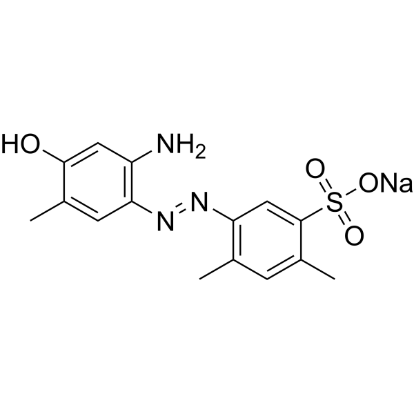
Ischemin sodium

Based on 1 Customer Validation
Ischemin sodium is a CBP bromodomain inhibitor that inhibits p53 interaction with CBP and transcriptional activity in cells. Ischemin sodium salt inhibits p53-induced p21 activation with an IC50 value of 5 µM. Ischemin sodium salt also prevents apoptosis in ischemic cardiomyocytes. Ischemin sodium salt can be used in the study of cardiovascular diseases (such as myocardial ischemia).
Nos produits utilisent uniquement pour la recherche. Nous ne vendons pas aux patients.
- Pureté : 99.32%
- Formule: C15H16N3NaO4S
- Masse moléculaire:357.36
-
Stockage:
4°C, sealed storage, away from moisture and light
* In solvent : -80°C, 6 months; -20°C, 1 month (sealed storage, away from moisture and light)

To place orders, for customer services and technical support, please contact: MedChemExpress USA
Tel: 609-228-6898 E-mail: [email protected] [email protected]


-
 Activité biologique
-
Chemical Information
-
Solvant et solubilité
- Protocole
- Pureté et documentation
- Références
-
 Help & FAQs




Activité biologique
Description
In Vitro
Ischemin sodium (50, 100 µM; 24 h) inhibits p53 activation on DNA damaging stress in U2OS cells[1].
Ischemin sodium (10 μM; 3 days) blocks apoptosis in cardiomyocytes by inhibiting caspase 3/7 activity[1].
Ischemin sodium salt inhibits p53 cellular signaling pathways in U2OS cells[1].
MedChemExpress (MCE) has not independently confirmed the accuracy of these methods. They are for reference only.
-
Cell Line:U2OS cells
-
Concentration:50, 100 μM
-
Incubation Time:24 h
-
Result:Inhibited the Doxorubicin-induced increased levels of p53 protein, its Ser15-phosphorylated (p53S15p) and Lys382-acetylated (p53K382ac).
-
Cell Line:U2OS cells
-
Concentration:10 μM
-
Incubation Time:3 days
-
Result:Functioned as a cellular protective agent againsted myocardial ischemic stress.


Chemical Information
-
Appearance Solid
-
Masse moléculaire 357.36
-
Formule C15H16N3NaO4S
-
Color Light brown to brown
-
SMILES
CC1=CC(/N=N/C2=C(C=C(C(S(=O)(O[Na])=O)=C2)C)C)=C(C=C1O)N
-
Livraison
Room temperature in continental US; may vary elsewhere.
-
Stockage
4°C, sealed storage, away from moisture and light
* In solvent : -80°C, 6 months; -20°C, 1 month (sealed storage, away from moisture and light)


Solvant et solubilité
In Vitro:
H2O : 25 mg/mL (69.96 mM; Need ultrasonic)
Please refer to the solubility information to select the appropriate solvent. Once prepared, please aliquot and store the solution to prevent product inactivation from repeated freeze-thaw cycles.
Storage method and period of stock solution: -80°C, 6 months; -20°C, 1 month (sealed storage, away from moisture and light). When stored at -80°C, please use it within 6 months. When stored at -20°C, please use it within 1 month.
* Note: If you choose water as the stock solution, please dilute it to the working solution, then filter and sterilize it with a 0.22 μm filter before use.
Please refer to the solubility information to select the appropriate solvent. Once prepared, please aliquot and store the solution to prevent product inactivation from repeated freeze-thaw cycles.
Storage method and period of stock solution: -80°C, 6 months; -20°C, 1 month (sealed storage, away from moisture and light). When stored at -80°C, please use it within 6 months. When stored at -20°C, please use it within 1 month.
* Note: If you choose water as the stock solution, please dilute it to the working solution, then filter and sterilize it with a 0.22 μm filter before use.
Concentration (start) × Volume (start) = Concentration (final) × Volume (final)


Pureté et documentation
-
Fiche technique (272 KB)
-
SDS (252 KB)
- English - EN (252 KB)
- Français - FR (252 KB)
- Deutsch - DE (252 KB)
- Norwegian - NO (252 KB)
- Español - ES (252 KB)
- Swedish - SV (252 KB)
- Italian - IT (252 KB)
- Korean - KR (252 KB)
- Portuguese - PT (252 KB)
-
Instruction de manipulation (2659 KB)
Références


Complete Stock Solution Preparation Table
Please refer to the solubility information to select the appropriate solvent. Once prepared, please aliquot and store the solution to prevent product inactivation from repeated freeze-thaw cycles.
Storage method and period of stock solution: -80°C, 6 months; -20°C, 1 month (sealed storage, away from moisture and light). When stored at -80°C, please use it within 6 months. When stored at -20°C, please use it within 1 month.
| Optional Solvent | Concentration Solvent Mass | 1 mg | 5 mg | 10 mg | 25 mg |
|---|---|---|---|---|---|
| H2O | 1 mM | 2.7983 mL | 13.9915 mL | 27.9830 mL | 69.9575 mL |
| 5 mM | 0.5597 mL | 2.7983 mL | 5.5966 mL | 13.9915 mL | |
| 10 mM | 0.2798 mL | 1.3991 mL | 2.7983 mL | 6.9957 mL | |
| 15 mM | 0.1866 mL | 0.9328 mL | 1.8655 mL | 4.6638 mL | |
| 20 mM | 0.1399 mL | 0.6996 mL | 1.3991 mL | 3.4979 mL | |
| 25 mM | 0.1119 mL | 0.5597 mL | 1.1193 mL | 2.7983 mL | |
| 30 mM | 0.0933 mL | 0.4664 mL | 0.9328 mL | 2.3319 mL | |
| 40 mM | 0.0700 mL | 0.3498 mL | 0.6996 mL | 1.7489 mL | |
| 50 mM | 0.0560 mL | 0.2798 mL | 0.5597 mL | 1.3991 mL | |
| 60 mM | 0.0466 mL | 0.2332 mL | 0.4664 mL | 1.1660 mL |
* Note: If you choose water as the stock solution, please dilute it to the working solution, then filter and sterilize it with a 0.22 μm filter before use.


Ischemin sodium Related Classifications
HY-110315 Related Classifications
 Powered by Bioz