



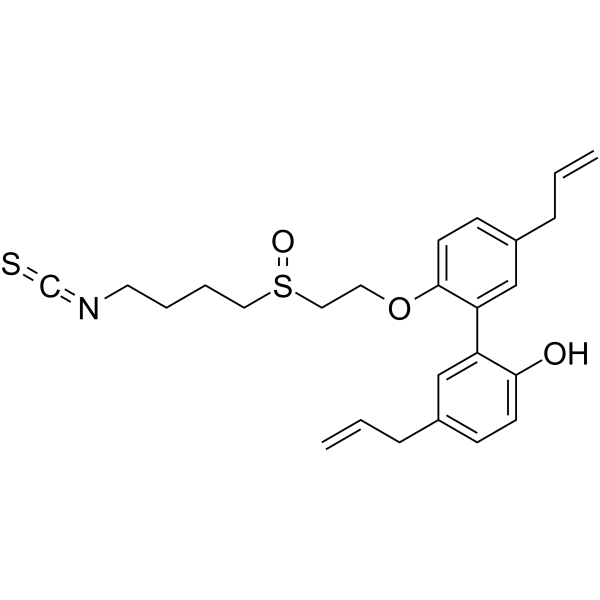
CT1-3
CT1-3 is a potent anticancer agent. CT1-3 induces mitochondria-mediated apoptosis by regulating JNK/Bcl-2/Bax/XIAP pathway. CT1-3 suppresses the epithelial mesenchymal transition (EMT) potential of human cancer cells (HCCs) via regulating the E-cadherin/Snail axis, thus inhibits tumorigenesis. CT1-3 has a strong antitumor effect in mice model and exhibits no significant hepatic and renal toxicity.
商品は「研究用試薬」です。人や動物の医療用・臨床診断用・食品用の製品ではありません。
研究用途以外に使用した場合、当社は一切の責任を負いかねます。
- CAS 番号: 2460738-32-1
- 分子式: C25H29NO3S2
- 分子量:455.63
-
保管条件:
Please store the product under the recommended conditions in the Certificate of Analysis.

To place orders, for customer services and technical support, please contact: MedChemExpress USA
Tel: 609-228-6898 E-mail: [email protected] [email protected]


-
 生物活性
生物活性
-
化学情報
- プロトコル
- 純度とドキュメンテーション
- 参考文献
-
 Help & FAQs




生物活性
製品説明
Cellular Effect
|
Cell Line

|
Type | Value | Description | References |
|---|---|---|---|---|
| A549 | IC50 |
9.07 μM
Compound: CT1-3
|
Antiproliferative activity against human A549 cells assessed as reduction in cell viability incubated for 24 hrs by CCK-8 assay
Antiproliferative activity against human A549 cells assessed as reduction in cell viability incubated for 24 hrs by CCK-8 assay
|
[PMID: 32416457] |
| AGS | IC50 |
8.02 μM
Compound: CT1-3
|
Antiproliferative activity against human AGS cells assessed as reduction in cell viability incubated for 24 hrs by CCK-8 assay
Antiproliferative activity against human AGS cells assessed as reduction in cell viability incubated for 24 hrs by CCK-8 assay
|
[PMID: 32416457] |
| HepG2 | IC50 |
6.24 μM
Compound: CT1-3
|
Antiproliferative activity against human HepG2 cells assessed as reduction in cell viability incubated for 24 hrs by CCK-8 assay
Antiproliferative activity against human HepG2 cells assessed as reduction in cell viability incubated for 24 hrs by CCK-8 assay
|
[PMID: 32416457] |
| HONE1 cell line | IC50 |
14.06 μM
Compound: CT1-3
|
Antiproliferative activity against human HONE1 cell line assessed as reduction in cell viability incubated for 24 hrs by CCK-8 assay
Antiproliferative activity against human HONE1 cell line assessed as reduction in cell viability incubated for 24 hrs by CCK-8 assay
|
[PMID: 32416457] |
| LoVo | IC50 |
5.1 μM
Compound: CT1-3
|
Antiproliferative activity against human LoVo cells assessed as reduction in cell viability incubated for 24 hrs by CCK-8 assay
Antiproliferative activity against human LoVo cells assessed as reduction in cell viability incubated for 24 hrs by CCK-8 assay
|
[PMID: 32416457] |
| MCF7 | IC50 |
12.72 μM
Compound: CT1-3
|
Antiproliferative activity against human MCF7 cells assessed as reduction in cell viability incubated for 24 hrs by CCK-8 assay
Antiproliferative activity against human MCF7 cells assessed as reduction in cell viability incubated for 24 hrs by CCK-8 assay
|
[PMID: 32416457] |
| MDA-MB-231 | IC50 |
7.48 μM
Compound: CT1-3
|
Antiproliferative activity against human MDA-MB-231 cells assessed as reduction in cell viability incubated for 24 hrs by CCK-8 assay
Antiproliferative activity against human MDA-MB-231 cells assessed as reduction in cell viability incubated for 24 hrs by CCK-8 assay
|
[PMID: 32416457] |
| NCI-H1299 | IC50 |
9.17 μM
Compound: CT1-3
|
Antiproliferative activity against human NCI-H1299 cells assessed as reduction in cell viability incubated for 24 hrs by CCK-8 assay
Antiproliferative activity against human NCI-H1299 cells assessed as reduction in cell viability incubated for 24 hrs by CCK-8 assay
|
[PMID: 32416457] |
| NCI-H1650 | IC50 |
6.92 μM
Compound: CT1-3
|
Antiproliferative activity against human NCI-H1650 cells assessed as reduction in cell viability incubated for 24 hrs by CCK-8 assay
Antiproliferative activity against human NCI-H1650 cells assessed as reduction in cell viability incubated for 24 hrs by CCK-8 assay
|
[PMID: 32416457] |
| NCI-H460 | IC50 |
6.15 μM
Compound: CT1-3
|
Antiproliferative activity against human NCI-H460 cells assessed as reduction in cell viability incubated for 24 hrs by CCK-8 assay
Antiproliferative activity against human NCI-H460 cells assessed as reduction in cell viability incubated for 24 hrs by CCK-8 assay
|
[PMID: 32416457] |
| OVCAR-3 | IC50 |
5.34 μM
Compound: CT1-3
|
Antiproliferative activity against human OVCAR-3 cells assessed as reduction in cell viability incubated for 24 hrs by CCK-8 assay
Antiproliferative activity against human OVCAR-3 cells assessed as reduction in cell viability incubated for 24 hrs by CCK-8 assay
|
[PMID: 32416457] |
| SGC-7901 | IC50 |
11.47 μM
Compound: CT1-3
|
Antiproliferative activity against human SGC-7901 cells assessed as reduction in cell viability incubated for 24 hrs by CCK-8 assay
Antiproliferative activity against human SGC-7901 cells assessed as reduction in cell viability incubated for 24 hrs by CCK-8 assay
|
[PMID: 32416457] |
| SW480 | IC50 |
6.07 μM
Compound: CT1-3
|
Antiproliferative activity against human SW480 cells assessed as reduction in cell viability incubated for 24 hrs by CCK-8 assay
Antiproliferative activity against human SW480 cells assessed as reduction in cell viability incubated for 24 hrs by CCK-8 assay
|
[PMID: 32416457] |
| U-251 | IC50 |
7.75 μM
Compound: CT1-3
|
Antiproliferative activity against human U-251 cells assessed as reduction in cell viability incubated for 24 hrs by CCK-8 assay
Antiproliferative activity against human U-251 cells assessed as reduction in cell viability incubated for 24 hrs by CCK-8 assay
|
[PMID: 32416457] |
体外実験
CT1-3 has excellent inhibitory activity against multiple cancer cells with IC50 range of 5.10~14.06 μM in LOVO, A549, HepG2, MDA-MB-231 and HONE1, et al[1].
CT1-3 (10 μM; 24 h) notably increases ROS production in HCCs, significantly decreases mitochondrial membrane potential, and reduces Bcl-2 and XIAP levels and increases phospho-JNK and Bax levels[1].
CT1-3 (7.5 μM; 24 h) reduces the capacity of migration and invasion of HCCs, significantly promotes the expression level of E-cadherin (E-cad), and markedly decreases the pro-metastatic and pro-invasive protein Snail[1].
MedChemExpress (MCE) has not independently confirmed the accuracy of these methods. They are for reference only.
-
Cell Line:A549, H460 and LOVO
-
Concentration:10 μM
-
Incubation Time:24 h
-
Result:Notably increased ROS production, significantly decreased mitochondrial membrane potential, and reduced Bcl-2 and XIAP levels and increased phospho-JNK and Bax levels.
体内実験
MedChemExpress (MCE) has not independently confirmed the accuracy of these methods. They are for reference only.
-
Animal Model:Male BALB/c-nu/nu mice (injected with A549, OVCAR3 and MDA-MB-231)[1]
-
Dosage:20 mg/kg
-
Administration:IP, for 28 days
-
Result:Significantly suppresses tumor growth and exhibited no hepatic and renal toxicity.


化学情報
-
CAS 番号 2460738-32-1
-
分子量 455.63
-
分子式 C25H29NO3S2
-
SMILES
OC1=CC=C(CC=C)C=C1C2=CC(CC=C)=CC=C2OCCS(CCCCN=C=S)=O
-
輸送条件
Room temperature in continental US; may vary elsewhere.
-
保管条件
Please store the product under the recommended conditions in the Certificate of Analysis.


純度とドキュメンテーション
参考文献

Calculators
濃度 (開始) × 体積 (開始) = 濃度 (終了) × 体積 (終了)


CT1-3 関連分類
HY-150596 関連分類
 Powered by Bioz