



Rosnilimab (Mouse IgG2a)

Based on 1 Customer Validation
Rosnilimab (Mouse IgG2a) is a mouse-derived IgG2a antibody, Rosnilimab. Rosnilimab is a PD-1 agonistic monoclonal antibody. Rosnilimab (Mouse IgG2a) optimizes the inhibitory PD-1 signaling pathway without blocking PD-L1 binding; it also activates moderately activated PD-1+ T cells and eliminates PD-1high cells, Tfh/Tph cells and Teff cells. Rosnilimab (Mouse IgG2a) reduces the secretion and gene expression of IFN-γ, CXCL13, IL-6 and TNF, decreases macrophage infiltration, and downregulates the gene expression of inflammatory myeloid cells. Rosnilimab (Mouse IgG2a) can be used in studies related to ulcerative colitis.
연구목적의 판매만을 진행합니다. 환자를 대상으로 한 판매는 하지 않습니다.
- Purity : 98.0%
- 분자량:147.527 kDa
-
보관:
Please store the product under the recommended conditions in the Certificate of Analysis.


To place orders, for customer services and technical support, please contact: MedChemExpress USA
Tel: 609-228-6898 E-mail: [email protected] [email protected]


-
 Biological Activity
-
Chemical Information
- Protocol
- 순도&문서
- References
-
 Help & FAQs




Biological Activity
제품 설명
Isotype
Mouse IgG2a kappa
Recommend Isotype Controls
Species Reactivity
Human
|
PD-1 |
IFN-γ |
CXCL13 |
IL-6 |
TNF |
In Vivo
Rosnilimab (Mouse IgG2a) (10 mg/kg; i.p.; twice weekly; 4 weeks) reduces pathogenic T cell and proinflammatory macrophage infiltration, and downregulates an inflammatory myeloid gene signature overlapping with human UC, in an adoptive transfer murine model of colitis[2].
MedChemExpress (MCE) has not independently confirmed the accuracy of these methods. They are for reference only.
-
Animal Model:Rag2-/- recipients, hPD-1 transgenic donor mice[1]
-
Dosage:10 mg/kg
-
Administration:i.p.; twice weekly; 4 weeks
-
Result:Maintained body weight compared to isotype control.
Reduced colonic inflammation measured by histological pathology score.
Reduced the number and percentage of CD4+ T cells in the colon.
Reduced colonic gene expression of Ifng and Cxcl13 compared to isotype control.
-
Animal Model:Rag2-/- (recipient); hPD-1 transgenic (donor, adoptive transfer of hPD1 CD4+CD45Rb+ T cells into Rag2-/- recipients to induce colitis)[2]
-
Dosage:10 mg/kg
-
Administration:i.p.; twice weekly; 4 weeks
-
Result:Reduced CD4+ T cell infiltration into the mouse colon.
Downregulated 399 myeloid genes that overlap with a human UC inflammatory myeloid gene signature and genes upregulated in mouse colitis colon tissue.
Reduced colon tissue expression of inflammatory cytokine genes Il6, Tnf, and Ifng.
Reduced counts of infiltrating M1 and M2B macrophages in colon tissue.
Decreased colon tissue expression of macrophage recruiting chemokine/receptor genes Ccl2 and Ccr2, inflammatory monocyte marker gene Cd14, pathogen detection genes Tlr2 and Tlr4, and immune activation/amplification genes Cd80, Cd86, Myd88, and Trem1.
Gene ID
Accession
Target
PDCD1/PD-1/CD279
Conjugated
Unconjugated
Reconsititution
The product can be reconstituted/diluted with sterile PBS or saline.
신청
ELISA, FACS, Functional assay
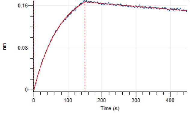
Verified Bioactivity


Chemical Information
-
Appearance Liquid
-
분자량 147.527 kDa
-
Color Colorless to light yellow
-
SMILES
[Rosnilimab (Mouse IgG2a)]
-
선적
Shipping with dry ice.
-
Formulation
Please refer to the lot-specific COA for specific buffer information.
-
보관
Please store the product under the recommended conditions in the Certificate of Analysis.

Protocol
-
RT-PCR
Reverse transcription technology uses RNA as a template to synthesize DNA. RT-PCR is simple, specific and sensitive, and can be used to detect gene expression levels and expression differences in cells; detect RNA virus content; clone cDNA sequences of specific genes.
-
Phagocytosis Functional Assay
A phagocytosis functional assay measures the ability of phagocytic cells, such as neutrophils, macrophages, monocytes, or microglia/macrophages, to bind and internalize particulate targets including bacteria, yeast particles, beads, or myelin particles. Fluorescent flow-cytometry assays detect target uptake as fluorescence associated with gated phagocytes, while pH-sensitive dyes such as pHrodo increase signal in acidic phagosomal compartments and therefore preferentially report internalized particles rather than particles remaining outside the cell. Microscopy or high-content imaging can be used to confirm intracellular localization and, in some protocols, to follow uptake kinetics.
-
RNA extraction experimental
By lysing cells, releasing RNA, and removing impurities such as proteins and DNA, high-purity RNA products are finally obtained. The commonly used traditional method is the guanidine isothiocyanate/phenol/chloroform method (Trizol), which is suitable for a variety of animal materials including animal tissues, microorganisms, cultured cells, etc., and most plant materials.
-
DSS-Induced Colitis
Dextran sulfate sodium (DSS)-induced colitis is generated by administering DSS in mouse drinking water, producing epithelial injury, barrier disruption, weight loss, diarrhea, fecal blood, colon shortening, histologic mucosal damage, and inflammatory mediator changes; the model is mainly used to study acute or chronic intestinal inflammation resembling selected features of ulcerative colitis. DSS injury is interpreted through clinical and tissue readouts rather than a single molecular endpoint: daily body weight, stool consistency, and bleeding are combined into a disease activity index, while colon length, histology, cytokines, myeloperoxidase activity, intestinal permeability, and tight-junction markers provide complementary measures of inflammation and barrier damage.
-
TNBS-Induced Colitis
TNBS-induced colitis is produced by intrarectal delivery of 2,4,6-trinitrobenzene sulfonic acid in ethanol, where ethanol disrupts the mucosal barrier and TNBS haptenates colonic proteins, generating immune-mediated colonic inflammation with weight loss, diarrhea, ulceration, transmural injury, inflammatory-cell infiltration, and cytokine responses. The model is used as an experimental intestinal inflammation model with Crohn’s disease–like features, especially when Th1-type responses, IL-12–dependent inflammation, chronic relapsing inflammation, or fibrosis-related endpoints are studied.
-
Real Time qPCR (Q-PCR)
Real-time quantitative PCR (qPCR) quantifies an amplifiable nucleic-acid target by monitoring fluorescence during PCR cycling rather than measuring product only after amplification. The increase in fluorescence tracks accumulation of PCR product, and the quantification cycle (Cq; historically also Ct/CP) is related to the initial amount of target: samples containing more starting target generally reach the defined fluorescence threshold in fewer cycles.


순도&문서
-
Data Sheet (275 KB)
-
SDS (251 KB)
- English - EN (251 KB)
- Français - FR (251 KB)
- Deutsch - DE (251 KB)
- Norwegian - NO (251 KB)
- Español - ES (251 KB)
- Swedish - SV (251 KB)
- Italian - IT (251 KB)
- Korean - KR (251 KB)
- Portuguese - PT (251 KB)
-
Inhibitory Antibodies User Guide (603 KB)
References

Calculators
Concentration (start) × Volume (start) = Concentration (final) × Volume (final)


Rosnilimab (Mouse IgG2a) Related Classifications
HY-P9S0062 Related Classifications
 Powered by Bioz