



SHP2-D26
Based on 1 publication(s) in Google Scholar
SHP2-D26 is a SHP2 PROTAC degrader with DC50 values of 6.0 nM (KYSE520 cells) and 2.6 nM (MV4;11 cells), respectively. SHP2-D26 inhibits ERK phosphorylation, upregulates Bim levels, downregulates Mcl-1 levels, and induces cell cycle arrest and apoptosis. SHP2-D26 can be used in studies related to esophageal cancer, acute myeloid leukemia and non-small cell lung cancer.
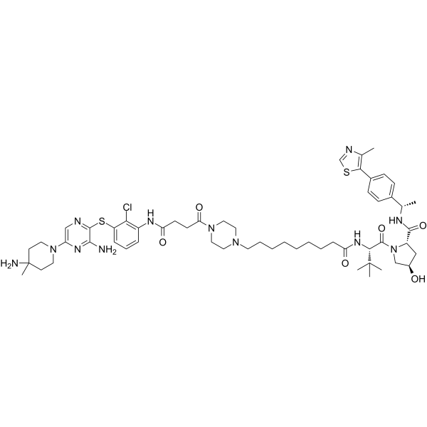
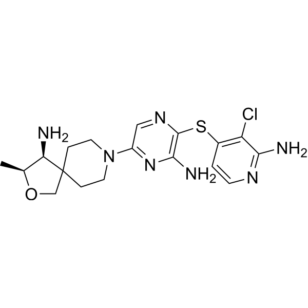
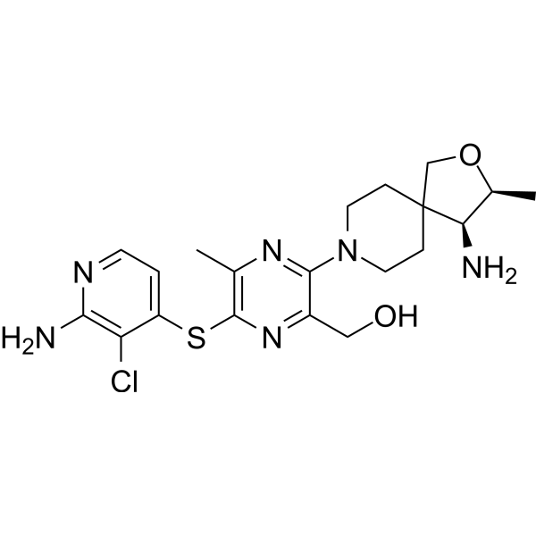
(Pink: SHP2 ligand (HY-176797); Blue: VHL ligand (HY-150803); Black: linker).
연구목적의 판매만을 진행합니다. 환자를 대상으로 한 판매는 하지 않습니다.
- Purity : 95.83%
- CAS No.: 2458219-65-1
- 화학식: C56H79ClN12O6S2
- 분자량:1115.89
-
보관:Powder -20°C, 3 years , 4°C, 2 years ; In solvent -80°C, 6 months , -20°C, 1 month

To place orders, for customer services and technical support, please contact: MedChemExpress USA
Tel: 609-228-6898 E-mail: [email protected] [email protected]


-
 Biological Activity
-
Chemical Information
-
용액&용해도
- Protocol
- 순도&문서
- References
-
 Help & FAQs


Publications Citing Use of MedChemExpress (MCE) SHP2-D26
MoreAll PROTACs Isoforms
More

Biological Activity
제품 설명
|
VHL |
Mcl-1 |
Bim |
MCE Validation Data
Cellular Effect
|
Cell Line

|
Type | Value | Description | References |
|---|---|---|---|---|
| KYSE-520 cell line | IC50 |
0.66 μM
Compound: 26; SHP2-D26
|
Growth inhibition of human KYSE520 cells measured after 4 days by WST8 assay
Growth inhibition of human KYSE520 cells measured after 4 days by WST8 assay
|
[PMID: 32437146] |
| MV4-11 | DC50 |
2.6 nM
Compound: 26; SHP2-D26
|
Protac activity at VHL1/SHP2 in human MV4-11 cells assessed as induction of SHP2 degradation measured after 12 hrs by Western blot analysis
Protac activity at VHL1/SHP2 in human MV4-11 cells assessed as induction of SHP2 degradation measured after 12 hrs by Western blot analysis
|
[PMID: 32437146] |
| MV4-11 | IC50 |
9.9 nM
Compound: 26; SHP2-D26
|
Growth inhibition of human MV4-11 cells measured after 4 days by WST8 assay
Growth inhibition of human MV4-11 cells measured after 4 days by WST8 assay
|
[PMID: 32437146] |
In Vitro
SHP2-D26 (100 nM; 2-24 h) induces rapid degradation of SHP2 protein in KYSE520 esophageal cancer cells and MV4;11 acute myeloid leukemia cells, with nearly complete depletion of the protein observed at 8 h of treatment[1].
SHP2-D26 (3 nM-1000 nM; 48 h) effectively reduces p-ERK levels in KYSE520 esophageal cancer cells and MV4;11 acute myeloid leukemia cells[1].
SHP2-D26 (0.001 μM-100 μM; 4 days) inhibits the proliferation of KYSE520 esophageal cancer cells and MV4;11 acute myeloid leukemia cells, with IC50 values of 0.66 μM and 9.9 nM[1].
SHP2-D26 (72 h) potently inhibits the growth of all tested human non-small cell lung cancer (NSCLC) cell lines, with IC50 values all < 8 μM; it exhibits stronger activity against PC-9, H1648, H1792 and HCC827 cell lines, with IC50 values ≤ 4 μM[2].
SHP2-D26 (0.1-4 μM; 2 h-24 h) sustainably inhibits the p70S6K/S6 signaling pathway in sensitive human non-small cell lung cancer cell lines (PC-9, H1792, HCC827), whereas exerts cell line-dependent effects on the ERK1/2 and Akt signaling pathways[2].
SHP2-D26 (1 μM; 6 h) increases the stability of Bim protein and decreases the stability of Mcl-1 protein in human non-small cell lung cancer cell lines HCC827 and PC-9[2].
SHP2-D26 (1-7.5 μM; 4-48 h) induces apoptosis in human non-small cell lung cancer cell lines (PC-9, H1792, HCC827) by upregulating Bim levels and downregulating Mcl-1 levels, and both the upregulation of Bim and downregulation of Mcl-1 are essential for its pro-apoptotic activity[2].
SHP2-D26 (0.075 μM-6 μM; 16 h-12 days) acts synergistically with Osimertinib (HY-15772) to reduce cell viability, inhibit colony formation and enhance apoptosis in osimertinib-resistant human non-small cell lung cancer cell lines PC-9/AR, PC-9/GR/AR, PC-9/3 M and HCC827/AR[2].
MedChemExpress (MCE) has not independently confirmed the accuracy of these methods. They are for reference only.
-
Cell Line:KYSE520 esophageal cancer cells and MV4;11 acute myeloid leukemia cells
-
Concentration:100 nM
-
Incubation Time:2 h, 4 h, 8 h, 12 h, 24 h
-
Result:Reduced SHP2 protein levels within 4 h, and achieved essentially complete SHP2 depletion after 8 h of treatment.
Reduced SHP2 protein levels within 4 h, and achieved essentially complete SHP2 depletion after 8 h of treatment, similar to kinetics observed in KYSE520 cells.
-
Cell Line:KYSE520 esophageal cancer cells
-
Concentration:10 nM, 30 nM, 100 nM, 300 nM, 1000 nM
-
Incubation Time:48 h
-
Result:Dose-dependently inhibited ERK phosphorylation, with 100 nM SHP2-D26 effectively reducing p-ERK levels; this potency was > 30-times higher than the SHP2 inhibitor SHP099 (HY-100388).
-
Cell Line:MV4;11 acute myeloid leukemia cells
-
Concentration:3 nM, 10 nM, 30 nM, 100 nM, 300 nM
-
Incubation Time:48 h
-
Result:Dose-dependently inhibited ERK phosphorylation, with 100 nM SHP2-D26 completely blocking p-ERK levels; this potency was > 30-times higher than the SHP2 inhibitor SHP099.
-
Cell Line:human NSCLC cell lines (PC-9, H1792, HCC827, H157, H1944, H1651, H1975)
-
Concentration:0.1, 0.5, 1, 2 and 4 μM
-
Incubation Time:2, 4, 8, 16 and 24 h
-
Result:Consistently decreased phosphorylated S6 (p-S6) levels in all sensitive NSCLC cell lines (PC-9, H1792, HCC827), and weakly in H1975, but not in other less sensitive lines.
Decreased phosphorylated p70S6K (p-p70S6K) in sensitive lines, with weak suppression of phosphorylated mTOR (p-mTOR) only in HCC827.
Reduced p-ERK1/2 in H1792 but only weakly in PC-9 and HCC827; minimally decreased p-Akt in PC-9 but increased p-Akt in H1792.
Decreased total ERK1/2 levels in PC-9 and H1792.
-
Cell Line:human NSCLC cell lines (PC-9, H1792, HCC827, Bim-knockout PC-9, Mcl-1-overexpressing PC-9)
-
Concentration:1, 2.5, 5, 7.5 μM
-
Incubation Time:4, 8, 12, 16 and 48 h
-
Result:Induced apoptosis in sensitive NSCLC cell lines in a concentration-dependent manner: increased Annexin V-positive cells and cleaved PARP/caspase-3 in PC-9 at 5 μM and 7.5 μM; induced similar effects in H1792 and HCC827 at 5 μM.
Increased Bim levels and decreased Mcl-1 levels in sensitive lines, with Bim elevation detected at concentrations as low as 1 μM and Mcl-1 reduction detected at 5 μM or higher in PC-9.
Reduced sensitivity to apoptosis and cell number reduction in Bim-knockout PC-9 cells; attenuated apoptosis and reduced sensitivity to cell survival inhibition in Mcl-1-overexpressing PC-9 cells.
In Vivo
SHP2-D26 (30 mg/kg; i.p.; once daily; for 24 consecutive days) exhibits only very low monotherapy activity against Osimertinib-resistant PC-9/AR non-small cell lung cancer xenografts in nude mice, but when combined with Osimertinib, it significantly inhibits tumor growth by enhancing SHP2 degradation, upregulating Bim, downregulating Mcl-1 and inducing apoptosis[2].
MedChemExpress (MCE) has not independently confirmed the accuracy of these methods. They are for reference only.
-
Animal Model:Nude mice[2]
-
Dosage:20 mg/kg; 40 mg/kg
-
Administration:i.p.; once daily for first 10 days, then i.v.; twice weekly until study end (20 mg/kg); i.p.; once daily throughout study (40 mg/kg)
-
Result:Significantly reduced PC-9 tumor volume to a mean end-of-study volume of ~520 mm3 compared to ~850 mm3 for vehicle.
Significantly reduced PC-9 tumor weight to a mean weight of ~0.27 g compared to ~0.55 g for vehicle.
Significantly reduced PC-9 tumor volume to a mean end-of-study volume of ~650 mm3 compared to ~850 mm3 for vehicle.
Significantly reduced PC-9 tumor weight to a mean weight of ~0.35 g compared to ~0.55 g for vehicle.
Induced SHP2 degradation in tumor tissue to a SHP2/GAPDH ratio of ~0.7 for 20 mg/kg and ~0.2 for 40 mg/kg compared to ~1.8 for vehicle.
Significantly increased Bim levels to a Bim/GAPDH ratio of ~0.6 compared to ~0.3 for vehicle.
Significantly reduced Mcl-1 levels to a Mcl-1/GAPDH ratio of ~0.2 compared to ~2.0 for vehicle.
Induced cleaved PARP.
Reduced p-S6 levels, with the 40 mg/kg dose causing a significant reduction to a p-S6/S6 ratio of ~0.2 compared to ~1.0 for vehicle.
Caused no apparent body weight loss in either treatment group.
-
Animal Model:Nude mice[2]
-
Dosage:30 mg/kg
-
Administration:i.p.; once daily; 24 consecutive days
-
Result:Minimally reduced PC-9/AR tumor volume to a mean end-of-study volume of ~820 mm3 compared to ~950 mm3 for vehicle.
Minimally reduced PC-9/AR tumor weight to a mean weight of ~0.5 g compared to ~0.7 g for vehicle.
When combined with Osimertinib, significantly reduced PC-9/AR tumor volume to a mean end-of-study volume of ~320 mm3 compared to ~950 mm3 for vehicle.
When combined with osimertinib, significantly reduced PC-9/AR tumor weight to a mean weight of ~0.3 g compared to ~0.7 g for vehicle.
Induced SHP2 degradation in tumor tissue to a SHP2/Actin ratio of ~0.2 for single agent and ~0.2 for combination compared to ~1.0 for vehicle.
When combined with Osimertinib, significantly elevated Bim levels to a Bim/GAPDH ratio of ~1.2 compared to ~0.5 for vehicle.
When combined with Osimertinib, significantly reduced Mcl-1 levels to a Mcl-1/GAPDH ratio of ~0.2 compared to ~1.1 for vehicle.
Induced cleaved PARP when combined with Osimertinib.
When combined with Osimertinib, significantly reduced p-S6 levels to a p-S6/S6 ratio of ~0 compared to ~0.8 for vehicle.
Caused no apparent body weight loss in the SHP2-D26 treatment group or combination group.
- Kinase activity and phosphorylation assays
- Western Blot
- Detection of Apoptosis
- TUNEL staining for apoptotic DNA fragmentation
- Flow cytometric DNA-content cell-cycle staining
- Annexin V plus membrane-impermeant dye apoptosis staining
- BrdU Incorporation Assay
- Apoptosis Solutions
- Protocol for Cell Cycle
- Protocol for Kinase activity and phosphorylation assays


Chemical Information
-
CAS No. 2458219-65-1
-
Appearance Solid
-
분자량 1115.89
-
화학식 C56H79ClN12O6S2
-
Color Off-white to light yellow
-
SMILES
ClC(C(SC1=NC=C(N2CCC(N)(CC2)C)N=C1N)=CC=C3)=C3NC(CCC(N(CC4)CCN4CCCCCCCCC(N[C@@H](C(C)(C)C)C(N5[C@@H](C[C@H](C5)O)C(N[C@H](C6=CC=C(C7=C(C)N=CS7)C=C6)C)=O)=O)=O)=O)=O
-
선적
Room temperature in continental US; may vary elsewhere.
-
보관
Powder -20°C 3 years 4°C 2 years In solvent -80°C 6 months -20°C 1 month


Publications (1)
-
Journal Impact Factor
-
Most Recent
-
J Biol Chem
2024 Sep;300(9):107616. PMID: 39089586


용액&용해도
In Vitro:
DMSO : 83.33 mg/mL (74.68 mM; Need ultrasonic; Hygroscopic DMSO has a significant impact on the solubility of product, please use newly opened DMSO)
Please refer to the solubility information to select the appropriate solvent. Once prepared, please aliquot and store the solution to prevent product inactivation from repeated freeze-thaw cycles.
Storage method and period of stock solution: -80°C, 6 months; -20°C, 1 month. When stored at -80°C, please use it within 6 months. When stored at -20°C, please use it within 1 month.
Please refer to the solubility information to select the appropriate solvent. Once prepared, please aliquot and store the solution to prevent product inactivation from repeated freeze-thaw cycles.
Storage method and period of stock solution: -80°C, 6 months; -20°C, 1 month. When stored at -80°C, please use it within 6 months. When stored at -20°C, please use it within 1 month.
Concentration (start) × Volume (start) = Concentration (final) × Volume (final)


순도&문서
-
Data Sheet (291 KB)
-
SDS (254 KB)
- English - EN (254 KB)
- Français - FR (254 KB)
- Deutsch - DE (254 KB)
- Norwegian - NO (254 KB)
- Español - ES (254 KB)
- Swedish - SV (254 KB)
- Italian - IT (254 KB)
- Korean - KR (254 KB)
- Portuguese - PT (254 KB)
-
Handling Instructions (2659 KB)
References
[1]. Wang M, et al. Discovery of SHP2-D26 as a First, Potent, and Effective PROTAC Degrader of SHP2 Protein. Journal of medicinal chemistry. 2020 Jul 23;63(14):7510-7528. [Content Brief]
[2]. Deng Y, et al. Therapeutic efficacy of the novel SHP2 degrader SHP2-D26, alone or in combination, against lung cancer is associated with modulation of p70S6K/S6, Bim and Mcl-1. Cancer gene therapy. 2022 Nov;29(11):1558-1569. [Content Brief]


Complete Stock Solution Preparation Table
Please refer to the solubility information to select the appropriate solvent. Once prepared, please aliquot and store the solution to prevent product inactivation from repeated freeze-thaw cycles.
Storage method and period of stock solution: -80°C, 6 months; -20°C, 1 month. When stored at -80°C, please use it within 6 months. When stored at -20°C, please use it within 1 month.
| Optional Solvent | Concentration Solvent Mass | 1 mg | 5 mg | 10 mg | 25 mg |
|---|---|---|---|---|---|
| DMSO | 1 mM | 0.8961 mL | 4.4807 mL | 8.9615 mL | 22.4036 mL |
| 5 mM | 0.1792 mL | 0.8961 mL | 1.7923 mL | 4.4807 mL | |
| 10 mM | 0.0896 mL | 0.4481 mL | 0.8961 mL | 2.2404 mL | |
| 15 mM | 0.0597 mL | 0.2987 mL | 0.5974 mL | 1.4936 mL | |
| 20 mM | 0.0448 mL | 0.2240 mL | 0.4481 mL | 1.1202 mL | |
| 25 mM | 0.0358 mL | 0.1792 mL | 0.3585 mL | 0.8961 mL | |
| 30 mM | 0.0299 mL | 0.1494 mL | 0.2987 mL | 0.7468 mL | |
| 40 mM | 0.0224 mL | 0.1120 mL | 0.2240 mL | 0.5601 mL | |
| 50 mM | 0.0179 mL | 0.0896 mL | 0.1792 mL | 0.4481 mL | |
| 60 mM | 0.0149 mL | 0.0747 mL | 0.1494 mL | 0.3734 mL |


SHP2-D26 Related Classifications
HY-145162 Related Classifications
 Powered by Bioz
Keywords