



m-PEG2000-NHS ester
Based on 1 publication(s) in Google Scholar
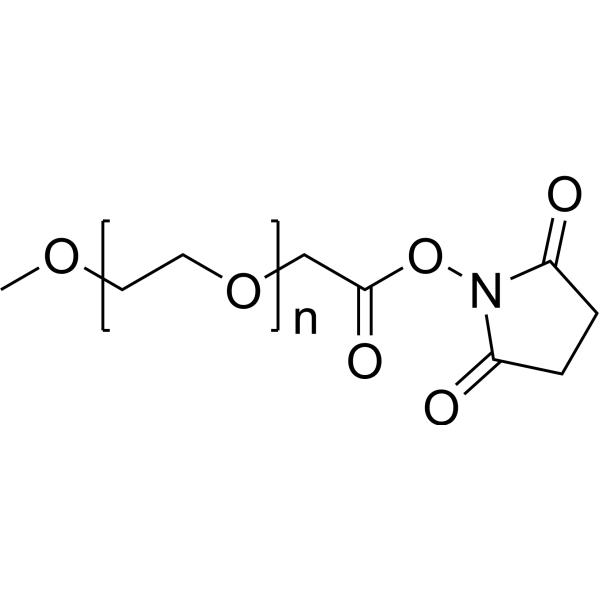
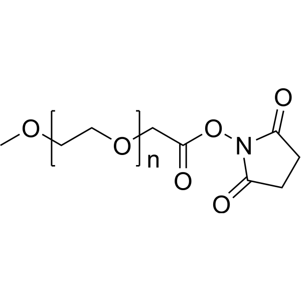
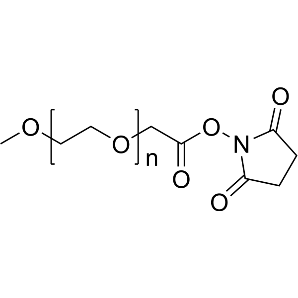
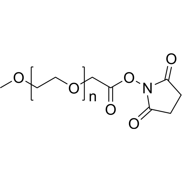
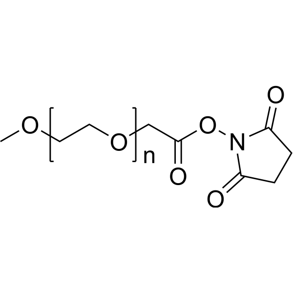
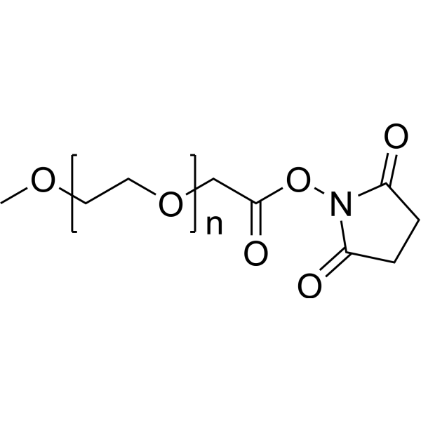
m-PEG2000-NHS ester (mPEG2000-SC) is a reagent with both cell adhesion inhibition and peptide conjugation functions. The NHS ester group of m-PEG2000-NHS ester forms stable amide bonds with primary amine-containing molecules (e.g., the N-terminus of MMP-2-cleavable octapeptide) to generate mPEG-peptide intermediates for liposome surface modification. When m-PEG2000-NHS ester is immobilized on a cystamine-modified gold surface, it can construct an in vitro model for cell adhesion kinetic studies, and higher PEG density and thicker layers correlate with lower cell adhesion rates. m-PEG2000-NHS ester can synthesize MMP-2-responsive PEGylated lipid conjugates to achieve MMP-triggered dePEGylation in the tumor microenvironment. m-PEG2000-NHS ester can be used in studies related to colon cancer and other conditions.
For research use only. We do not sell to patients.
- Purity : 95.89%
- CAS No.: 92451-01-9
- Formula: (C2H4O)nC7H9NO5
- Molecular Weight:2000 (Average)
-
Storage:
-20°C, sealed storage, away from moisture and light
* In solvent : -80°C, 6 months; -20°C, 1 month (sealed storage, away from moisture and light)

To place orders, for customer services and technical support, please contact: MedChemExpress USA
Tel: 609-228-6898 E-mail: [email protected] [email protected]


-
 Biological Activity
-
Chemical Information
-
Solvent & Solubility
- Protocols
- Purity & Documentation
- References
-
 Help & FAQs


-
HY-140698
m-PEG5000-NHS esterPEG Derivative -
HY-155909A
m-PEG1000-NHS esterBiochemical Assay Reagents -
HY-140699
m-PEG10000-NHS esterPEG Derivative -
HY-155909C
m-PEG350-NHS esterBiochemical Assay Reagents -
HY-157745
m-PEG40000-NHS esterPolyethylene Glycol Derivative -
HY-140700
m-PEG20000-NHS esterPEG Derivative -
HY-155909
m-PEG3400-NHS esterBiochemical Assay Reagents -
HY-155909B
m-PEG550-NHS esterBiochemical Assay Reagents -

HY-172352
PEG900 tosylateBiochemical Assay Reagents -

HY-172352A
PEG2000 tosylateBiochemical Assay Reagents
Publications Citing Use of MedChemExpress (MCE) m-PEG2000-NHS ester
More

Biological Activity
Description
IC50 & Target
|
MMP-2 |
In Vitro
m-PEG2000-NHS ester (10 μL/h; 8 h) creates a PEG thickness gradient (0 to 16 nm) that reduces the adhesion rate of BAVEC-1 cells, with near-zero adhesion rate at the 16 nm thick gradient center when delivered into a 3% agarose gel overlay on cystamine-coated gold substrates[1].
m-PEG2000-NHS ester (1:5 molar ratio; overnight, 2 h, 48 h) successfully forms an amide bond with the N-terminus of an MMP-2-cleavable octapeptide, which is then conjugated to DOPE to create an MMP-2-responsive PEG-lipid conjugate[2].
MedChemExpress (MCE) has not independently confirmed the accuracy of these methods. They are for reference only. Further protocols information, click here.


Chemical Information
-
CAS No. 92451-01-9
-
Appearance Solid
-
Molecular Weight 2000 (Average)
-
Formula (C2H4O)nC7H9NO5
-
Color White to off-white
-
SMILES
COCCOCC(ON1C(CCC1=O)=O)=O.[n]
-
Synonyms
mPEG2000-SC; mPEG2000-Succinimidyl ester
-
Shipping
Room temperature in continental US; may vary elsewhere.
-
Storage
-20°C, sealed storage, away from moisture and light
* In solvent : -80°C, 6 months; -20°C, 1 month (sealed storage, away from moisture and light)


Publications (1)
-
Journal Impact Factor
-
Most Recent


Solvent & Solubility
In Vitro:
DMSO : 100 mg/mL (Need ultrasonic; Hygroscopic DMSO has a significant impact on the solubility of product, please use newly opened DMSO)

Protocols
-
Multiplex immunofluorescence IHC
Multiplex immunofluorescence IHC detects multiple protein biomarkers in one tissue section by sequential antibody staining, HRP-mediated tyramide fluorophore deposition, heat-mediated antibody stripping, nuclear counterstaining, multispectral imaging, spectral unmixing, and digital cell phenotyping; TSA deposits fluorophore near the antigen so the fluorescence signal remains after primary and secondary antibodies are removed, enabling repeated staining cycles, including with antibodies from the same host species. Classic FFPE tumor immune-profiling applications use panels such as CD3, CD8, CD68/CD163, FOXP3, PD-1, PD-L1, pancytokeratin, Ki67, and DAPI to identify tumor cells, immune-cell subsets, checkpoint-marker expression, co-expression phenotypes, cell density, and spatial relationships in the tumor microenvironment.
-
Mitochondrial membrane-potential fluorescent assay
Mitochondrial membrane potential fluorescent assays estimate ΔΨm in living cells using lipophilic cationic dyes such as TMRM, TMRE, rhodamine 123, and JC-1, which accumulate in mitochondria according to membrane polarization; loss of signal after FCCP or CCCP treatment is interpreted as mitochondrial depolarization. TMRM/TMRE and rhodamine 123 are commonly used for semi-quantitative live-cell microscopy or flow cytometry, while JC-1 can report a shift from red aggregate fluorescence to green monomer fluorescence during depolarization; interpretation requires controls because dye concentration, quenching mode, cell type, dye efflux, and mitochondrial mass can affect fluorescence independently of ΔΨm.
-
Mitochondrial membrane-potential and mitochondrial mass staining
Mitochondrial membrane potential staining measures the electrochemical polarization across the mitochondrial inner membrane in live cells using lipophilic cationic fluorescent probes; early rhodamine-based work showed that selective mitochondrial dye accumulation is lost when the mitochondrial transmembrane potential is dissipated. JC-1 reports mitochondrial polarization by shifting from green monomer fluorescence to red J-aggregate fluorescence as dye concentration increases within energized mitochondria; therefore, the red/green fluorescence ratio is used as a relative readout of mitochondrial membrane potential. TMRE or TMRM staining provides a single-channel relative readout because these cationic rhodamine esters accumulate in polarized mitochondria, and lower fluorescence indicates reduced mitochondrial polarization when acquisition and dye-loading conditions are controlled. Mitochondrial mass staining is commonly performed with MitoTracker Green FM or related MitoTracker dyes as
-
Multiplex immunohistochemistry
Multiplex immunohistochemistry (mIHC), also known as tyramide dignal amplification (TSA), is an enzymatic detection method that uses horseradish peroxidase (HRP) to perform high-density in-situ labeling of target proteins or nucleic acids.
-
Research Protocol for Cancer Immunology
Cancer immunology studies how the immune system recognizes, suppresses, edits, or fails to eliminate malignant cells through tumor antigen release, antigen presentation, T-cell priming, immune trafficking, tumor-cell killing, and feedback inhibition in the tumor microenvironment. The cancer-immunity cycle links tumor antigenicity, dendritic-cell priming, CD8+ T-cell infiltration, cytotoxic function, and immune-checkpoint regulation to tumor rejection or immune escape. Immune-checkpoint pathways such as PD-1/PD-L1 and CTLA-4 suppress antitumor T-cell activity and can be therapeutically blocked, but many tumors remain resistant because of poor antigen presentation, weak T-cell infiltration, suppressive myeloid cells, regulatory T cells, and tumor-intrinsic immune-exclusion programs. Unresolved questions include which immune-cell states predict response, how tumor-intrinsic pathways exclude immune cells, how myeloid suppression limits checkpoint blockade, and which combination strategies


Purity & Documentation
-
Data Sheet (271 KB)
-
SDS (393 KB)
- English - EN (393 KB)
- Français - FR (393 KB)
- Deutsch - DE (393 KB)
- Norwegian - NO (393 KB)
- Español - ES (393 KB)
- Swedish - SV (393 KB)
- Italian - IT (393 KB)
- Korean - KR (393 KB)
- Portuguese - PT (393 KB)
-
Handling Instructions (2659 KB)
References
[1]. Mougin K, et al. Construction of a tethered poly(ethylene glycol) surface gradient for studies of cell adhesion kinetics. Langmuir. 2005;21(11):4809-4812. [Content Brief]
[2]. Askarizadeh A, et al. Surface-modified cationic liposomes with a matrix metalloproteinase-degradable polyethylene glycol derivative improved doxorubicin delivery in murine colon cancer. J Liposome Res. 2024;34(2):221-238. [Content Brief]

Calculators
Concentration (start) × Volume (start) = Concentration (final) × Volume (final)


m-PEG2000-NHS ester Related Classifications
HY-W591424 Related Classifications
 Powered by Bioz