



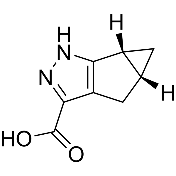
MK-1903
Based on 1 publication(s) in Google Scholar
MK-1903 is an orally active full agonist of GPR109a/HCAR2, with an EC50 of 12.9 nM. MK-1903 activates antilipolytic and vasodilatory pathways, reduces plasma free fatty acid levels, and induces skin flushing. MK-1903 stimulates the expression of HCAR2 protein and regulates the inflammatory response of microglia. MK-1903 prevents the enhanced firing activity of spinal nociceptive neurons. MK-1903 triggers the release of MMP-9 and the formation of NET. MK-1903 can be used in the research of dyslipidemia and neuroinflammation-based central nervous system diseases.
For research use only. We do not sell to patients.
- Purity : 99.68%
- CAS No.: 1268882-43-4
- Formula: C8H8N2O2
- Molecular Weight:164.16
-
Storage:Powder -20°C, 3 years ; In solvent -80°C, 6 months , -20°C, 1 month

To place orders, for customer services and technical support, please contact: MedChemExpress USA
Tel: 609-228-6898 E-mail: [email protected] [email protected]


-
 Biological Activity
-
Chemical Information
-
Solvent & Solubility
- Protocols
- Purity & Documentation
- References
-
 Help & FAQs


-
HY-17394
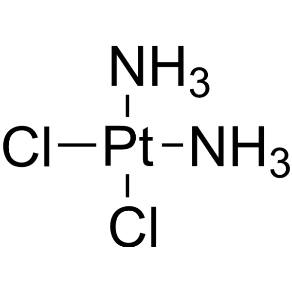
CisplatinDNA Alkylator -
HY-13008
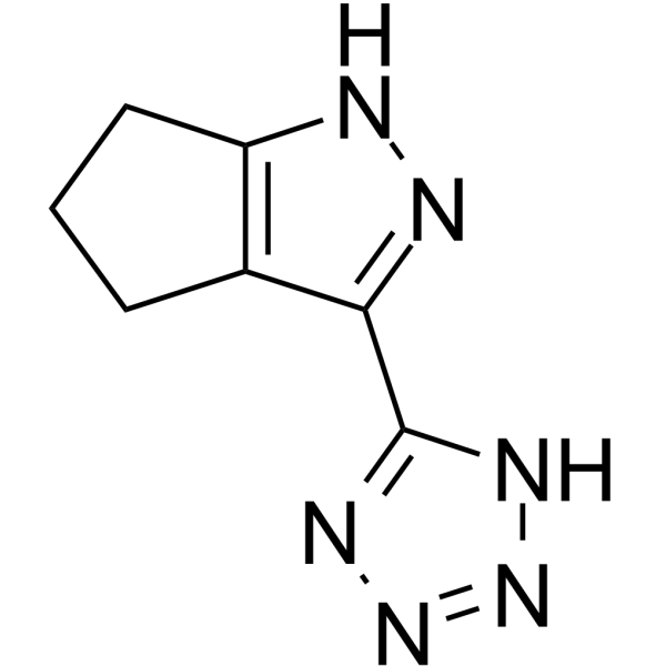
MK-0354GPR109A Agonist -
HY-107579
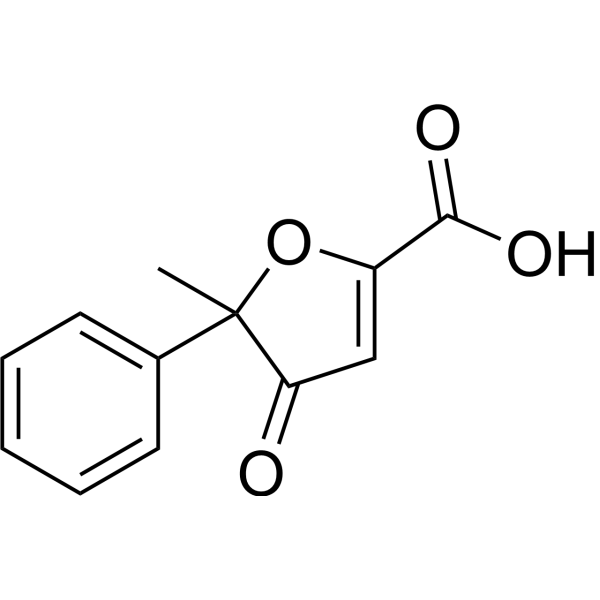
AcifranHypolipidemic Agent -
HY-W015560
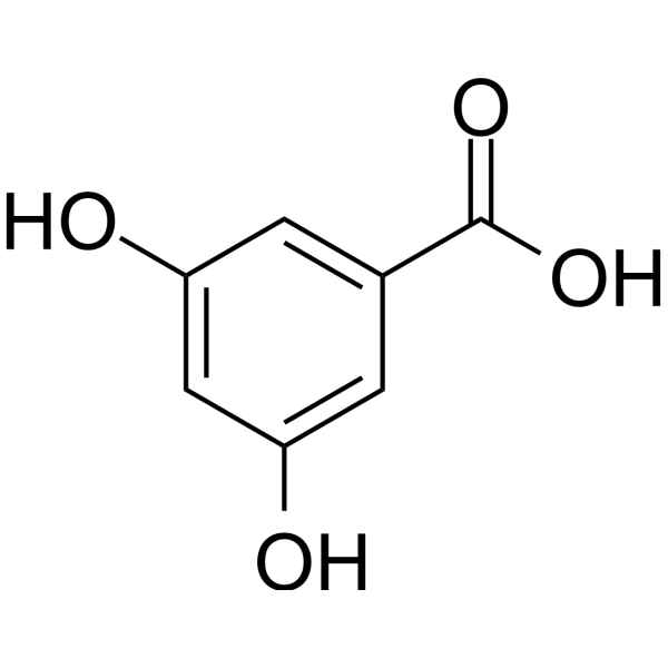
3,5-Dihydroxybenzoic acidGPR81 Agonist -
HY-107580
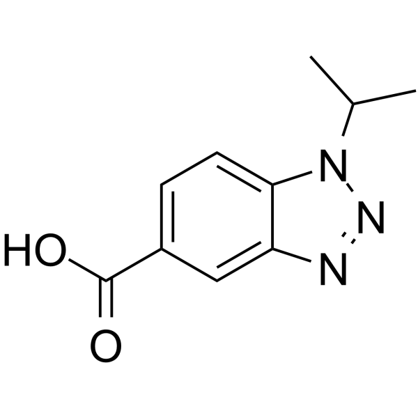
GPR109 receptor agonist-1GPR109b Receptor Agonist -

HY-107581R
MK-1903 (Standard)Reference Standard -
HY-100976
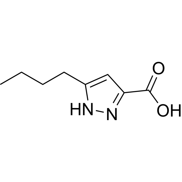
LUF6283HCA(2) Agonist -

HY-107579R
Acifran (Standard)Reference Standard -

HY-W015560R
3,5-Dihydroxybenzoic acid (Standard)GPR81 Agonist
Publications Citing Use of MedChemExpress (MCE) MK-1903
More

Biological Activity
Description
Cellular Effect
|
Cell Line

|
Type | Value | Description | References |
|---|---|---|---|---|
| CHO | EC50 |
12.9 nM
Compound: R,R-19a, MK-1903
|
Agonist activity at human GPR109a expressed in CHO cells assessed as decrease in forskolin-stimulated cAMP production by HTRF assay
Agonist activity at human GPR109a expressed in CHO cells assessed as decrease in forskolin-stimulated cAMP production by HTRF assay
|
[PMID: 22435740] |
In Vitro
MK-1903 (0.25-4 μM; 24 h) does not alter the viability of primary rat microglia, and can partially inhibit LPS (HY-D1056)-induced cytotoxicity in these cells[2].
MK-1903 (1 μM; 24 h) upregulates HCAR2 protein expression in primary rat microglia, and this effect is enhanced when the cells are subsequently treated with LPS[2].
MK-1903 (1 μM; 24 h) inhibits LPS-induced morphological activation of primary rat microglia, but induces an activated morphology in microglia when used alone[2].
MK-1903 (1 μM; 24 h) inhibits the LPS-induced upregulation of pro-inflammatory markers (COX-2, IL-1β) and restores the protein expression of the anti-inflammatory factor IL-10 in primary rat microglia[2].
MK-1903 (1 μM; 24 h) alone increases extracellular IL-10 levels in primary rat microglia, without altering the LPS-induced reduction in extracellular IL-10 levels in these cells[2].
MK-1903 (1 μM; 24 h) inhibits FKN-induced pro-inflammatory gene expression in mouse BV-2 microglial cells, and upregulates the expression of anti-inflammatory marker genes in these cells when applied alone[2].
MK-1903 (0.1-10 μM; 10 min) induces the release of MMP-9 from bovine polymorphonuclear leukocytes (PMN)[3].
MK-1903 (10 μM; 30 min) has no effect on the expression of CD11b or CD47 on the surface of bovine polymorphonuclear neutrophils (PMN)[3].
MK-1903 (1-10 μM; 60 min) induces the formation of neutrophil extracellular traps (NETs) in bovine polymorphonuclear neutrophils (PMNs), and the NETs colocalize with citrullinated histone H4[3].
MK-1903 (0.1-10 μM; 2 h) does not induce apoptosis or cell death in bovine polymorphonuclear neutrophils (PMN)[3].
MedChemExpress (MCE) has not independently confirmed the accuracy of these methods. They are for reference only. Further protocols information, click here.
-
Cell Line:murine BV-2 microglial cells
-
Concentration:1 μM (pretreatment)
-
Incubation Time:24 h (pretreatment) followed by FKN 24 h
-
Result:Significantly reduced FKN-induced increases in pro-inflammatory marker gene expression relative to FKN-only treated cells.
Significantly increased gene expression of anti-inflammatory markers IL-10 and ARG-1 relative to vehicle when used alone.
Increased gene expression of pro-inflammatory markers IL-1β, IL-6, iNOS, TNF-α, and TG2 relative to vehicle when treated with FKN alone.
In Vivo
MK-1903 (1-100 mg/kg; p.o.; single dose) induces a dose-dependent flushing response in anesthetized male C57BL/6 mice, with a ~30% peak increase in ear cutaneous blood flow at the 100 mg/kg oral dose[1].
MK-1903 (2.62 nmol/5 μL; spinal; single dose) prevents fractalkine-induced spinal nociceptive specific neuron hyperexcitability in healthy male Wistar rats, with no significant changes in NS neuron activity relative to baseline observed post-treatment[2].
MedChemExpress (MCE) has not independently confirmed the accuracy of these methods. They are for reference only.
-
Animal Model:Sprague-Dawley (male, fasted)[1]
-
Dosage:0.001 mg/kg; 0.01 mg/kg; 0.1 mg/kg; 1 mg/kg; 10 mg/kg; 100 mg/kg
-
Administration:p.o.; single dose
-
Result:Lowered plasma FFAs by approximately 90% of baseline levels at doses ≥ 0.01 mg/kg.
Produced a more sustained decrease in FFAs at higher doses.
Showed no distinguishable effect from vehicle at 0.001 mg/kg.
-
Animal Model:C57BL/6 (male, anesthetized)[1]
-
Dosage:1 mg/kg; 10 mg/kg; 100 mg/kg
-
Administration:p.o.; single dose
-
Result:Induced a peak increase of approximately 30% in cutaneous blood flow over baseline 5 minutes after administration at 100 mg/kg.
Produced smaller increases in blood flow relative to vehicle at 1 mg/kg and 10 mg/kg doses.
-
Animal Model:Wistar (male, 270-280 g)[2]
-
Dosage:2.62 nmol/5 μL
-
Administration:spinal; single dose
-
Result:Completely prevented the fractalkine-induced increase in spinal nociceptive specific neuron hyperexcitability.
Showed no significant changes in mean percentage of spontaneous activity (96.65%), frequency of excitation (99.54%), or duration of excitation (98.34%) following fractalkine administration relative to baseline.
Did not alter spontaneous or evoked nociceptive specific neuron activity, with mean percentage spontaneous activity (95.29%), frequency of excitation (95.13%), and duration of excitation (95.33%) relative to baseline.
Reduced fractalkine-induced microglial morphological activation in the spinal cord.
Clinical Trial
| NCT Number | Sponsor | Condition | Start Date |
Phase
|
|---|---|---|---|---|
| NCT01329991 | Plexxikon| | 2011-05 | PHASE1 |


Chemical Information
-
CAS No. 1268882-43-4
-
Appearance Solid
-
Molecular Weight 164.16
-
Formula C8H8N2O2
-
Color Off-white to light yellow
-
SMILES
OC(C1=NNC2=C1C[C@]3([H])[C@@]2([H])C3)=O
-
Shipping
Room temperature in continental US; may vary elsewhere.
-
Storage
Powder -20°C 3 years In solvent -80°C 6 months -20°C 1 month


Publications (1)
-
Journal Impact Factor
-
Most Recent
-
PLoS Biol
Structures of G-protein coupled receptor HCAR1 in complex with Gi1 protein reveal the mechanistic basis for ligand recognition and agonist selectivity. [Abstract]2025 Apr 15;23(4):e3003126. PMID: 40233099


Solvent & Solubility
In Vitro:
DMSO : 125 mg/mL (761.45 mM; Need ultrasonic; Hygroscopic DMSO has a significant impact on the solubility of product, please use newly opened DMSO)
Please refer to the solubility information to select the appropriate solvent. Once prepared, please aliquot and store the solution to prevent product inactivation from repeated freeze-thaw cycles.
Storage method and period of stock solution: -80°C, 6 months; -20°C, 1 month. When stored at -80°C, please use it within 6 months. When stored at -20°C, please use it within 1 month.
Please refer to the solubility information to select the appropriate solvent. Once prepared, please aliquot and store the solution to prevent product inactivation from repeated freeze-thaw cycles.
Storage method and period of stock solution: -80°C, 6 months; -20°C, 1 month. When stored at -80°C, please use it within 6 months. When stored at -20°C, please use it within 1 month.
Concentration (start) × Volume (start) = Concentration (final) × Volume (final)
In Vivo:
Select the appropriate dissolution method based on your experimental animal and administration route.
- For the following dissolution methods, please ensure to first prepare a clear stock solution using an In Vitro approach and then sequentially add co-solvents:
- To ensure reliable experimental results, the clarified stock solution can be appropriately stored based on storage conditions. As for the working solution for In Vivo experiments, it is recommended to prepare freshly and use it on the same day.
- The percentages shown for the solvents indicate their volumetric ratio in the final prepared solution. If precipitation or phase separation occurs during preparation, heat and/or sonication can be used to aid dissolution.
Add each solvent one by one: 10% DMSO 40% PEG300 5% Tween-80 45% Saline
Solubility: ≥ 2.08 mg/mL (12.67 mM); Clear solution
This protocol yields a clear solution of ≥ 2.08 mg/mL (saturation unknown).
Taking 1 mL working solution as an example, add 100 μL DMSO stock solution (20.8 mg/mL) to 400 μL PEG300, and mix evenly; then add 50 μL Tween-80 and mix evenly; then add 450 μL Saline to adjust the volume to 1 mL.
Preparation of Saline: Dissolve 0.9 g sodium chloride in ddH₂O and dilute to 100 mL to obtain a clear Saline solution.
Add each solvent one by one: 10% DMSO 90% (20% SBE-β-CD in Saline)
Solubility: ≥ 2.08 mg/mL (12.67 mM); Clear solution
This protocol yields a clear solution of ≥ 2.08 mg/mL (saturation unknown).
Taking 1 mL working solution as an example, add 100 μL DMSO stock solution (20.8 mg/mL) to 900 μL 20% SBE-β-CD in Saline, and mix evenly.
Preparation of 20% SBE-β-CD in Saline (4°C, storage for one week): 2 g SBE-β-CD powder is dissolved in 10 mL Saline, completely dissolve until clear.
In Vivo Dissolution Calculator
Please enter the basic information of animal experiments:
-
-
-
-
Recommended: Prepare an additional quantity of animals to account for potential losses during experiments.
Please enter your animal formula composition:
-
%DMSO +
Recommended: Keep the proportion of DMSO in working solution below 2% if your animal is weak.
-
%+
-
+%Tween-80 + +
-
%Saline +
The co-solvents required include: DMSO, . All of co-solvents are available by MedChemExpress (MCE). , Tween 80. All of co-solvents are available by MedChemExpress (MCE).
Working solution concentration: 0.22 mg/mL
Method for preparing stock solution: mg drug dissolved in μL DMSO. Stock solution concentration: mg/mL.
1. Take μL DMSO stock solution;
2. Add μL .
μL , mix evenly;
3. Then add μL Tween 80, mix evenly;
4. Then add μL
Please ensure that the stock solution in the first step is dissolved to a clear state, and add co-solvents in sequence. You can use ultrasonic heating (ultrasonic cleaner, recommended frequency 20-40 kHz), vortexing, etc. to assist dissolution.

Protocols
-
RNA extraction experimental
By lysing cells, releasing RNA, and removing impurities such as proteins and DNA, high-purity RNA products are finally obtained. The commonly used traditional method is the guanidine isothiocyanate/phenol/chloroform method (Trizol), which is suitable for a variety of animal materials including animal tissues, microorganisms, cultured cells, etc., and most plant materials.
-
Mitochondrial membrane-potential fluorescent assay
Mitochondrial membrane potential fluorescent assays estimate ΔΨm in living cells using lipophilic cationic dyes such as TMRM, TMRE, rhodamine 123, and JC-1, which accumulate in mitochondria according to membrane polarization; loss of signal after FCCP or CCCP treatment is interpreted as mitochondrial depolarization. TMRM/TMRE and rhodamine 123 are commonly used for semi-quantitative live-cell microscopy or flow cytometry, while JC-1 can report a shift from red aggregate fluorescence to green monomer fluorescence during depolarization; interpretation requires controls because dye concentration, quenching mode, cell type, dye efflux, and mitochondrial mass can affect fluorescence independently of ΔΨm.
-
Mitochondrial membrane-potential and mitochondrial mass staining
Mitochondrial membrane potential staining measures the electrochemical polarization across the mitochondrial inner membrane in live cells using lipophilic cationic fluorescent probes; early rhodamine-based work showed that selective mitochondrial dye accumulation is lost when the mitochondrial transmembrane potential is dissipated. JC-1 reports mitochondrial polarization by shifting from green monomer fluorescence to red J-aggregate fluorescence as dye concentration increases within energized mitochondria; therefore, the red/green fluorescence ratio is used as a relative readout of mitochondrial membrane potential. TMRE or TMRM staining provides a single-channel relative readout because these cationic rhodamine esters accumulate in polarized mitochondria, and lower fluorescence indicates reduced mitochondrial polarization when acquisition and dye-loading conditions are controlled. Mitochondrial mass staining is commonly performed with MitoTracker Green FM or related MitoTracker dyes as
-
Research Protocol for Inflammation-related Diseases
The NLRP3 inflammasome is a cytosolic innate immune signaling platform that integrates priming signals and danger-signal activation to promote caspase-1 activation, maturation of IL-1β and IL-18, and gasdermin D-mediated pyroptotic cell death. The core experimental logic is to determine whether inflammatory disease phenotypes are driven by increased NLRP3 expression, ASC-containing inflammasome assembly, caspase-1 cleavage, GSDMD cleavage, and extracellular release of IL-1β/IL-18 rather than by nonspecific cell injury alone. The pathway is strongly linked to inflammation-related disease phenotypes because monosodium urate crystals activate NALP3/NLRP3 inflammasome signaling in gout-like crystal inflammation, cholesterol crystals activate NLRP3 inflammasomes in atherogenesis models, and DSS-induced intestinal inflammation has been reported to involve NLRP3 inflammasome activity. However, experimental colitis studies also show context-dependent protective effects of NLRP3 inflammasome co
-
Research Protocol for Neurological Diseases
PINK1/Parkin-mediated mitophagy pathway is a mitochondrial quality-control signaling axis in which mitochondrial depolarization stabilizes PINK1 on damaged mitochondria, activates Parkin recruitment and E3 ubiquitin ligase activity, promotes ubiquitination of outer mitochondrial membrane proteins, recruits selective autophagy adaptors, and drives lysosomal degradation of damaged mitochondria. In neurological disease research, this pathway is experimentally important because neurons, especially dopaminergic neurons, are highly dependent on mitochondrial integrity, and defective mitochondrial turnover can lead to mitochondrial dysfunction, oxidative stress, impaired neuronal survival, α-synuclein accumulation, and neuroinflammatory damage-associated signals. The genetic disease link is strongest in Parkinson’s disease because mutations in PRKN/parkin cause autosomal recessive juvenile parkinsonism, mutations in PINK1 cause hereditary early-onset Parkinson’s disease, and Drosophila studie
-
Research Protocol for Metabolic Diseases
AMP-activated protein kinase, AMPK, is a conserved cellular energy sensor that responds to reduced cellular energy status and coordinates metabolism by increasing ATP-generating catabolic pathways while suppressing ATP-consuming anabolic processes. In metabolic disease research, the AMPK pathway is experimentally relevant because it regulates hepatic lipid synthesis, fatty acid oxidation, glucose production, skeletal-muscle glucose disposal, mTORC1-linked biosynthesis, autophagy, mitochondrial homeostasis, and whole-body energy balance. The central pathway logic is that energy stress, metformin, exercise-like stimulation, or direct AMPK activators increase AMPKα Thr172 phosphorylation and downstream substrate phosphorylation, including ACC and RAPTOR. Phosphorylation of ACC suppresses lipogenesis and supports fatty acid oxidation, whereas phosphorylation of RAPTOR suppresses mTORC1 signaling and links cellular energy status to growth and protein synthesis control. The pathway is linked


Purity & Documentation
-
Data Sheet (284 KB)
-
SDS (394 KB)
- English - EN (394 KB)
- Français - FR (394 KB)
- Deutsch - DE (394 KB)
- Norwegian - NO (394 KB)
- Español - ES (394 KB)
- Swedish - SV (394 KB)
- Italian - IT (394 KB)
- Korean - KR (394 KB)
- Portuguese - PT (394 KB)
-
Handling Instructions (2659 KB)
References
[2]. Perrone M, et al. Potential role of the hydroxyl carboxylic acid receptor type 2 (HCAR2) in microglia pathophysiology: A possible cross-talk with C-X-C chemokine receptor 1 (CXCR1). Neuropharmacology. 2023;228:109456. [Content Brief]
[3]. Carretta MD, et al. Hydroxycarboxylic acid receptor 2 (HCA2) agonists induce NET formation and MMP-9 release from bovine polymorphonuclear leukocytes. Dev Comp Immunol. 2023;139:104562. [Content Brief]


Complete Stock Solution Preparation Table
Please refer to the solubility information to select the appropriate solvent. Once prepared, please aliquot and store the solution to prevent product inactivation from repeated freeze-thaw cycles.
Storage method and period of stock solution: -80°C, 6 months; -20°C, 1 month. When stored at -80°C, please use it within 6 months. When stored at -20°C, please use it within 1 month.
| Optional Solvent | Concentration Solvent Mass | 1 mg | 5 mg | 10 mg | 25 mg |
|---|---|---|---|---|---|
| DMSO | 1 mM | 6.0916 mL | 30.4581 mL | 60.9162 mL | 152.2904 mL |
| 5 mM | 1.2183 mL | 6.0916 mL | 12.1832 mL | 30.4581 mL | |
| 10 mM | 0.6092 mL | 3.0458 mL | 6.0916 mL | 15.2290 mL | |
| 15 mM | 0.4061 mL | 2.0305 mL | 4.0611 mL | 10.1527 mL | |
| 20 mM | 0.3046 mL | 1.5229 mL | 3.0458 mL | 7.6145 mL | |
| 25 mM | 0.2437 mL | 1.2183 mL | 2.4366 mL | 6.0916 mL | |
| 30 mM | 0.2031 mL | 1.0153 mL | 2.0305 mL | 5.0763 mL | |
| 40 mM | 0.1523 mL | 0.7615 mL | 1.5229 mL | 3.8073 mL | |
| 50 mM | 0.1218 mL | 0.6092 mL | 1.2183 mL | 3.0458 mL | |
| 60 mM | 0.1015 mL | 0.5076 mL | 1.0153 mL | 2.5382 mL | |
| 80 mM | 0.0761 mL | 0.3807 mL | 0.7615 mL | 1.9036 mL | |
| 100 mM | 0.0609 mL | 0.3046 mL | 0.6092 mL | 1.5229 mL |


MK-1903 Related Classifications
HY-107581 Related Classifications
 Powered by Bioz