



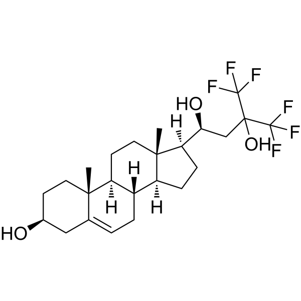
NSC12
NSC12 is an orally active pan-FGF trap. NSC12 inhibits the interaction between FGF2/FGFR. NSC12 suppresses the phosphorylation of FGFR3. NSC12 reduces c-Myc levels, induces DNA damage, triggers the cleavage of Caspase 3, and promotes ROS production. NSC12 exhibits anticancer activity against lung cancer and multiple myeloma.
For research use only. We do not sell to patients.
- CAS No.: 102586-30-1
- Formula: C24H34F6O3
- Molecular Weight:484.52
-
Storage:Powder -20°C, 3 years ; In solvent -80°C, 6 months , -20°C, 1 month

To place orders, for customer services and technical support, please contact: MedChemExpress USA
Tel: 609-228-6898 E-mail: [email protected] [email protected]


-
 Biological Activity
-
Chemical Information
-
Solvent & Solubility
- Protocols
- Purity & Documentation
- References
-
 Help & FAQs


-
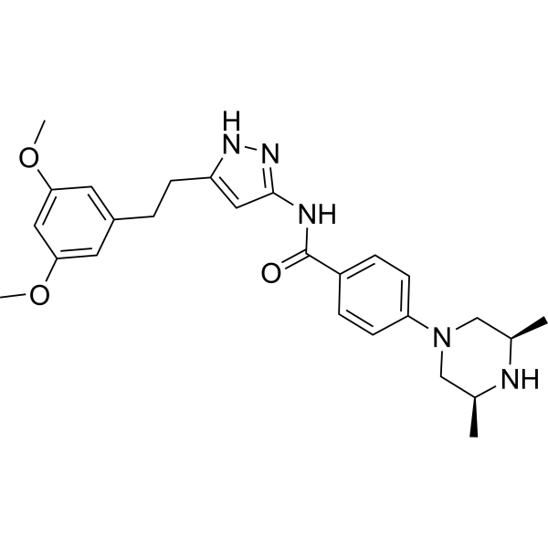
HY-13330
FexagratinibFGFR Inhibitor -
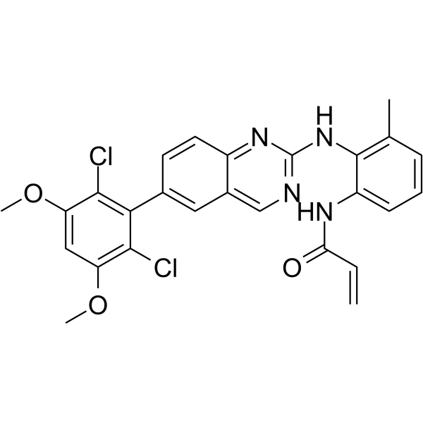
HY-12823
BLU9931FGFR4 Inhibitor -
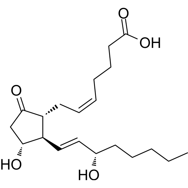
HY-101952
Prostaglandin E2Prostaglandin Receptor Agonist -
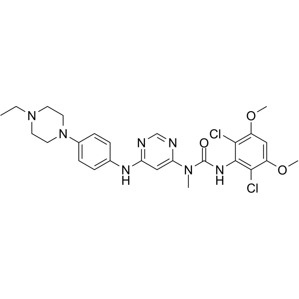
HY-13311
InfigratinibFGFR Inhibitor -
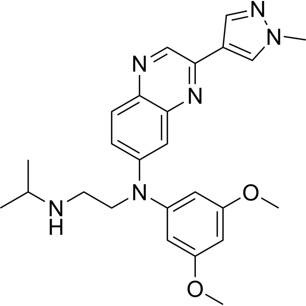
HY-18708
ErdafitinibFGFR Inhibitor -
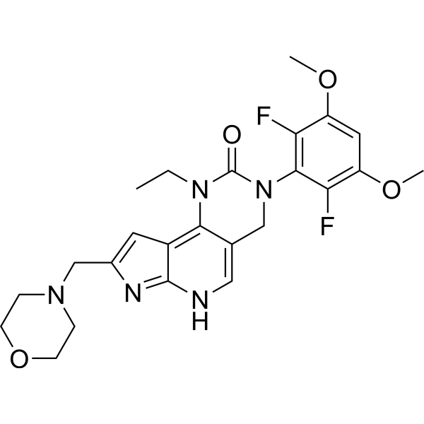
HY-109099
PemigatinibFGFR Inhibitor -
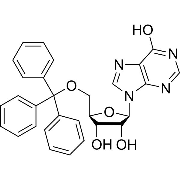
HY-102071
KIN59Thymidine Phosphorylase Inhibitor -
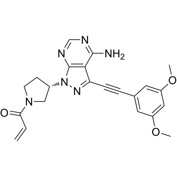
HY-100818
FutibatinibFGFR Inhibitor -
HY-15599
SSR128129EFGFR Inhibitor -
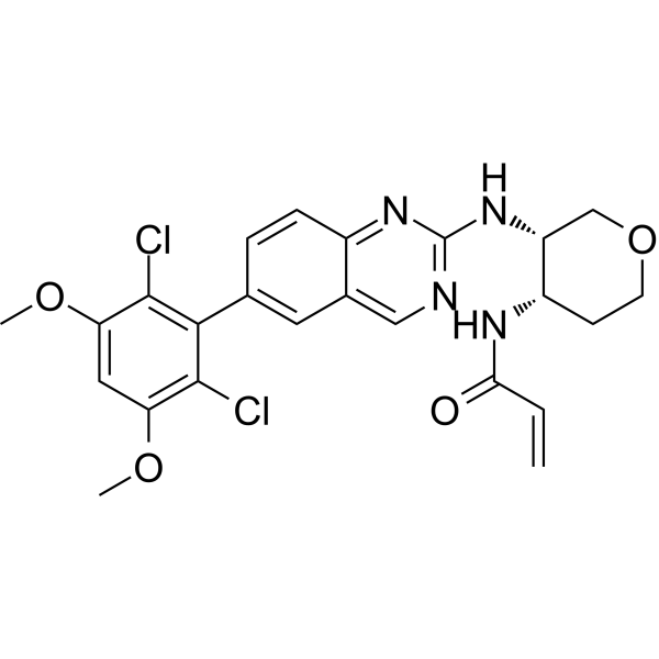
HY-100492
FisogatinibFGFR4 Inhibitor -
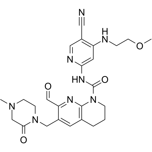
HY-101568
RoblitinibFGFR4 Inhibitor -
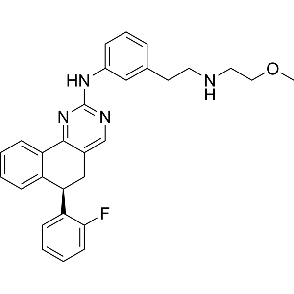
HY-19981
Derazantinib


Biological Activity
Description
IC50 & Target
[1]|
Caspase 3 |
FGFR3 |
Cellular Effect
|
Cell Line

|
Type | Value | Description | References |
|---|---|---|---|---|
| CHO-K1 | IC50 |
10 μM
Compound: 11
|
Inhibition of human recombinant FGF2 assessed as reduction in HSPG/FGF2/FGDR interaction by measuring FGF2 mediated A475-CHO-flg-1A-luc cell to CHO-K1 cell adhesion incubated for 2 hrs by luciferase assay
Inhibition of human recombinant FGF2 assessed as reduction in HSPG/FGF2/FGDR interaction by measuring FGF2 mediated A475-CHO-flg-1A-luc cell to CHO-K1 cell adhesion incubated for 2 hrs by luciferase assay
|
[PMID: 27138345] |
| KMS-11 | IC50 |
3.4 μM
Compound: 1; NSC12
|
Antiproliferative activity against human KMS-11 cells assessed as reduction in cell viability incubated for 48 hrs by propidium iodide staining-based flow cytometric analysis
Antiproliferative activity against human KMS-11 cells assessed as reduction in cell viability incubated for 48 hrs by propidium iodide staining-based flow cytometric analysis
|
[PMID: 34004471] |
| Lewis lung carcinoma cell line | IC50 |
2 μM
Compound: 11
|
Antiproliferative activity against mouse LLC cells incubated for 48 hrs by PI-staining based flow cytometry
Antiproliferative activity against mouse LLC cells incubated for 48 hrs by PI-staining based flow cytometry
|
[PMID: 27138345] |
| NCI-H520 | IC50 |
4.1 μM
Compound: 11
|
Antiproliferative activity against human NCI-H520 cells incubated for 72 hrs by PI-staining based flow cytometry
Antiproliferative activity against human NCI-H520 cells incubated for 72 hrs by PI-staining based flow cytometry
|
[PMID: 27138345] |
In Vitro
NSC12 (0.3-10.0 μM; 48 h for LLC cells, 72 h for H520 cells) inhibits the proliferation of mouse Lewis lung carcinoma (LLC) cells and H520 lung cancer cells, with an IC50 of 2.0 μM for the former and 4.1 μM for the latter[1].
NSC12 (6 μM; 6 h) inhibits FGFR3 phosphorylation by 75% in multiple myeloma KMS-11 cells treated with 6 μM NSC12 for 6 h[2].
NSC12 (6 μM; 6 h) inhibits FGFR phosphorylation in multiple myeloma cell lines KMS-11, OPM-2, U-266, and the Bortezomib-resistant strain KMS-11/BTZ[2].
NSC12 (6 μM; 6 h) reduces c-Myc levels and induces DNA damage in multiple myeloma KMS-11 cells[2].
MedChemExpress (MCE) has not independently confirmed the accuracy of these methods. They are for reference only. Further protocols information, click here.
-
Cell Line:murine Lewis lung carcinoma (LLC) cells, human H520 lung carcinoma cells
-
Concentration:0.3-10.0 μM
-
Incubation Time:48 h (LLC cells); 72 h (H520 cells)
-
Result:Efficiently impaired proliferation of both FGF-dependent cell lines, with an IC50 of 2.0 μM for LLC cells and 4.1 μM for H520 cells.
-
Cell Line:human multiple myeloma KMS-11 cells (t(4;14) translocation, FGFR3 overexpression)
-
Concentration:6 μM
-
Incubation Time:6 h
-
Result:Inhibited FGFR3 phosphorylation by 75%.
-
Cell Line:human multiple myeloma cell lines KMS-11, OPM-2, U-266, and bortezomib-resistant KMS-11/BTZ
-
Concentration:0.1-10.0 μM
-
Incubation Time:48 h
-
Result:Inhibited cell viability with the following half-maximal inhibitory concentrations (IC50): 3.6 μM (KMS-11), 4.4 μM (OPM-2), 3.4 μM (U-266), 3.6 μM (KMS-11/BTZ).
-
Cell Line:human multiple myeloma KMS-11 cells
-
Concentration:6 μM
-
Incubation Time:6 h
-
Result:Strongly reduced c-Myc protein levels.
Increased γH2AX (DNA damage marker) levels.
Induced cleavage of caspase 3 (apoptosis marker).
In Vivo
MedChemExpress (MCE) has not independently confirmed the accuracy of these methods. They are for reference only.
-
Animal Model:NOD/SCID (female, 6-8-week-old)[2]
-
Dosage:7.5 mg/kg
-
Administration:i.p.; every other day
-
Result:Reduced tumor growth by approximately 40% compared to vehicle control.
Lowered tumor weight significantly compared to vehicle control.
Inhibited tumor FGFR phosphorylation.
Reduced c-Myc protein levels.
Increased tissue oxidative stress (nitrotyrosine staining).
Increased DNA damage (γH2AX staining).
Reduced tumor cell proliferation (pHH3 staining).
Increased apoptotic cell death (cleaved caspase 3 staining).
Caused no significant changes in mouse body weight, blood cell composition, blood biochemical parameters, or blood phosphorus/calcium levels.


Chemical Information
-
CAS No. 102586-30-1
-
Appearance Solid
-
Molecular Weight 484.52
-
Formula C24H34F6O3
-
Color White to light yellow
-
SMILES
C[C@@]12[C@](CC[C@]2([H])[C@@H](O)CC(C(F)(F)F)(O)C(F)(F)F)([H])[C@@]3([H])[C@@](CC1)([H])[C@@]4(C(C[C@H](CC4)O)=CC3)C
-
Shipping
Room temperature in continental US; may vary elsewhere.
-
Storage
Powder -20°C 3 years In solvent -80°C 6 months -20°C 1 month


Solvent & Solubility
In Vitro:
DMSO : 10 mg/mL (20.64 mM; ultrasonic and warming and heat to 60°C; Hygroscopic DMSO has a significant impact on the solubility of product, please use newly opened DMSO)
Please refer to the solubility information to select the appropriate solvent. Once prepared, please aliquot and store the solution to prevent product inactivation from repeated freeze-thaw cycles.
Storage method and period of stock solution: -80°C, 6 months; -20°C, 1 month. When stored at -80°C, please use it within 6 months. When stored at -20°C, please use it within 1 month.
Please refer to the solubility information to select the appropriate solvent. Once prepared, please aliquot and store the solution to prevent product inactivation from repeated freeze-thaw cycles.
Storage method and period of stock solution: -80°C, 6 months; -20°C, 1 month. When stored at -80°C, please use it within 6 months. When stored at -20°C, please use it within 1 month.
Concentration (start) × Volume (start) = Concentration (final) × Volume (final)

Protocols
-
Kinase activity and phosphorylation assays
Kinase activity assays measure the ability of kinases to transfer phosphate groups from ATP to specific substrates, while phosphorylation assays detect the presence and levels of phosphorylated proteins. Common methods include radiolabeled ATP incorporation (e. g. ,), ADP release detection via bioluminescence (e. g. ,[3]), enzyme-linked immunosorbent assays (ELISA) for phospho-specific epitopes (e. g. ,[6]), and microtiter-based formats for high-throughput screening (e. g. ,[8]). The ADP-Glo assay quantifies kinase activity by measuring ADP produced during phosphorylation using a luciferase-based system. Radiometric assays involve autoradiography or scintillation counting after incorporation of 32P-labeled ATP into substrate proteins. ELISA-based approaches rely on phospho-specific antibodies to detect activated kinases in cell lysates or purified samples.
-
Western Blot
Western blotting (WB) is a commonly used experimental method in molecular biology, biochemistry, and immunogenetics for identifying and quantifying target proteins. It combines gel electrophoresis with immunoassay, enabling researchers to analyze protein expression, post-translational modifications, and molecular weight.
-
ROS/oxidative-stress fluorescent staining
ROS/oxidative-stress fluorescent staining uses cell-permeant fluorogenic probes that become fluorescent after oxidation inside cells or tissues; commonly used examples include DCFH-DA/DCFDA for broad cellular oxidant detection, DHE for superoxide-related signal detection, MitoSOX for mitochondrial superoxide-related signal detection, and CellROX probes for oxidative-stress-associated fluorescence readouts. The assay detects probe oxidation rather than a single ROS species unless the probe and analysis method have been chemically validated for that species. DCFH-DA enters cells, is deacetylated by intracellular esterases to DCFH, and produces fluorescent DCF after oxidation, so the readout is used as an operational measure of total cellular oxidative stress rather than a species-specific ROS measurement. DHE and MitoSOX can report superoxide-related oxidation, but red fluorescence alone can include non-specific ethidium-like oxidation products; HPLC or optimized spectral approaches are
-
Apoptosis Solutions
Apoptosis is a regulated, generally non-lytic cell-death pathway that removes unwanted, damaged, infected, or abnormal cells through coordinated morphological changes, caspase activation, DNA fragmentation, and membrane remodeling. The intrinsic apoptosis pathway is controlled mainly by mitochondrial outer membrane permeabilization, BCL-2 family proteins, cytochrome c release, apoptosome formation, caspase-9 activation, and downstream executioner caspase-3/7 activation. The extrinsic apoptosis pathway is initiated by death receptors such as Fas, TNFR, and TRAIL receptors, which recruit adaptor proteins and activate caspase-8 before engaging executioner caspases or mitochondrial amplification through BID cleavage. Apoptosis is linked to many phenotypes, including cancer cell killing, tissue homeostasis, immune regulation, neurodegeneration, infection response, and treatment-induced cytotoxicity; unresolved questions include how apoptosis interacts with necroptosis, pyroptosis, ferroptos
-
Protocol for Kinase activity and phosphorylation assays
Kinase activity assays measure transfer of phosphate from ATP to a protein or peptide substrate, generating phosphorylated substrate, ADP, or incorporated radiolabeled phosphate as the readout; phosphorylation assays measure site-specific phosphorylation in cells or tissues as a proxy for kinase-pathway activation, inhibition, or substrate regulation. Phosphorylation can be detected by phospho-specific Western blot, immunoprecipitation kinase assay, phospho-immunofluorescence, phospho-flow cytometry, luminescent ADP detection, radiolabeled ATP incorporation, or reporter-based pathway assays, and these readouts can be applied to cancer cells, primary neurons, mouse tumors, organoids, inflammatory macrophages, ferroptosis studies, and mitophagy studies when the kinase target is biologically relevant.
-
Genotoxicity/Mutagenicity Study
The bacterial reverse mutation assay detects point mutations that restore amino-acid prototrophy in auxotrophic Salmonella typhimurium or Escherichia coli tester strains; after exposure to a test article, mutagenic activity is read out as an increased number of revertant colonies on minimal agar compared with the vehicle control. The assay uses tester strains with different mutation targets so that base-substitution and frameshift mutagens can be detected, and testing is performed with and without exogenous mammalian metabolic activation because some chemicals require biotransformation to become mutagenic.


Purity & Documentation
-
Data Sheet (280 KB)
-
SDS (536 KB)
- English - EN (536 KB)
- Français - FR (536 KB)
- Deutsch - DE (536 KB)
- Norwegian - NO (536 KB)
- Español - ES (536 KB)
- Swedish - SV (536 KB)
- Italian - IT (536 KB)
- Korean - KR (536 KB)
- Portuguese - PT (536 KB)
-
Handling Instructions (2659 KB)
References
[1]. Castelli R, et al. Synthesis, Structural Elucidation, and Biological Evaluation of NSC12, an Orally Available Fibroblast Growth Factor (FGF) Ligand Trap for the Treatment of FGF-Dependent Lung Tumors. J Med Chem. 2016;59(10):4651-4663. [Content Brief]
[2]. Taranto S, et al. Discovery of novel FGF trap small molecules endowed with anti-myeloma activity. Pharmacol Res. 2024;206:107291. [Content Brief]


Complete Stock Solution Preparation Table
Please refer to the solubility information to select the appropriate solvent. Once prepared, please aliquot and store the solution to prevent product inactivation from repeated freeze-thaw cycles.
Storage method and period of stock solution: -80°C, 6 months; -20°C, 1 month. When stored at -80°C, please use it within 6 months. When stored at -20°C, please use it within 1 month.
| Optional Solvent | Concentration Solvent Mass | 1 mg | 5 mg | 10 mg | 25 mg |
|---|---|---|---|---|---|
| DMSO | 1 mM | 2.0639 mL | 10.3195 mL | 20.6390 mL | 51.5975 mL |
| 5 mM | 0.4128 mL | 2.0639 mL | 4.1278 mL | 10.3195 mL | |
| 10 mM | 0.2064 mL | 1.0319 mL | 2.0639 mL | 5.1597 mL | |
| 15 mM | 0.1376 mL | 0.6880 mL | 1.3759 mL | 3.4398 mL | |
| 20 mM | 0.1032 mL | 0.5160 mL | 1.0319 mL | 2.5799 mL |


NSC12 Related Classifications
HY-107194 Related Classifications
 Powered by Bioz