



PTG-0861
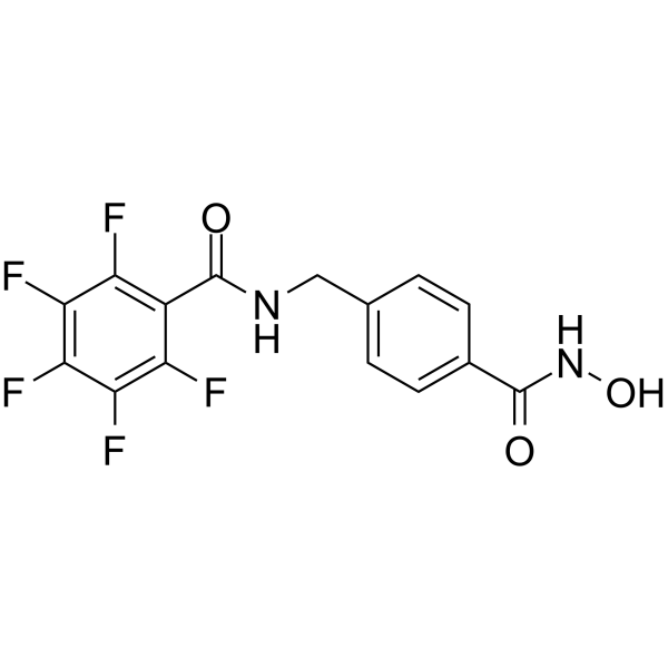
PTG-0861 is a selective histone deacetylase 6 (HDAC6) inhibitor with the IC50 value of 5.92 nM. PTG-0861 induces apoptosis and can be used in the study of acute myeloid leukemia, multiple myeloma and other hematological cancers.
Nur für Forschungszwecke. Wir verkaufen nicht an Patienten.
- CAS. Nr.: 2494082-34-5
- Formel: C15H9F5N2O3
- Molecular Weight:360.24
-
Speicherung:
Please store the product under the recommended conditions in the Certificate of Analysis.

To place orders, for customer services and technical support, please contact: MedChemExpress USA
Tel: 609-228-6898 E-mail: [email protected] [email protected]


-
 Biologische Aktivität
-
Chemical Information
- Protokoll
- Reinheit & Dokumentation
- Verweise
-
 Help & FAQs




Biologische Aktivität
Beschreibung
IC50 & Target
[1]|
HDAC6 5.92 nM (IC50) |
Cellular Effect
|
Cell Line

|
Type | Value | Description | References |
|---|---|---|---|---|
| Fibroblast | IC50 |
11.5 μM
Compound: 54; PTG-0861
|
Cytotoxicity against human fibroblasts measured after 72 hrs by spectrophotometer method
Cytotoxicity against human fibroblasts measured after 72 hrs by spectrophotometer method
|
[PMID: 32615502] |
| Fibroblast | IC50 |
14.6 μM
Compound: 54; PTG-0861
|
Cytotoxicity against pooled human fibroblasts measured after 72 hrs by spectrophotometer method
Cytotoxicity against pooled human fibroblasts measured after 72 hrs by spectrophotometer method
|
[PMID: 32615502] |
| HUVEC | IC50 |
10.3 μM
Compound: 54; PTG-0861
|
Cytotoxicity against human HUVEC cells measured after 72 hrs by spectrophotometer method
Cytotoxicity against human HUVEC cells measured after 72 hrs by spectrophotometer method
|
[PMID: 32615502] |
| MM1.S | IC50 |
1.9 μM
Compound: 54; PTG-0861
|
Cytotoxicity against human MM1.S cells measured after 72 hrs by spectrophotometer method
Cytotoxicity against human MM1.S cells measured after 72 hrs by spectrophotometer method
|
[PMID: 32615502] |
| MV4-11 | IC50 |
1.85 μM
Compound: 1
|
Cytotoxicity against human MV4-11 cells assessed as cell viability incubated for 72 hrs by Cell titer-blue assay
Cytotoxicity against human MV4-11 cells assessed as cell viability incubated for 72 hrs by Cell titer-blue assay
|
[PMID: 34101461] |
| MV4-11 | IC50 |
1.85 μM
Compound: 54; PTG-0861
|
Cytotoxicity against human MV4-11 cells measured after 72 hrs by spectrophotometer method
Cytotoxicity against human MV4-11 cells measured after 72 hrs by spectrophotometer method
|
[PMID: 32615502] |
| RPMI-8226 | IC50 |
4.94 μM
Compound: 54; PTG-0861
|
Cytotoxicity against human RPMI-8226 cells measured after 72 hrs by spectrophotometer method
Cytotoxicity against human RPMI-8226 cells measured after 72 hrs by spectrophotometer method
|
[PMID: 32615502] |
In Vitro
PTG-0861 (compound 54) (0.1-5 μM, 6 hours) stimulates the expression of acetylated a-tubulin and has inhibitory activity against HDAC6 with the IC50 value of 0.59 μM[1].
PTG-0861 (compound 54) (0-4 μM, 18 hours) can induce apoptosis in a dose-dependent manner and has some cytotoxic effect[1].
MedChemExpress (MCE) has not independently confirmed the accuracy of these methods. They are for reference only.
-
Cell Line:MV4-11 cells
-
Concentration:0.1-5 μM
-
Incubation Time:6 hours
-
Result:Induced the accumulation of acetylated a-tubulin expression at 500 nM.
-
Cell Line:HeLa cells
-
Concentration:0-2 μM
-
Incubation Time:6 hours
-
Result:Increased levels of acetylated a-tubulin at 0.1 μM.
-
Cell Line:MV4-11 cells
-
Concentration:0-4 μM
-
Incubation Time:18 hours
-
Result:Induced about 18% cells late apoptosis at 4 μM while at low dose 0.25 μM only about 5%.
-
Cell Line:Hematological cancer cell lines MV4-11, MM.1S, and RPMI 8226
-
Concentration:1.24-4.94 μM
-
Incubation Time:72 hours
-
Result:Showed cytotoxic effects on MV4-11, MM.1S, and RPMI 8226 with the IC50 value of 1.85 μM,1.9 μM,4.94 μM, respectively.
-
Cell Line:
-
Concentration:
-
Incubation Time:
-
Result:Result: The pharmacokinetic parameters of PTG-0861 in vitro
Parameter PTG-0861 Percent Remaining (%) 0 min 100.00 Percent Remaining (%) 30 min 96.41 Percent Remaining (%) 60 min 97.98 Percent Remaining (%) 120 min 97.08 T1/2 (min) ∞ T1/2 (min) 50.85 ± 3.37 CLint (mL/min/106 cells) 27.32 ± 1.81 -Log Pe 5.66 ± 0.02 Papp (A-B) (10 6, cm/s) 1.33 ± 0.03 Papp (B-A) (10 6, cm/s) 0.94 ± 0.13 Efflux Ratio 0.71 ± 0.08
In Vivo
MedChemExpress (MCE) has not independently confirmed the accuracy of these methods. They are for reference only.
-
Animal Model:CD1 mice[1]
-
Dosage:20 mg/kg
-
Administration:Oral administration; everyday; 5 days
-
Result:No weight loss in mice and no obvious toxicity.
-
Animal Model:Male CD1 mice[1]
-
Dosage:20 mg/kg
-
Administration:Intraperitoneal injection; once
-
Result:The pharmacokinetic parameters of PTG-0861 in vivo
Parameter PTG-0861 half-life 0.25 h Cmax 526 ng/mL AUClast 190 h∗ng/mL AUCinf 219 h∗ng/mL AUC Extr(%) 0.324 MRT(h) 0.350 AUC/D(h∗mg/mL) 9.5


Chemical Information
-
CAS. Nr. 2494082-34-5
-
Molecular Weight 360.24
-
Formel C15H9F5N2O3
-
SMILES
O=C(NCC1=CC=C(C(NO)=O)C=C1)C2=C(F)C(F)=C(F)C(F)=C2F
-
Versand
Room temperature in continental US; may vary elsewhere.
-
Speicherung
Please store the product under the recommended conditions in the Certificate of Analysis.


Reinheit & Dokumentation
Verweise

Calculators
Konzentration (Stammlösung) × Volumen (Stammlösung) = Konzentration (Ziellösung) × Volumen (Ziellösung)


PTG-0861 Related Classifications
HY-150586 Related Classifications
 Powered by Bioz