



Vorinostat-d5

Based on 1 Customer Validation
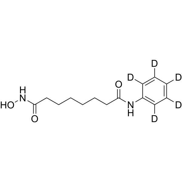
Vorinostat-d5 (SAHA-d5) is the deuterium labeled Vorinostat. Vorinostat (SAHA) is a potent and orally active pan-inhibitor of HDAC1, HDAC2 and HDAC3 (Class I), HDAC7 (Class II) and HDAC11 (Class IV), with ID50 values of 10 nM and 20 nM for HDAC1 and HDAC3, respectively. Vorinostat induces cell apoptosis. Vorinostat is also an effective inhibitor of human papillomaviruse (HPV)-18 DNA amplification.
商品は「研究用試薬」です。人や動物の医療用・臨床診断用・食品用の製品ではありません。
研究用途以外に使用した場合、当社は一切の責任を負いかねます。
- 純度 : 99.0%
- CAS 番号: 1132749-48-4
- 分子式: C14H15D5N2O3
- 分子量:269.35
-
保管条件:Powder -20°C, 3 years ; In solvent -80°C, 6 months , -20°C, 1 month

To place orders, for customer services and technical support, please contact: MedChemExpress USA
Tel: 609-228-6898 E-mail: [email protected] [email protected]


-
 生物活性
生物活性
-
化学情報
- プロトコル
- 純度とドキュメンテーション
- 参考文献
-
 Help & FAQs




生物活性
製品説明
体外実験
Stable heavy isotopes of hydrogen, carbon, and other elements have been incorporated into drug molecules, largely as tracers for quantitation during the drug development process. Deuteration has gained attention because of its potential to affect the pharmacokinetic and metabolic profiles of drugs[1].
MedChemExpress (MCE) has not independently confirmed the accuracy of these methods. They are for reference only.
アプリケーション
1. This compound can be used as a tracer
2. This compound can be used as an internal standard for quantitative analysis by NMR, GC-MS, or LC-MS.


化学情報
-
CAS 番号 1132749-48-4
-
非標識Cas 149647-78-9
-
性状 Solid
-
分子量 269.35
-
分子式 C14H15D5N2O3
-
Color White to off-white
-
SMILES
O=C(NO)CCCCCCC(NC1=C([2H])C([2H])=C([2H])C([2H])=C1[2H])=O
-
別名
SAHA-d5; Suberoylanilide hydroxamic acid-d5
-
輸送条件
Room temperature in continental US; may vary elsewhere.
-
保管条件
Powder -20°C 3 years In solvent -80°C 6 months -20°C 1 month


純度とドキュメンテーション
-
データシート (266 KB)
-
SDS (252 KB)
- English - EN (252 KB)
- Français - FR (252 KB)
- Deutsch - DE (252 KB)
- Norwegian - NO (252 KB)
- Español - ES (252 KB)
- Swedish - SV (252 KB)
- Italian - IT (252 KB)
- Korean - KR (252 KB)
- Portuguese - PT (252 KB)
-
取扱説明書 (2659 KB)
参考文献
[1]. Russak EM, et al. Impact of Deuterium Substitution on the Pharmacokinetics of Pharmaceuticals. Ann Pharmacother. 2019;53(2):211-216. [Content Brief]
[2]. Hrzenjak A et al. Histone deacetylase inhibitor vorinostat suppresses the growth of uterine sarcomas in vitro and in vivo. Mol Cancer. 2010 Mar 4;9:49.;Lautz TB, et al. The effect of vorinostat on the development of resistance to NSC 123127 in neuroblastoma. PLoS One. 2012;7(7):e40816.;Richon VM, et al. A class of hybrid polar inducers of transformed cell differentiation inhibits histone deacetylases. Proc Natl Acad Sci U S A. 1998 Mar 17;95(6):3003-7.;Xu WS, et al. Histone deacetylase inhibitors: molecular mechanisms of action. Oncogene. 2007 Aug 13;26(37):5541-52.;Pérez-Cañamás A, et al. Sphingomyelin-induced inhibition of the plasma membrane calcium ATPase causes neurodegeneration in type A Niemann-Pick disease. Mol Psychiatry. 2017 May;22(5):711-723.;Wang J, et al. Snail determines the therapeutic response to mTOR kinase inhibitors by transcriptional repression of 4E-BP1. Nat Commun. 2017 Dec 20;8(1):2207.;Banerjee NS, et al. Vorinostat, a pan-HDAC inhibitor, abrogates productive HPV-18 DNA amplification. Proc Natl Acad Sci U S A. 2018 Nov 20;115(47):E11138-E11147. [Content Brief]

Calculators
濃度 (開始) × 体積 (開始) = 濃度 (終了) × 体積 (終了)


Vorinostat-d5 関連分類
HY-115412 関連分類
 Powered by Bioz