



LL-K9-3

Based on 1 Customer Validation
LL-K9-3 is a selective CDK9-cyclin T1 HyT degrader, with DC50 values of 0.662 μM and 0.589 μM for CDK9 and Cyclin T1, respectively. LL-K9-3 lacks an E3 ligand moiety and induces polyubiquitination and proteasome-mediated synchronous degradation of CDK9 and cyclin T1 via a hydrophobic tag-mediated mechanism. LL-K9-3 downregulates the protein levels of androgen receptor (AR) and c-Myc, and exhibits antiproliferative and pro-apoptotic (apoptosis) activity in prostate cancer cells. LL-K9-3 can be used in studies related to prostate cancer.
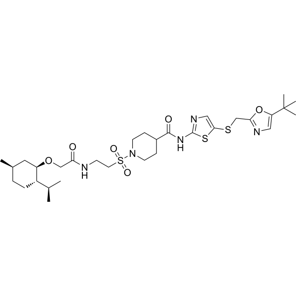
(Pink: CDK9/cyclinT1 ligand (HY-10008); Blue: hydrophobic tag ligand (HY-W093149); Black: linker).
For research use only. We do not sell to patients.
- Purity : 98.90%
- CAS No.: 2809353-52-2
- Formula: C31H49N5O6S3
- Molecular Weight:683.95
-
Storage:Powder -20°C, 3 years ; In solvent -80°C, 6 months , -20°C, 1 month

To place orders, for customer services and technical support, please contact: MedChemExpress USA
Tel: 609-228-6898 E-mail: [email protected] [email protected]


-
 Biological Activity
-
Chemical Information
- Protocols
- Purity & Documentation
- References
-
 Help & FAQs




Biological Activity
Description
IC50 & Target
[1]|
CDK9 662 nM (DC50) |
cyclin T1 589 nM (DC50) |
Cellular Effect
|
Cell Line

|
Type | Value | Description | References |
|---|---|---|---|---|
| CWR22R | IC50 |
0.095 μM
Compound: LL-K9-3
|
Antiproliferative activity against human 22Rv1 cells assessed as reduction in cell viability measured after 5 days by CellTiter-Glo assay
Antiproliferative activity against human 22Rv1 cells assessed as reduction in cell viability measured after 5 days by CellTiter-Glo assay
|
[PMID: 35925880] |
In Vitro
LL-K9-3 (3 μM; 1-48 h) induces synchronous, time-dependent degradation of CDK9 and Cyclin T1 in 22RV1 cells, with a significant reduction starting at 5-7 h and near-complete depletion observed by 48 h[1].
LL-K9-3 (5 days) potently inhibits the proliferation of 22RV1 cells, with IC50 values of 95 nM (Ref. 1) and 224.85 nM (Ref. 2)[1][2].
LL-K9-3 (100-400 nM; 14 days) potently inhibits colony formation of 22RV1 cells at nanomolar concentrations[1].
LL-K9-3 (0.3-1 μM; 24 h) inhibits CDK9 and AR downstream signaling pathways in 22RV1 cells, including reducing the levels of cMyc, MCL-1, AR and AR target proteins[1].
LL-K9-3 (2 μM; 24 h) inhibits MYC- and AR-mediated oncogenic transcriptional programs in 22RV1 cells[1].
LL-K9-3 (0.3-1 μM; 24 h) induces G1-phase cell cycle arrest and significant apoptosis in 22RV1 cells[1].
MedChemExpress (MCE) has not independently confirmed the accuracy of these methods. They are for reference only.
-
Cell Line:22RV1
-
Concentration:100, 200, 400 nM
-
Incubation Time:14 days
-
Result:Inhibited colony formation of 22RV1 cells.
-
Cell Line:22RV1
-
Concentration:0.3 and 1 μM
-
Incubation Time:24 h
-
Result:Induced cell apoptosis.
-
Cell Line:22RV1
-
Concentration:0.3 and 1 μM
-
Incubation Time:24 h
-
Result:Induced G1-phase cell cycle arrest.
-
Cell Line:22RV1
-
Concentration:0.3 and 1 μM
-
Incubation Time:24 h
-
Result:Caused a pronounced decrease in phospho-Rpb1 CTD (Ser2) levels, indicating inhibition of CDK9 downstream signaling.
Reduced MCL-1, full-length AR, AR splice variant ARV7, NKX3-1, and KLK3 levels.


Chemical Information
-
CAS No. 2809353-52-2
-
Appearance Solid
-
Molecular Weight 683.95
-
Formula C31H49N5O6S3
-
Color White to off-white
-
SMILES
O=S(CCNC(CO[C@H]1[C@@H](CC[C@H](C1)C)C(C)C)=O)(N2CCC(CC2)C(NC3=NC=C(SCC4=NC=C(C(C)(C)C)O4)S3)=O)=O
-
Shipping
Room temperature in continental US; may vary elsewhere.
-
Storage
Powder -20°C 3 years In solvent -80°C 6 months -20°C 1 month


Purity & Documentation
-
Data Sheet (270 KB)
-
SDS (252 KB)
- English - EN (252 KB)
- Français - FR (252 KB)
- Deutsch - DE (252 KB)
- Norwegian - NO (252 KB)
- Español - ES (252 KB)
- Swedish - SV (252 KB)
- Italian - IT (252 KB)
- Korean - KR (252 KB)
- Portuguese - PT (252 KB)
-
Handling Instructions (2659 KB)
References
[1]. Li J, et al. Discovery of Small-Molecule Degraders of the CDK9-Cyclin T1 Complex for Targeting Transcriptional Addiction in Prostate Cancer. Journal of medicinal chemistry. 2022 Aug 25;65(16):11034-11057. [Content Brief]
[2]. Lin R, et al. Discovery of HyT-Based Degraders of CDK9-Cyclin T1 Complex. Chemistry & biodiversity. 2023 Aug;20(8):e202300769. [Content Brief]

Calculators
Concentration (start) × Volume (start) = Concentration (final) × Volume (final)


LL-K9-3 Related Classifications
HY-156084 Related Classifications
 Powered by Bioz