



Atazanavir sulfate
Based on 13 publication(s) in Google Scholar
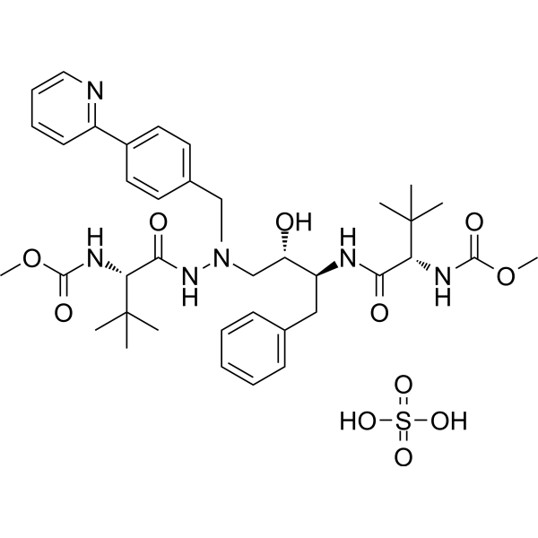
Atazanavir (BMS-232632) sulfate is a highly selective and orally active HIV-1 protease inhibitor with blood-brain barrier permeability. Atazanavir sulfate is a substrate and inhibitor of CYP3A4, and an inhibitor of P-glycoprotein (P-gp). Atazanavir sulfate is also a SARS-CoV 3CLpro inhibitor with an IC50 of 3.49 μM. Atazanavir sulfate inhibits cardiac fibrosis, hyperlipidemia and induces malignant glioma death.
For research use only. We do not sell to patients.
- Purity : 99.68%
- CAS No.: 229975-97-7
- Formula: C38H54N6O11S
- Molecular Weight:802.93
-
Storage:
4°C, sealed storage, away from moisture
* In solvent : -80°C, 6 months; -20°C, 1 month (sealed storage, away from moisture)

To place orders, for customer services and technical support, please contact: MedChemExpress USA
Tel: 609-228-6898 E-mail: [email protected] [email protected]


-
 Biological Activity
-
Chemical Information
-
Solvent & Solubility
- Protocols
- Purity & Documentation
- References
-
 Help & FAQs


-
HY-90001
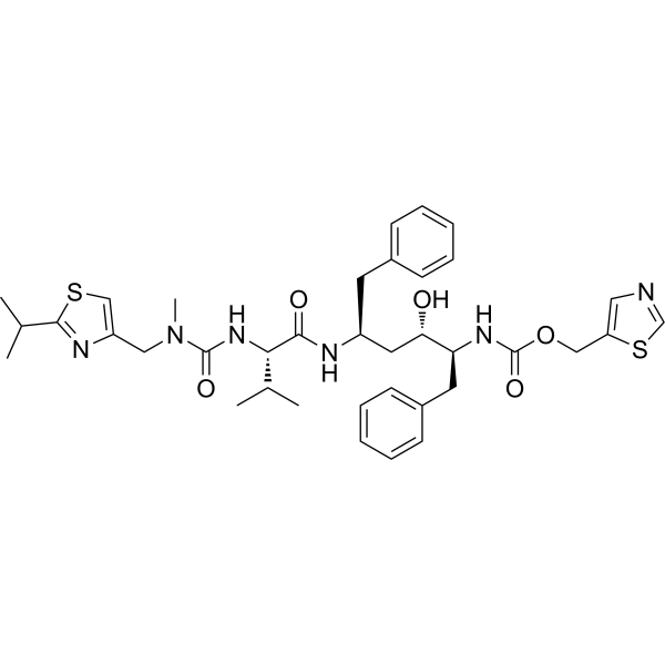
RitonavirHIV Protease Inhibitor -
HY-17041
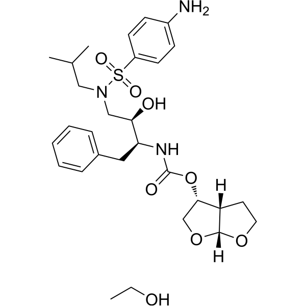
Darunavir EthanolateHIV Inhibitor -
HY-17007
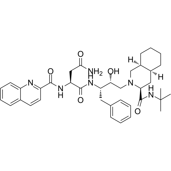
SaquinavirHIV Protease Inhibitor -
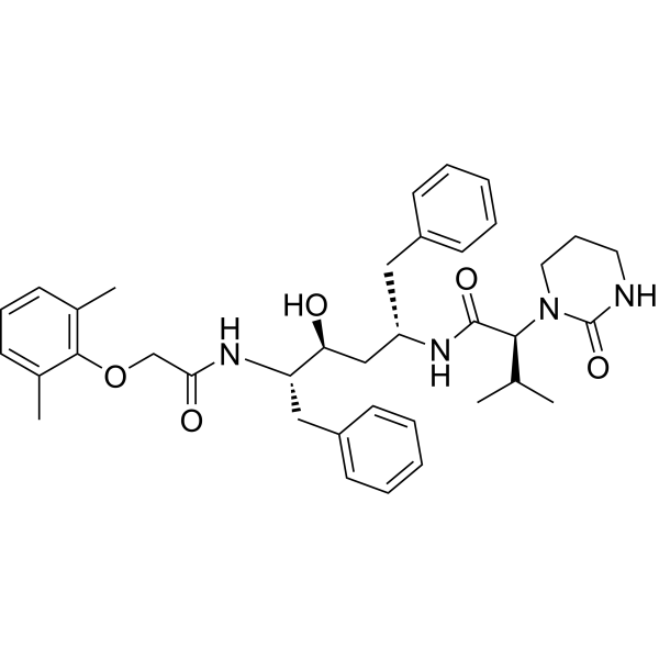
HY-14588
LopinavirHIV-1 Protease Inhibitor -
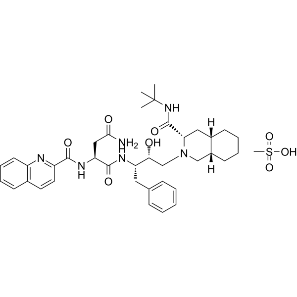
HY-17003
Saquinavir mesylateHIV Protease Inhibitor -
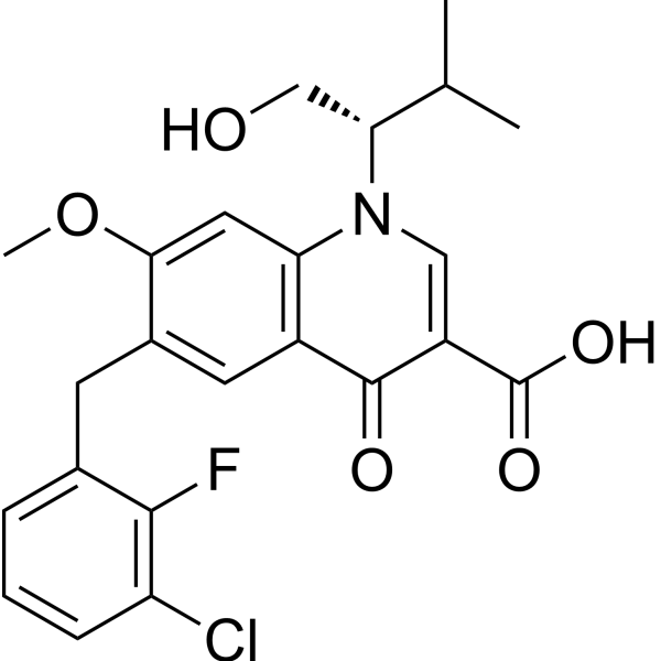
HY-14740
ElvitegravirHIV Integrase Inhibitor -
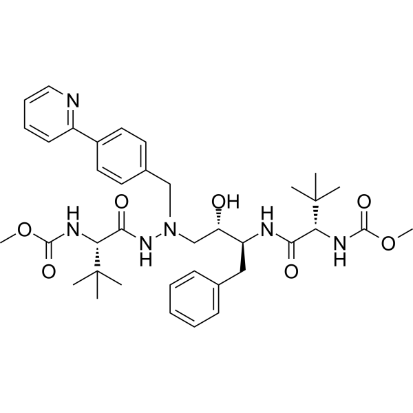
HY-17367
AtazanavirHIV Protease Inhibitor -
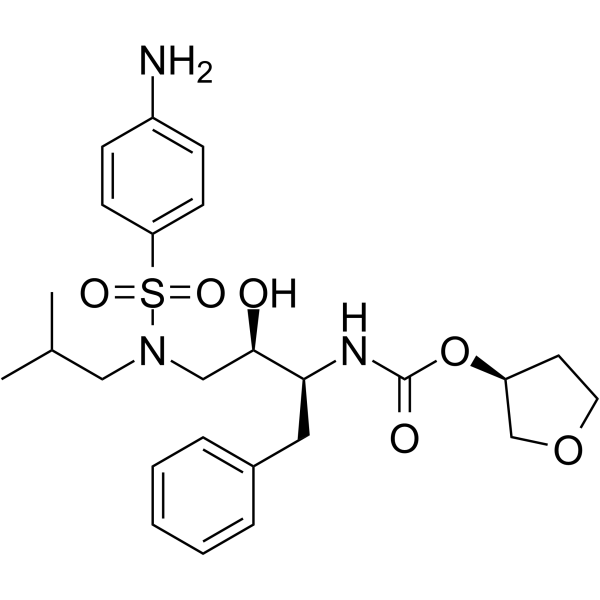
HY-17430
AmprenavirHIV Protease Inhibitor -
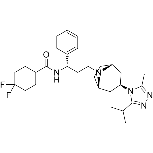
HY-13004
MaravirocCCR5 Antagonist -
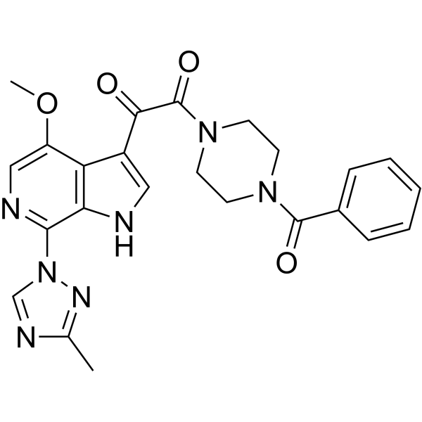
HY-15440
TemsavirHIV Inhibitor -
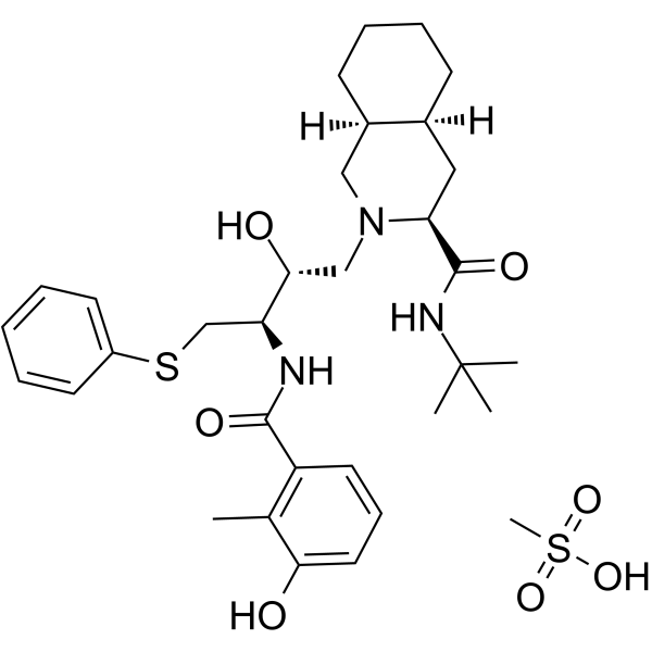
HY-15287A
Nelfinavir MesylateHIV-1 Protease Inhibitor -
HY-10353
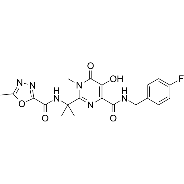
RaltegravirIntegrase Inhibitor
Publications Citing Use of MedChemExpress (MCE) Atazanavir sulfate
More- Signal Transduct Target Ther. 2025 Dec 15;10(1):406. [Abstract]
- Signal Transduct Target Ther. 2021 May 29;6(1):212. [Abstract]
- Nat Commun. 2020 Sep 4;11(1):4417. [Abstract]
- Nat Commun. 2020 Apr 14;11(1):1792. [Abstract]
- Aging Cell. 2023 Jan;22(1):e13750. [Abstract]
- PLoS Biol. 2020 Jan 16;18(1):e3000599. [Abstract]
- Int J Antimicrob Agents. 2019 Dec;54(6):814-819. [Abstract]
- J Photochem Photobiol A Chem. 2025 Nov 15;473:116930.
- Antimicrob Agents Chemother. 2020 Aug 20;64(9):e00872-20. [Abstract]
- Curr Protoc. 2021 Feb;1(2):e32. [Abstract]
- Research Square Print. 2022 May.
- bioRxiv. 2020 Apr.
- University of Oxford. 2019 Jul.


Biological Activity
Description
IC50 & Target
|
CYP3 |
HIV-1 |
In Vitro
Atazanavir (1-10 μM, 72 h) attenuates hypoxia induced rat cardiac fibroblasts (rCFs) s proliferation by modulating the HMGB1/TLR 9 pathway[4].
Atazanavir (1-10 μM, 72 h) inhibits collagen I, collagen III, HMGB1, p-IκBα and p-NF-κB expression levels in rat cardiac fibroblasts[4].
Atazanavir (1-10 μM, 48 h) inhibits the growth of A, U251, T98G, LN229 cells and induces ESR (endoplasmic reticulum stress response) in U251, T98G, and LN229 cells[6].
Atazanavir (6.0 μM) elicites Ca2+ transients and blebbing of the plasma membranes of C2C12 skeletal muscle myotubes[7].
MedChemExpress (MCE) has not independently confirmed the accuracy of these methods. They are for reference only. Further protocols information, click here.
-
Cell Line:Rat cardiac fibroblasts (rCFs)
-
Concentration:0, 1, 3, 10 μM
-
Incubation Time:72 h
-
Result:Inhibited rCFs proliferation in a concentration-dependent manner.
-
Cell Line:Rat cardiac fibroblasts (rCFs)
-
Concentration:0, 1, 3, 10 μM
-
Incubation Time:72 h
-
Result:Reduced collagen I and collagen III levels in a concentration-dependent manner.
Inhibited HMGB1, p-IκBα and p-NF-κB expression.
-
Cell Line:A, U251, T98G, LN229 glioblastoma cell
-
Concentration:0, 12, 25, 50, 75, 100 μM
-
Incubation Time:48 h
-
Result:Reduced cell growth and survival.
-
Cell Line:U251, T98G, and LN229 glioblastoma cell lines
-
Concentration:0, 12, 25, 50, 75, 100 μM
-
Incubation Time:48 h
-
Result:Increased GRP78 and CHOP protein levels dose-dependently and activated caspase-4 in all cell lines.
Parmacokinetics
| Species | Dose | Route | T1/2 | AUC | Clearance (CL) | Vd | Tmax |
|---|---|---|---|---|---|---|---|
| Rat | 7 mg/kg | i.p. | 1.22 h | 3.38 μg·h/mL | / | / | / |
| Rat | 7 mg/kg | i.v. | 1.13 h | 4.09 μg·h/mL | 1.77 L/h/kg | 2.23 L/kg | / |
| Rat | 7 mg/kg | p.o. | 1.04 h | 1.70 μg·h/mL | 0.54 μg/mL | / | 2.25 h |
In Vivo
Atazanavir sulfate has poor blood-brain barrier (BBB) ??permeability, but it can be verified in vitro using human BBB models[8][9].
MedChemExpress (MCE) has not independently confirmed the accuracy of these methods. They are for reference only.
-
Animal Model:The myocardial infarction (MI) model was established in adult male Sprague-Dawley rats through surgical procedures[4].
-
Dosage:30 mg/kg
-
Administration:i.g., daily, 4 weeks
-
Result:Significantly improved systolic pressure (LVSP), +dp/dtmax and −dp/dtmax after MI.
Decreased the non-infarcted LV and hypertrophy of the myocytes.
Significantly attenuated an increase in morphometrical collagen volume fraction in the border left ventricle.
Lowered the heart index (heart-weight to body-weight ratio).
Decreased expressions of α-SMA, HMGB1, TLR 9, p-NF-κB, collagen I and collagen III, and the content of Hyp.
Clinical Trial
| NCT Number | Sponsor | Condition | Start Date |
Phase

|
|---|---|---|---|---|
| NCT01329991 | Plexxikon| | 2011-05 | PHASE1 |


Chemical Information
-
CAS No. 229975-97-7
-
Appearance Solid
-
Molecular Weight 802.93
-
Formula C38H54N6O11S
-
Color White to light yellow
-
SMILES
O=C(OC)N[C@@H](C(C)(C)C)C(NN(CC1=CC=C(C2=NC=CC=C2)C=C1)C[C@H](O)[C@H](CC3=CC=CC=C3)NC([C@H](C(C)(C)C)NC(OC)=O)=O)=O.O=S(O)(O)=O
-
Synonyms
BMS-232632 sulfate
-
Shipping
Room temperature in continental US; may vary elsewhere.
-
Storage
4°C, sealed storage, away from moisture
* In solvent : -80°C, 6 months; -20°C, 1 month (sealed storage, away from moisture)


Publications (13)
-
Journal Impact Factor
-
Most Recent
-
Signal Transduct Target Ther
Selective depletion of tumor-associated SAMHD1 enhances chemotherapeutic efficacy and antitumor immune responses. [Abstract]2025 Dec 15;10(1):406. PMID: 41392286 -
Signal Transduct Target Ther
Bardoxolone and bardoxolone methyl, two Nrf2 activators in clinical trials, inhibit SARS-CoV-2 replication and its 3C-like protease. [Abstract]2021 May 29;6(1):212. PMID: 34052830 -
Nat Commun
Both Boceprevir and GC376 efficaciously inhibit SARS-CoV-2 by targeting its main protease. [Abstract]2020 Sep 4;11(1):4417. PMID: 32887884 -
Nat Commun
Selective inhibition of cancer cell self-renewal through a Quisinostat-histone H1.0 axis. [Abstract]2020 Apr 14;11(1):1792. PMID: 32286289 -
Aging Cell
Antiretroviral protease inhibitors induce features of cellular senescence that are reversible upon drug removal. [Abstract]2023 Jan;22(1):e13750. PMID: 36539941 -
PLoS Biol
2020 Jan 16;18(1):e3000599. PMID: 31945054 -
Int J Antimicrob Agents
2019 Dec;54(6):814-819. PMID: 31479744 -
-
Antimicrob Agents Chemother
2020 Aug 20;64(9):e00872-20. PMID: 32669265 -
Curr Protoc
Quantitative Proteomic Analysis of the Senescence-Associated Secretory Phenotype by Data-Independent Acquisition. [Abstract]2021 Feb;1(2):e32. PMID: 33524224 -
-
-


Solvent & Solubility
In Vitro:
DMSO : 166 mg/mL (206.74 mM; Need ultrasonic and warming; Hygroscopic DMSO has a significant impact on the solubility of product, please use newly opened DMSO)
H2O : < 0.1 mg/mL (insoluble)
Please refer to the solubility information to select the appropriate solvent. Once prepared, please aliquot and store the solution to prevent product inactivation from repeated freeze-thaw cycles.
Storage method and period of stock solution: -80°C, 6 months; -20°C, 1 month (sealed storage, away from moisture). When stored at -80°C, please use it within 6 months. When stored at -20°C, please use it within 1 month.
Please refer to the solubility information to select the appropriate solvent. Once prepared, please aliquot and store the solution to prevent product inactivation from repeated freeze-thaw cycles.
Storage method and period of stock solution: -80°C, 6 months; -20°C, 1 month (sealed storage, away from moisture). When stored at -80°C, please use it within 6 months. When stored at -20°C, please use it within 1 month.
Concentration (start) × Volume (start) = Concentration (final) × Volume (final)
In Vivo:
Select the appropriate dissolution method based on your experimental animal and administration route.
- For the following dissolution methods, please ensure to first prepare a clear stock solution using an In Vitro approach and then sequentially add co-solvents:
- To ensure reliable experimental results, the clarified stock solution can be appropriately stored based on storage conditions. As for the working solution for In Vivo experiments, it is recommended to prepare freshly and use it on the same day.
- The percentages shown for the solvents indicate their volumetric ratio in the final prepared solution. If precipitation or phase separation occurs during preparation, heat and/or sonication can be used to aid dissolution.
Add each solvent one by one: 10% DMSO 40% PEG300 5% Tween-80 45% Saline
Solubility: ≥ 2.5 mg/mL (3.11 mM); Clear solution
This protocol yields a clear solution of ≥ 2.5 mg/mL (saturation unknown).
Taking 1 mL working solution as an example, add 100 μL DMSO stock solution (25.0 mg/mL) to 400 μL PEG300, and mix evenly; then add 50 μL Tween-80 and mix evenly; then add 450 μL Saline to adjust the volume to 1 mL.
Preparation of Saline: Dissolve 0.9 g sodium chloride in ddH₂O and dilute to 100 mL to obtain a clear Saline solution.
Add each solvent one by one: 10% DMSO 90% (20% SBE-β-CD in Saline)
Solubility: ≥ 2.5 mg/mL (3.11 mM); Clear solution
This protocol yields a clear solution of ≥ 2.5 mg/mL (saturation unknown).
Taking 1 mL working solution as an example, add 100 μL DMSO stock solution (25.0 mg/mL) to 900 μL 20% SBE-β-CD in Saline, and mix evenly.
Preparation of 20% SBE-β-CD in Saline (4°C, storage for one week): 2 g SBE-β-CD powder is dissolved in 10 mL Saline, completely dissolve until clear.
In Vivo Dissolution Calculator
Please enter the basic information of animal experiments:
-
-
-
-
Recommended: Prepare an additional quantity of animals to account for potential losses during experiments.
Please enter your animal formula composition:
-
%DMSO +
Recommended: Keep the proportion of DMSO in working solution below 2% if your animal is weak.
-
%+
-
+%Tween-80 + +
-
%Saline +
The co-solvents required include: DMSO, . All of co-solvents are available by MedChemExpress (MCE). , Tween 80. All of co-solvents are available by MedChemExpress (MCE).
Working solution concentration: 0.22 mg/mL
Method for preparing stock solution: mg drug dissolved in μL DMSO. Stock solution concentration: mg/mL. * In solvent : -80°C, 6 months; -20°C, 1 month (sealed storage, away from moisture)
1. Take μL DMSO stock solution;
2. Add μL .
μL , mix evenly;
3. Then add μL Tween 80, mix evenly;
4. Then add μL
Please ensure that the stock solution in the first step is dissolved to a clear state, and add co-solvents in sequence. You can use ultrasonic heating (ultrasonic cleaner, recommended frequency 20-40 kHz), vortexing, etc. to assist dissolution.

Protocols
-
Fibrosis/Collagen Morphometry
Fibrosis and collagen morphometry is based on the quantitative visualization of fibrillar collagen deposition in tissue sections using histochemical stains such as Sirius Red (Picrosirius Red) or Masson's trichrome, followed by image-based or polarization-enhanced analysis to estimate collagen proportional area as a surrogate of extracellular matrix accumulation during fibrotic remodeling. Sirius Red combined with polarized light microscopy enhances detection of collagen fibers due to birefringence properties, enabling more specific visualization of collagen type I and III fibrils compared to conventional bright-field histology, while whole-section or region-restricted digital morphometry reduces field-selection bias in fibrosis assessment. Alternative quantitative approaches include second harmonic generation (SHG) and two-photon excited fluorescence microscopy, which enable label-free detection of fibrillar collagen and have been validated against histological staining and biochemica
-
Collagen: Sirius Red Staining
Sirius Red or picrosirius red staining is a histochemical method for visualizing collagen-rich extracellular matrix in tissue sections, and collagen fibers are detected as red-stained structures under bright-field microscopy with enhanced birefringence under polarized light. Picrosirius red is useful for assessing total collagen organization, distribution, and fibrosis burden, but polarized color should not be interpreted as a definitive collagen type I versus type III readout because color is affected by fiber orientation, thickness, and packing.
-
Cell Cytotoxicity Assay
Cytotoxicity assays are usually based on the assessment of cell membrane damage, which can also be indirectly detected by measuring cell viability. Detection methods include MTT assay, CKK-8 assay, LDH assay and ATP assay, etc.
-
Connective Tissue: Masson's Trichrome/Collagen Trichrome Staining
Masson’s Trichrome (collagen/trichrome staining) is a histological technique that differentially stains tissue compartments using sequential acidic dyes to distinguish collagen from muscle and cytoplasmic components based on dye affinity and tissue permeability differences, enabling visualization of fibrosis and connective tissue architecture in histological sections. The classical formulation typically uses Weigert's iron hematoxylin for nuclear staining, Biebrich scarlet-acid fuchsin for cytoplasm and muscle, and aniline blue (or light green variants) for collagen, producing a characteristic blue/green collagen signal contrasted against red cytoplasm and dark nuclei. The staining principle relies on selective displacement of smaller dye molecules by larger anionic dyes in collagen-rich regions under controlled acidified conditions, which enhances collagen-specific dye retention. This property makes the method widely used for fibrosis assessment in organs such as heart, liver, lung, a
-
Transepithelial/transendothelial electrical resistance assay
TEER measures electrical resistance across epithelial or endothelial monolayers cultured on permeable supports, and the readout reflects ionic conductance through the cell barrier, especially the paracellular pathway regulated by junctional integrity. TEER can be measured without destroying the monolayer and is commonly used before or during transport, permeability, barrier-disruption, and barrier-maturation experiments. TEER values are influenced by biological maturation and technical conditions; reported factors include temperature, medium formulation, passage number, electrode geometry, membrane properties, and junctional length during early monolayer maturation. Therefore, TEER should be interpreted with blank-insert subtraction, area normalization, repeated readings, and, when possible, orthogonal barrier readouts such as FITC-dextran flux or tight-junction staining.


Purity & Documentation
-
Data Sheet (300 KB)
-
SDS (640 KB)
- English - EN (640 KB)
- Français - FR (640 KB)
- Deutsch - DE (640 KB)
- Norwegian - NO (640 KB)
- Español - ES (640 KB)
- Swedish - SV (640 KB)
- Italian - IT (640 KB)
- Korean - KR (640 KB)
- Portuguese - PT (640 KB)
-
Handling Instructions (2659 KB)
References
[1]. Havlir DV, et al. Atazanavir: new option for treatment of HIV infection. Clin Infect Dis. 2004 Jun 1;38(11):1599-604. [Content Brief]
[2]. Wood R. Atazanavir: its role in HIV treatment. Expert Rev Anti Infect Ther. 2008 Dec;6(6):785-96. [Content Brief]
[3]. Qi Sun, et al. Bardoxolone and bardoxolone methyl, two Nrf2 activators in clinical trials, inhibit SARS-CoV-2 replication and its 3C-like protease. Signal Transduct Target Ther. 2021 May 29;6(1):212. [Content Brief]
[4]. Zhang G, et al. Long-term oral atazanavir attenuates myocardial infarction-induced cardiac fibrosis. Eur J Pharmacol. 2018 Jun 5;828:97-102. [Content Brief]
[5]. Fukushima K, et al. Effect of serum lipids on the pharmacokinetics of atazanavir in hyperlipidemic rats. Biomed Pharmacother. 2009 Nov;63(9):635-42. [Content Brief]
[6]. Pyrko P, et al. HIV-1 protease inhibitors nelfinavir and atazanavir induce malignant glioma death by triggering endoplasmic reticulum stress. Cancer Res. 2007 Nov 15;67(22):10920-8. [Content Brief]
[7]. Alomar FA, et al. Efavirenz, atazanavir, and ritonavir disrupt sarcoplasmic reticulum Ca2+ homeostasis in skeletal muscles. Antiviral Res. 2021 Mar;187:104975. [Content Brief]
[8]. Robillard KR, et al. Role of P-glycoprotein in the distribution of the HIV protease inhibitor atazanavir in the brain and male genital tract. Antimicrob Agents Chemother. 2014;58(3):1713-22. [Content Brief]
[9]. Bousquet L, et al. Comparison of ABC transporter modulation by atazanavir in lymphocytes and human brain endothelial cells: ABC transporters are involved in the atazanavir-limited passage across an in vitro human model of the blood-brain barrier. AIDS Res Hum Retroviruses. 2008 Sep;24(9):1147-54. [Content Brief]
[10]. Chattopadhyay N, et al. Solid lipid nanoparticles enhance the delivery of the HIV protease inhibitor, atazanavir, by a human brain endothelial cell line. Pharm Res. 2008 Oct;25(10):2262-71. [Content Brief]


Complete Stock Solution Preparation Table
Please refer to the solubility information to select the appropriate solvent. Once prepared, please aliquot and store the solution to prevent product inactivation from repeated freeze-thaw cycles.
Storage method and period of stock solution: -80°C, 6 months; -20°C, 1 month (sealed storage, away from moisture). When stored at -80°C, please use it within 6 months. When stored at -20°C, please use it within 1 month.
| Optional Solvent | Concentration Solvent Mass | 1 mg | 5 mg | 10 mg | 25 mg |
|---|---|---|---|---|---|
| DMSO | 1 mM | 1.2454 mL | 6.2272 mL | 12.4544 mL | 31.1360 mL |
| 5 mM | 0.2491 mL | 1.2454 mL | 2.4909 mL | 6.2272 mL | |
| 10 mM | 0.1245 mL | 0.6227 mL | 1.2454 mL | 3.1136 mL | |
| 15 mM | 0.0830 mL | 0.4151 mL | 0.8303 mL | 2.0757 mL | |
| 20 mM | 0.0623 mL | 0.3114 mL | 0.6227 mL | 1.5568 mL | |
| 25 mM | 0.0498 mL | 0.2491 mL | 0.4982 mL | 1.2454 mL | |
| 30 mM | 0.0415 mL | 0.2076 mL | 0.4151 mL | 1.0379 mL | |
| 40 mM | 0.0311 mL | 0.1557 mL | 0.3114 mL | 0.7784 mL | |
| 50 mM | 0.0249 mL | 0.1245 mL | 0.2491 mL | 0.6227 mL | |
| 60 mM | 0.0208 mL | 0.1038 mL | 0.2076 mL | 0.5189 mL | |
| 80 mM | 0.0156 mL | 0.0778 mL | 0.1557 mL | 0.3892 mL | |
| 100 mM | 0.0125 mL | 0.0623 mL | 0.1245 mL | 0.3114 mL |


Atazanavir sulfate Related Classifications
HY-17367A Related Classifications
 Powered by Bioz
Keywords