



Diethylcarbamazine citrate
Based on 1 publication(s) in Google Scholar
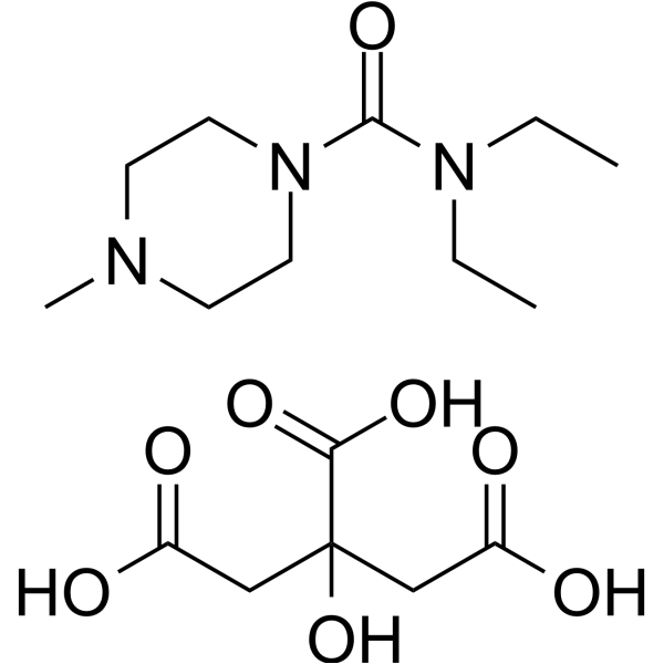
Diethylcarbamazine citrate is an orally active microfilaricidal agent used originally in onchocerciasis and lymphatic filiariasis. Diethylcarbamazine citrate reduces eosinophil trafficking to the lung tissue and exerts anti-allergic effects. Diethylcarbamazine citrate reduces serum levels of leptin, TNF-α, IL-6, MCP-1, glucose, insulin, and triglycerides, and ameliorates insulin resistance without altering body, liver, or adipose tissue weights. Diethylcarbamazine citrate enhances reactive oxygen intermediate expression by polymorphonuclear neutrophils, increases lymphocyte proliferation, and inhibits actinomycetoma lesion development. Diethylcarbamazine citrate can be used for the researches of bronchial asthma, insulin resistance and infection.
For research use only. We do not sell to patients.
- Purity : 98.01%
- CAS No.: 1642-54-2
- Formula: C16H29N3O8
- Molecular Weight:391.42
-
Storage:
4°C, sealed storage, away from moisture
* In solvent : -80°C, 6 months; -20°C, 1 month (sealed storage, away from moisture)

To place orders, for customer services and technical support, please contact: MedChemExpress USA
Tel: 609-228-6898 E-mail: [email protected] [email protected]


-
 Biological Activity
-
Chemical Information
-
Solvent & Solubility
- Protocols
- Purity & Documentation
- References
-
 Help & FAQs


-
HY-W050154
Kojic acidTyrosinase Inhibitor -

HY-12642R
Diethylcarbamazine citrate (Standard)Arachidonic Acid Metabolism Inhibitor -
HY-B0094
ArtemisininParasite Inhibitor -
HY-B0770
ArtemotilParasite Inhibitor -
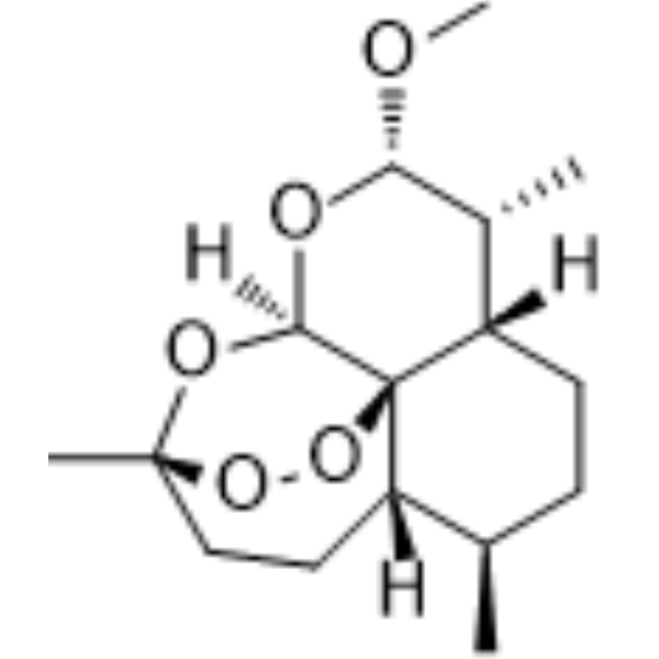
HY-N0402
ArtemetherParasite Inhibitor -
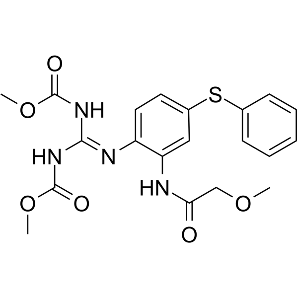
HY-17597
FebantelParasite Inhibitor -
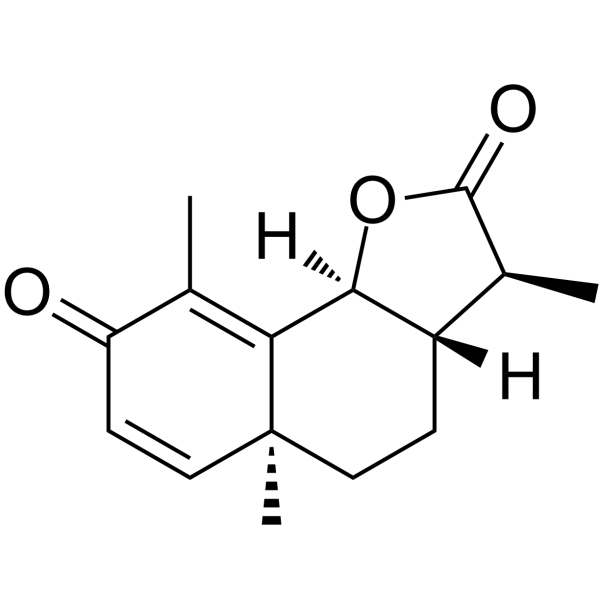
HY-B1761
SantoninParasite Inhibitor -
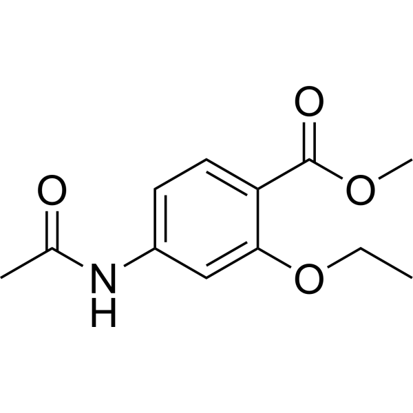
HY-B2138
EthopabateAntiparasitic Agent -
HY-101476
EmodepsideAnthelmintic Agent -
HY-13419
U-73122Phospholipase C Inhibitor -

HY-B1761R
Santonin (Standard)Parasite Inhibitor -
HY-17035
DoramectinAntibiotic
Publications Citing Use of MedChemExpress (MCE) Diethylcarbamazine citrate
MoreAll Parasite Isoforms
More

Biological Activity
Description
IC50 & Target
[1]|
IL-6 |
Cellular Effect
|
Cell Line

|
Type | Value | Description | References |
|---|---|---|---|---|
| Vero | CC50 |
9103 μg/mL
Compound: DEC-C
|
Cytotoxicity against african green monkey Vero cells
Cytotoxicity against african green monkey Vero cells
|
[PMID: 23541646] |
In Vitro
In Vivo
Diethylcarbamazine (12-200 mg/kg; p.o.; twice weekly; 12 weeks) citrate significantly ameliorates high-fat diet-induced insulin resistance in male Swiss mice via suppression of adipose tissue inflammation at the optimal 50 mg/kg dose, without affecting body or tissue weights, with significant reductions in serum glucose, insulin, triglycerides, proinflammatory mediators, liver cyclooxygenase activity, and NF-κBp65 nuclear translocation[2].
Diethylcarbamazine (6 mg/kg; p.o.; daily; 1 week) citrate inhibits actinomycetoma development in BALB/c mice, with a statistically significant reduction in lesion size, and enhances cellular immune responses including neutrophil reactive oxygen intermediate production and lymphocyte proliferation[3].
MedChemExpress (MCE) has not independently confirmed the accuracy of these methods. They are for reference only.
-
Animal Model:white albino mice (6 to 8-week-old female, 18-20 g, intradermal sensitization + aerosol exposure Ovalbumin-induced asthma)[1]
-
Dosage:12 mg/kg
-
Administration:p.o.;
-
Result:Reduced serum IL-4 and IL-5, total BALF IgE and specific anti-ovalbumin IgE, lung tissue EPO and eotaxin2, and reduced inflammatory cell infiltration and thickened alveolar walls compared to untreated asthmatic mice.
Showed no significant changes in serum IL-4, IL-5, BALF IgE, lung EPO, or eotaxin2, and similar histopathological improvement compared to DEC alone when preceded by anti-DEC antibody.
Showed no significant changes in serum IL-4, IL-5, lung EPO, or eotaxin2, and partial reduction in inflammatory cell infiltration and alveolar wall thickening compared to DEC alone when preceded by 10 mg/kg Quercetin.
Significantly reduced serum IL-4 and IL-5, increased IFN-γ/IL-4 ratio to near control levels, decreased total BALF IgE, and reduced inflammatory cell infiltration compared to DEC alone when preceded by 20 mg/kg and 40 mg/kg Quercetin.
-
Animal Model:Swiss mice (male, 5 weeks old, initial 10 g body weight, high-fat diet-induced insulin resistance)[2]
-
Dosage:12 mg/kg; 50 mg/kg; 200 mg/kg
-
Administration:p.o.; twice weekly; 12 weeks (from 6 to 18 week)
-
Result:Reduced serum glucose levels at all oral glucose tolerance test time points (0, 30, 60, 120 min) with a statistically significant reduction in area under the curve at 50 mg/kg.
Reduced serum triglyceride levels at 50 mg/kg.
Reduced serum insulin levels and HOMA-IR score at 50 mg/kg.
Reduced serum leptin levels at 50 mg/kg.
Reduced serum levels of TNF-α, IL-6, and MCP-1 at 50 mg/kg.
Significantly reduced liver cyclooxygenase activity at 50 mg/kg.
Significantly inhibited nuclear localization of NF-κBp65 with increased cytoplasmic retention at 50 mg/kg.
Showed no statistically significant reductions in serum glucose, triglycerides, insulin, HOMA-IR, leptin, TNF-α, IL-6, MCP-1, or liver cyclooxygenase activity relative to untreated high-fat diet-fed mice at 12 mg/kg and 200 mg/kg.
Exhibited less potent effects on NF-κBp65 nuclear localization at 12 mg/kg and 200 mg/kg compared to 50 mg/kg.
Caused no significant changes to body, adipose tissue, or liver weights at 50 mg/kg.
-
Animal Model:BALB/c (male, 10-12 weeks old, 37-45 g, actinomycetoma model via footpad inoculation with Nocardia brasiliensis)[3]
-
Dosage:6 mg/kg
-
Administration:p.o.; daily; 1 week
-
Result:Reduced actinomycetoma lesion size with statistical significance compared to untreated infected controls.
Resolved fistulae, granule discharge, hyperemia, and erythema by day 20 post-infection, and prevented mycetoma establishment through day 90.
Reduced cellular infiltrate, eliminated granuloma layer formation, restored epidermal integrity, and reorganized damaged striated muscle fibers by day 28 post-infection.
Enhanced polymorphonuclear neutrophil reactive oxygen intermediate production with statistical significance at day 3 post-infection.
Increased lymphocyte proliferation in response to N.
brasiliensis cellular crude extract with statistical significance at day 21 post-infection.
Increased lymphocyte proliferation in response to concanavalin A with statistical significance at day 28 post-infection.
Caused no significant changes in IgG or IgM antibody production against N.
brasiliensis P24 antigen.
Clinical Trial
| NCT Number | Sponsor | Condition | Start Date |
Phase
|
|---|---|---|---|---|
| NCT01329991 | Plexxikon| | 2011-05 | PHASE1 |


Chemical Information
-
CAS No. 1642-54-2
-
Appearance Solid
-
Molecular Weight 391.42
-
Formula C16H29N3O8
-
Color White to off-white
-
SMILES
O=C(N1CCN(C)CC1)N(CC)CC.O=C(CC(C(O)=O)(O)CC(O)=O)O
-
Shipping
Room temperature in continental US; may vary elsewhere.
-
Storage
4°C, sealed storage, away from moisture
* In solvent : -80°C, 6 months; -20°C, 1 month (sealed storage, away from moisture)


Publications (1)
-
Journal Impact Factor
-
Most Recent
-
Cancer Sci
PSMD14/E2F1 Axis-Mediated CENPF Promotes the Metastasis of Triple-Negative Breast Cancer Through Inhibiting Ferroptosis. [Abstract]2025 Aug;116(8):2281-2295. PMID: 40365861


Solvent & Solubility
In Vitro:
H2O : 100 mg/mL (255.48 mM; Need ultrasonic)
DMSO : ≥ 39 mg/mL (99.64 mM; Hygroscopic DMSO has a significant impact on the solubility of product, please use newly opened DMSO)
* "≥" means soluble, but saturation unknown.
Please refer to the solubility information to select the appropriate solvent. Once prepared, please aliquot and store the solution to prevent product inactivation from repeated freeze-thaw cycles.
Storage method and period of stock solution: -80°C, 6 months; -20°C, 1 month (sealed storage, away from moisture). When stored at -80°C, please use it within 6 months. When stored at -20°C, please use it within 1 month.
* Note: If you choose water as the stock solution, please dilute it to the working solution, then filter and sterilize it with a 0.22 μm filter before use.
Please refer to the solubility information to select the appropriate solvent. Once prepared, please aliquot and store the solution to prevent product inactivation from repeated freeze-thaw cycles.
Storage method and period of stock solution: -80°C, 6 months; -20°C, 1 month (sealed storage, away from moisture). When stored at -80°C, please use it within 6 months. When stored at -20°C, please use it within 1 month.
* Note: If you choose water as the stock solution, please dilute it to the working solution, then filter and sterilize it with a 0.22 μm filter before use.
Concentration (start) × Volume (start) = Concentration (final) × Volume (final)
In Vivo:
Select the appropriate dissolution method based on your experimental animal and administration route.
- For the following dissolution methods, please ensure to first prepare a clear stock solution using an In Vitro approach and then sequentially add co-solvents:
- To ensure reliable experimental results, the clarified stock solution can be appropriately stored based on storage conditions. As for the working solution for In Vivo experiments, it is recommended to prepare freshly and use it on the same day.
- The percentages shown for the solvents indicate their volumetric ratio in the final prepared solution. If precipitation or phase separation occurs during preparation, heat and/or sonication can be used to aid dissolution.
Add each solvent one by one: 10% DMSO 40% PEG300 5% Tween-80 45% Saline
Solubility: ≥ 2.5 mg/mL (6.39 mM); Clear solution
This protocol yields a clear solution of ≥ 2.5 mg/mL (saturation unknown).
Taking 1 mL working solution as an example, add 100 μL DMSO stock solution (25.0 mg/mL) to 400 μL PEG300, and mix evenly; then add 50 μL Tween-80 and mix evenly; then add 450 μL Saline to adjust the volume to 1 mL.
Preparation of Saline: Dissolve 0.9 g sodium chloride in ddH₂O and dilute to 100 mL to obtain a clear Saline solution.
Add each solvent one by one: 10% DMSO 90% (20% SBE-β-CD in Saline)
Solubility: ≥ 2.5 mg/mL (6.39 mM); Clear solution
This protocol yields a clear solution of ≥ 2.5 mg/mL (saturation unknown).
Taking 1 mL working solution as an example, add 100 μL DMSO stock solution (25.0 mg/mL) to 900 μL 20% SBE-β-CD in Saline, and mix evenly.
Preparation of 20% SBE-β-CD in Saline (4°C, storage for one week): 2 g SBE-β-CD powder is dissolved in 10 mL Saline, completely dissolve until clear.
In Vivo Dissolution Calculator
Please enter the basic information of animal experiments:
-
-
-
-
Recommended: Prepare an additional quantity of animals to account for potential losses during experiments.
Working solution concentration: 0.22 mg/mL
This product has good water solubility, please refer to the measured solubility data in water/PBS/Saline for details.

Protocols
-
RNA extraction experimental
By lysing cells, releasing RNA, and removing impurities such as proteins and DNA, high-purity RNA products are finally obtained. The commonly used traditional method is the guanidine isothiocyanate/phenol/chloroform method (Trizol), which is suitable for a variety of animal materials including animal tissues, microorganisms, cultured cells, etc., and most plant materials.
-
Research Protocol for Infectious Diseases
Infectious-disease experiments test how pathogens interact with host barriers, innate immune receptors, inflammatory signaling, pathogen replication, and tissue injury; pattern-recognition receptors such as TLRs, RIG-I-like receptors, NOD-like receptors, and inflammasomes detect microbial molecules and activate NF-κB, interferon, and cytokine responses. The central hypothesis is that infection severity reflects the balance between pathogen burden and host response: protective inflammation restricts pathogen growth, whereas excessive or mislocalized inflammation contributes to tissue damage and disease phenotype. Unresolved questions include which host pathways are protective versus pathogenic, why some infection models fail to translate to human disease, and which combined readouts best predict clinically relevant infection outcomes.
-
Research Protocol for Endocrine Diseases
Endocrine diseases often arise from disrupted hormone production, hormone signaling, or target-tissue responsiveness; for diabetes-focused endocrine disease models, insulin signaling regulates glucose uptake, hepatic glucose output, lipid metabolism, and β-cell compensation. Type 2 diabetes develops through interacting defects in insulin resistance, β-cell dysfunction, adipose inflammation, hepatic glucose overproduction, altered incretin signaling, and ectopic lipid metabolism. A major unresolved question is whether endocrine dysfunction is driven primarily by target-tissue insulin resistance, intrinsic β-cell failure, immune/inflammatory stress, or combined multi-organ failure that differs by disease stage.
-
Large-size fat particle sorting
Large-size fat particle sorting is widely used to isolate cells up to 200 μm in diameter. Single-cell flow sorting will allow greater insight into adipocyte heterogeneity by identifying gene expression, protein composition, and metabolic signatures at the single-cell level.
-
CFSE Dye Dilution Proliferation Assay
The CFSE (carboxyfluorescein diacetate succinimidyl ester) dye dilution proliferation assay is based on the covalent labeling of intracellular proteins by a cell-permeant fluorescent dye that becomes fluorescent upon intracellular ester cleavage and then is stably retained within cells. As labeled cells divide, the dye is partitioned equally between daughter cells, resulting in a stepwise halving of fluorescence intensity that can be quantified by flow cytometry to determine the number of cell divisions undergone by each cell population. This fluorescence dilution approach enables quantitative tracking of lymphocyte proliferation at the single-cell level over multiple rounds of division. CFSE-based proliferation analysis has been widely applied to measure antigen-driven lymphocyte expansion in vitro, where discrete fluorescence peaks correspond to successive cell divisions and allow reconstruction of proliferative history within heterogeneous populations.
-
Research Protocol for Metabolic Diseases
AMP-activated protein kinase, AMPK, is a conserved cellular energy sensor that responds to reduced cellular energy status and coordinates metabolism by increasing ATP-generating catabolic pathways while suppressing ATP-consuming anabolic processes. In metabolic disease research, the AMPK pathway is experimentally relevant because it regulates hepatic lipid synthesis, fatty acid oxidation, glucose production, skeletal-muscle glucose disposal, mTORC1-linked biosynthesis, autophagy, mitochondrial homeostasis, and whole-body energy balance. The central pathway logic is that energy stress, metformin, exercise-like stimulation, or direct AMPK activators increase AMPKα Thr172 phosphorylation and downstream substrate phosphorylation, including ACC and RAPTOR. Phosphorylation of ACC suppresses lipogenesis and supports fatty acid oxidation, whereas phosphorylation of RAPTOR suppresses mTORC1 signaling and links cellular energy status to growth and protein synthesis control. The pathway is linked
-
Cell Viability Determination by MTT Colorimetric Assay
The following protocol uses the MTT colorimetric assay as a classic literature-established method for assessing cell viability/metabolic activity in cultured mammalian cells. MTT[3-(4,5-dimethylthiazol-2-yl)-2,5-diphenyltetrazolium bromide] is reduced by metabolically active cells to a colored formazan product; the amount of formazan is quantified spectrophotometrically and provides an indirect measure of metabolically active viable cells. Importantly, MTT reduction reflects cellular oxidoreductase/metabolic activity rather than an absolute direct count of living cells, so changes in cellular metabolism can alter the signal independently of cell number.


Purity & Documentation
-
Data Sheet (289 KB)
-
SDS (420 KB)
- English - EN (420 KB)
- Français - FR (420 KB)
- Deutsch - DE (420 KB)
- Norwegian - NO (420 KB)
- Español - ES (420 KB)
- Swedish - SV (420 KB)
- Italian - IT (420 KB)
- Korean - KR (420 KB)
- Portuguese - PT (420 KB)
-
Handling Instructions (2659 KB)
References
[1]. Abdul-Razek N, et al. Enhancement of Anti-allergic Effect of Diethylcarbamazine Citrate in Asthmatic Mouse Model: Testing of Anti-drug Antibodies and Quercetin. Iran J Allergy Asthma Immunol. 2020 Aug 25;19(4):373-385. [Content Brief]
[2]. Abdel-Latif M. Diethylcarbamazine citrate ameliorates insulin resistance in high-fat diet-induced obese mice via modulation of adipose tissue inflammation. Int Immunopharmacol. 2015 Dec;29(2):607-612. [Content Brief]
[3]. García-Hernández M, et al. Immunomodulatory effect of diethylcarbamazine in mice infected with Nocardia brasiliensis. Int Immunopharmacol. 2014;23(1):113-120. [Content Brief]


Complete Stock Solution Preparation Table
Please refer to the solubility information to select the appropriate solvent. Once prepared, please aliquot and store the solution to prevent product inactivation from repeated freeze-thaw cycles.
Storage method and period of stock solution: -80°C, 6 months; -20°C, 1 month (sealed storage, away from moisture). When stored at -80°C, please use it within 6 months. When stored at -20°C, please use it within 1 month.
| Optional Solvent | Concentration Solvent Mass | 1 mg | 5 mg | 10 mg | 25 mg |
|---|---|---|---|---|---|
| DMSO / H2O | 1 mM | 2.5548 mL | 12.7740 mL | 25.5480 mL | 63.8700 mL |
| 5 mM | 0.5110 mL | 2.5548 mL | 5.1096 mL | 12.7740 mL | |
| 10 mM | 0.2555 mL | 1.2774 mL | 2.5548 mL | 6.3870 mL | |
| 15 mM | 0.1703 mL | 0.8516 mL | 1.7032 mL | 4.2580 mL | |
| 20 mM | 0.1277 mL | 0.6387 mL | 1.2774 mL | 3.1935 mL | |
| 25 mM | 0.1022 mL | 0.5110 mL | 1.0219 mL | 2.5548 mL | |
| 30 mM | 0.0852 mL | 0.4258 mL | 0.8516 mL | 2.1290 mL | |
| 40 mM | 0.0639 mL | 0.3194 mL | 0.6387 mL | 1.5968 mL | |
| 50 mM | 0.0511 mL | 0.2555 mL | 0.5110 mL | 1.2774 mL | |
| 60 mM | 0.0426 mL | 0.2129 mL | 0.4258 mL | 1.0645 mL | |
| 80 mM | 0.0319 mL | 0.1597 mL | 0.3194 mL | 0.7984 mL | |
| H2O | 100 mM | 0.0255 mL | 0.1277 mL | 0.2555 mL | 0.6387 mL |
* Note: If you choose water as the stock solution, please dilute it to the working solution, then filter and sterilize it with a 0.22 μm filter before use.


Diethylcarbamazine citrate Related Classifications
HY-12642 Related Classifications
 Powered by Bioz