



Furazolidone
Based on 3 publication(s) in Google Scholar
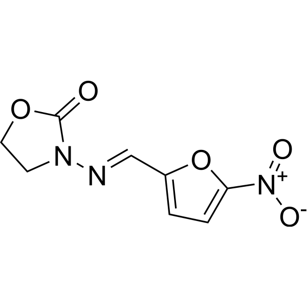
Furazolidone is a monoamine oxidase (MAO) inhibitor with antiproliferative, apoptosis-inducing and differentiation-promoting activities. Furazolidone may inhibit leukemia fusion protein-mediated bone marrow transformation by upregulating the stability of the tumor suppressor protein p53. Furazolidone exhibits anti-leukemic activity in acute myeloid leukemia (AML) cell lines and can be used for anti-AML research
For research use only. We do not sell to patients.
- Purity : 99.92%
- CAS No.: 67-45-8
- Formula: C8H7N3O5
- Molecular Weight:225.16
-
Storage:Powder -20°C, 3 years , 4°C, 2 years ; In solvent -80°C, 2 years , -20°C, 1 year

To place orders, for customer services and technical support, please contact: MedChemExpress USA
Tel: 609-228-6898 E-mail: [email protected] [email protected]


-
 Biological Activity
-
Chemical Information
-
Solvent & Solubility
- Protocols
- Purity & Documentation
- References
-
 Help & FAQs


-
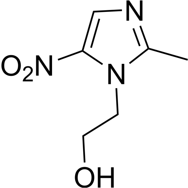
HY-B0318
MetronidazoleAntibiotic -
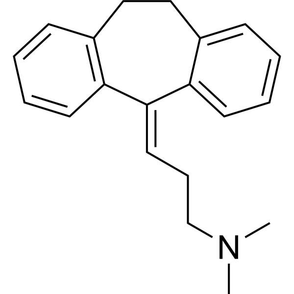
HY-B0527
AmitriptylineSERT/NET Inhibitor -
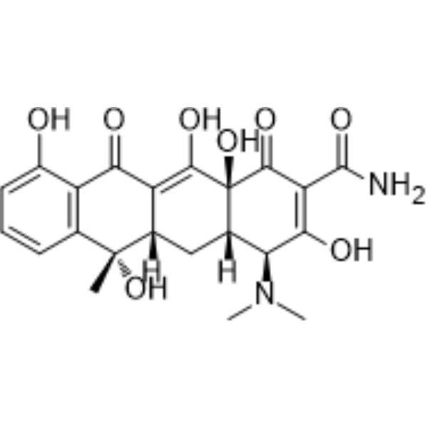
HY-A0107
TetracyclineAntibiotic -
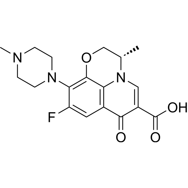
HY-B0330
LevofloxacinDNA Gyrase/Topoisomerase Inhibitor -
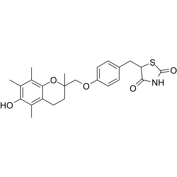
HY-50935
TroglitazonePPARγ Agonist -
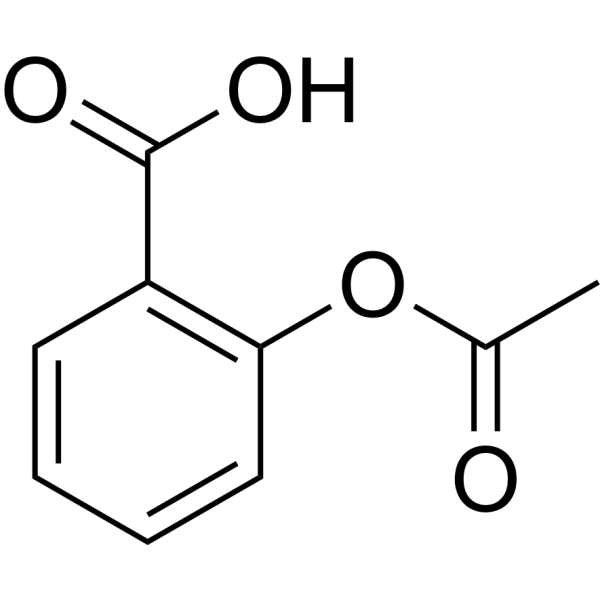
HY-14654
AspirinCOX Inhibitor -
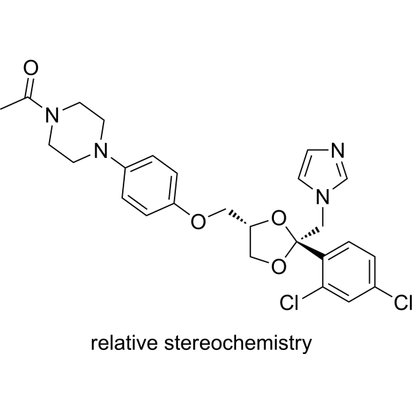
HY-B0105
KetoconazoleCYP3A4 Inhibitor -
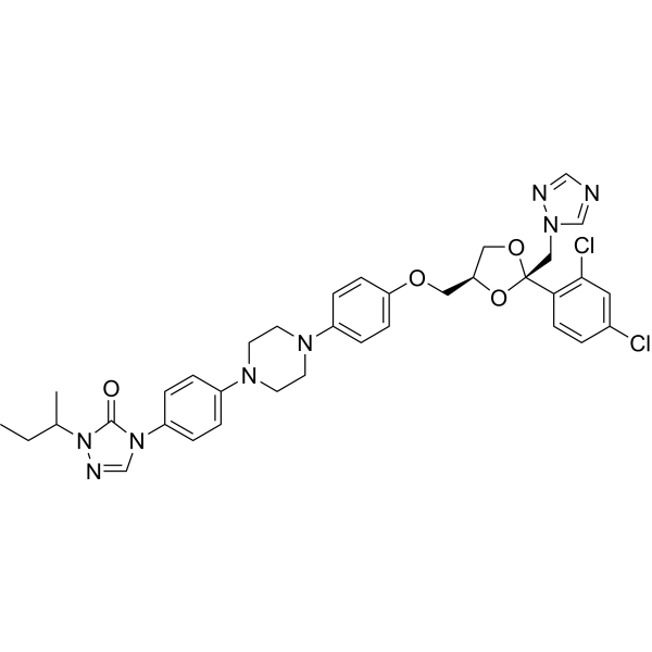
HY-17514
ItraconazoleAntifungal Agent -
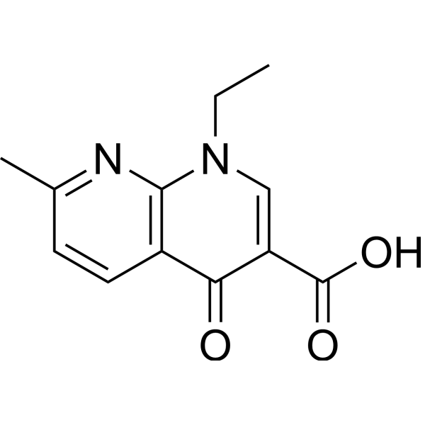
HY-B0398
Nalidixic acidAntibiotic -
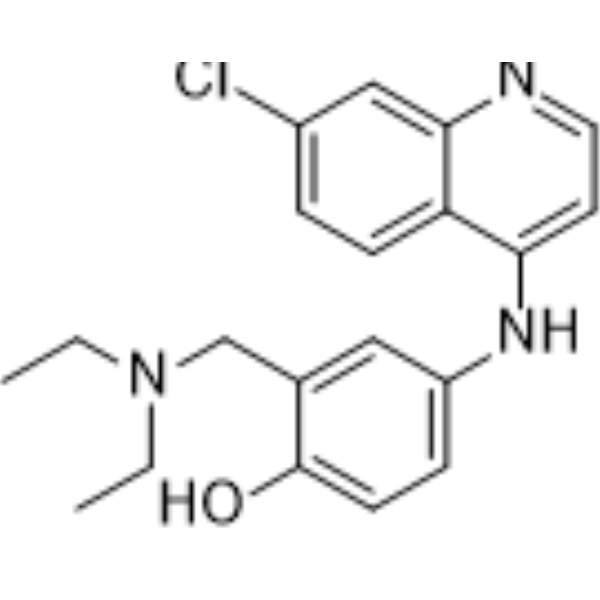
HY-B1322A
AmodiaquineAntimalarial Agent -
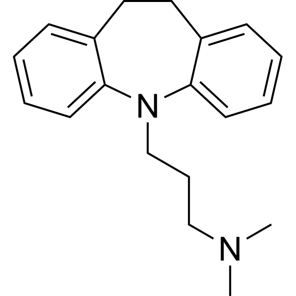
HY-B1490A
ImipramineAntidepressant -
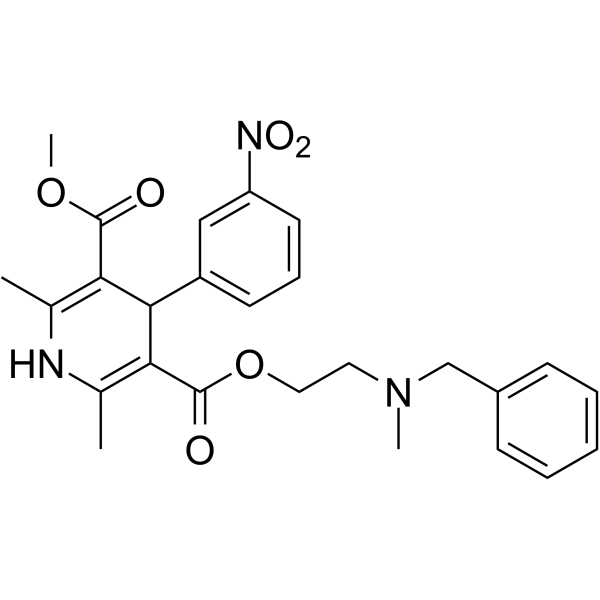
HY-12515
NicardipineCalcium Channel Blocker
Publications Citing Use of MedChemExpress (MCE) Furazolidone
MoreAll Antibiotic Isoforms
MoreAll Parasite Isoforms
More

Biological Activity
Description
Cellular Effect
|
Cell Line

|
Type | Value | Description | References |
|---|---|---|---|---|
| THP-1 | IC50 |
>100 μM
Compound: FZD
|
Cytotoxicity against human THP-1 cells assessed as inhibition of cell growth incubated for 48 hrs by resazurin dye based assay
Cytotoxicity against human THP-1 cells assessed as inhibition of cell growth incubated for 48 hrs by resazurin dye based assay
|
[PMID: 36516584] |
| THP-1 | IC50 |
>100 μM
Compound: FZD
|
Cytotoxicity against human THP-1 cells assessed as inhibition of cell growth incubated for 72 hrs by resazurin staining based analysis
Cytotoxicity against human THP-1 cells assessed as inhibition of cell growth incubated for 72 hrs by resazurin staining based analysis
|
[PMID: 37859713] |
| Vero | CC50 |
19 μg/mL
Compound: furazolidone
|
Cytotoxicity against african green monkey Vero cells after 72 hrs by CellTiterGlo assay
Cytotoxicity against african green monkey Vero cells after 72 hrs by CellTiterGlo assay
|
[PMID: 22691154] |
| Vero | IC50 |
>100 μM
Compound: FZD
|
Cytotoxicity against African green monkey Vero cells assessed as inhibition of cell growth incubated for 48 hrs by resazurin dye based assay
Cytotoxicity against African green monkey Vero cells assessed as inhibition of cell growth incubated for 48 hrs by resazurin dye based assay
|
[PMID: 36516584] |
| Vero | IC50 |
>100 μM
Compound: FZD
|
Cytotoxicity against African green monkey Vero cells assessed as inhibition of cell growth incubated for 48 hrs by resazurin staining based analysis
Cytotoxicity against African green monkey Vero cells assessed as inhibition of cell growth incubated for 48 hrs by resazurin staining based analysis
|
[PMID: 37859713] |
In Vitro
Furazolidone (50 μM; in the RTTA assay) inhibits bone marrow transformation mediated by a series of leukemia fusion proteins, including AML1-ETO, MLL-ENL, MLL-AF9, and R1A-RAR-RIIa, and shows a relatively higher IC50 value for normal murine bone-marrow c-Kit positive cells compared to AML1-ETO transformed cells[1].
Furazolidone (1-50 μM; 24 h-72 h) inhibits the proliferation of AML cell lines (Kasumi-1, NB4, MolM13, MV4-11, U937, and HL-60) in a dose-and time-dependent manner, with IC50 values ranging from 10-20 μM, and also compromises the ability of leukemic cells (Kasumi-1, NB4, MolM-13) to form colonies[1].
Furazolidone (IC50 values for each cell line; 72 h) induces apoptosis in AML cells (Kasumi-1, NB4, MV4-11, MolM13), with a 2.1-fold increase in %Annexin V+/7-AAD+ in Kasumi-1, 1.7-fold in NB4, 2.0-fold in MV4-11, and 1.6-fold in MolM13 compared with the vehicle-treated control (DMSO), but has no evidence of increased apoptosis in U937 and HL-60 leukemic cells[1].
Furazolidone (IC50 values for each cell line; 72 h) induces the differentiation of AML cell lines (Kasumi-1, NB4, MV4-11), as indicated by the increased expression of the myeloid differentiation marker CD11b, evident morphologic changes characteristic of differentiation (such as the appearance of granules and condensation of the nucleus), and an increased NBT-positive rate in Kasumi-1 and NB4 cells[1].
Furazolidone (IC50 values for each cell line; 72 h) up-regulates the protein level, but not the mRNA level, of p53 in AML cells (Kasumi-1, NB4, MV4-11, MolM13)[1].
MedChemExpress (MCE) has not independently confirmed the accuracy of these methods. They are for reference only. Further protocols information, click here.
-
Cell Line:Kasumi-1, NB4, MolM13, MV4-11, U937, HL-60
-
Concentration:1-50 μM
-
Incubation Time:24 h, 48 h, 72 h
-
Result:The number of viable cells in these AML cell lines decreased in a dose-and time-dependent manner when treated with Furazolidone.
The IC50 values, measured when cells were treated with Furazolidone for 72 h, ranged from 10-20 μM.
Furazolidone also compromised the ability of Kasumi-1, NB4, and MolM-13 cells to form colonies.
-
Cell Line:Kasumi-1, NB4, MV4-11, MolM13, U937, HL-60
-
Concentration:IC50 values for each cell line (Kasumi-1: 20 μM; NB4: 20 μM; MV4-11: 20 μM; MolM13: 15 μM; U937: 20 μM; HL-60: 10 μM)
-
Incubation Time:72 h
-
Result:Significantly increased the apoptosis rate in Kasumi-1, NB4, MV4-11, and MolM13 cells. The %Annexin V+/7-AAD+ increased by 2.1-fold in Kasumi-1, 1.7-fold in NB4, 2.0-fold in MV4-11, and 1.6-fold in MolM13 compared with the DMSO-treated control.
There was no evidence of increased apoptosis in U937 and HL-60 cells.
-
Cell Line:AML cell lines
-
Concentration:IC50 values for each cell line (Kasumi-1: 20 μM; NB4: 20 μM; MV4-11: 20 μM; MolM13: 15 μM; U937: 20 μM; HL-60: 10 μM)
-
Incubation Time:24 h
-
Result:Did not modulate the cell-cycle distribution in AML cells, suggesting that it prolonged cell-doubling time rather than inducing cell-cycle arrest[1].
-
Cell Line:Kasumi-1, NB4, MV4-11, MolM13
-
Concentration:IC50 values for each cell line (Kasumi-1: 20 μM; NB4: 20 μM; MV4-11: 20 μM; MolM13: 15 μM; U937: 20 μM; HL-60: 10 μM)
-
Incubation Time:72 h
-
Result:Up-regulated the protein level of p53 in these cells, while the relative intensity of β-actin was used as a control. The relative protein levels of p53, normalized to β-actin, were 3.2 in Kasumi-1, 2.1 in NB4, 4.0 in MV4-11, and 2.7 in MolM13. qPCR: Cell Line: Kasumi-1, NB4, MV4-11, MolM13, U937, HL-60 Concentration: Predetermined IC50 value for each cell line Incubation Time: 72 h Result: Furazolidone treatment did not up-regulate the mRNA level of p53 in these AML cell lines. However, it significantly increased the p21 mRNA level in all the cell lines tested, but the impact of this increase on the anti-leukemic effects of Furazolidone was unknown[1].
Clinical Trial
| NCT Number | Sponsor | Condition | Start Date |
Phase
|
|---|---|---|---|---|
| NCT01329991 | Plexxikon| | 2011-05 | PHASE1 |


Chemical Information
-
CAS No. 67-45-8
-
Appearance Solid
-
Molecular Weight 225.16
-
Formula C8H7N3O5
-
Color Light yellow to yellow
-
SMILES
O=C1OCCN1/N=C/C2=CC=C([N+]([O-])=O)O2
-
Shipping
Room temperature in continental US; may vary elsewhere.
-
Storage
Powder -20°C 3 years 4°C 2 years In solvent -80°C 2 years -20°C 1 year


Publications (3)
-
Journal Impact Factor
-
Most Recent
-
Zebrafish
Ronidazole Is a Superior Prodrug to Metronidazole for Nitroreductase-Mediated Hepatocytes Ablation in Zebrafish Larvae. [Abstract]2023 Jun;20(3):95-102. PMID: 37229597 -
Heliyon
Furazolidone reduces the pathogenesis of Trueperella pyogenes and Pseudomonas aeruginosa co-infection in a mouse model. [Abstract]2024 Oct 19;10(20):e39629. PMID: 39506932 -


Solvent & Solubility
In Vitro:
DMSO : 5.56 mg/mL (24.69 mM; ultrasonic and warming and heat to 60°C; Hygroscopic DMSO has a significant impact on the solubility of product, please use newly opened DMSO)
H2O : 0.67 mg/mL (2.98 mM; Need ultrasonic)
Please refer to the solubility information to select the appropriate solvent. Once prepared, please aliquot and store the solution to prevent product inactivation from repeated freeze-thaw cycles.
Storage method and period of stock solution: -80°C, 2 years; -20°C, 1 year. When stored at -80°C, please use it within 2 years. When stored at -20°C, please use it within 1 year.
* Note: If you choose water as the stock solution, please dilute it to the working solution, then filter and sterilize it with a 0.22 μm filter before use.
Please refer to the solubility information to select the appropriate solvent. Once prepared, please aliquot and store the solution to prevent product inactivation from repeated freeze-thaw cycles.
Storage method and period of stock solution: -80°C, 2 years; -20°C, 1 year. When stored at -80°C, please use it within 2 years. When stored at -20°C, please use it within 1 year.
* Note: If you choose water as the stock solution, please dilute it to the working solution, then filter and sterilize it with a 0.22 μm filter before use.
Concentration (start) × Volume (start) = Concentration (final) × Volume (final)

Protocols
-
Apoptosis
Apoptosis, also called programmed cell death, is generally characterized by distinct morphological characteristics.
-
TUNEL staining for apoptotic DNA fragmentation
TUNEL staining detects DNA strand breaks by using terminal deoxynucleotidyl transferase to add labeled nucleotides to exposed 3′-OH DNA termini, generating either microscopic staining in fixed cells or tissue sections, or fluorescence/cytometric signal in cell suspensions. TUNEL positivity reflects DNA fragmentation but should not be interpreted alone as definitive apoptosis, because TUNEL can also label necrotic, autolytic, mechanically damaged, or DNA-repair-associated DNA breaks.
-
Annexin V plus membrane-impermeant dye apoptosis staining
Annexin V-based apoptosis assays rely on the detection of phosphatidylserine (PS) externalization from the inner leaflet of the plasma membrane to the outer leaflet, an early biochemical hallmark of apoptosis. Fluorescently labeled Annexin V binds PS in a calcium-dependent manner, enabling identification of early apoptotic cells by flow cytometry or fluorescence microscopy. When combined with a membrane-impermeant DNA-binding dye (e. g. , propidium iodide), this approach allows discrimination between viable (Annexin V−/dye−), early apoptotic (Annexin V+/dye−), and late apoptotic or necrotic (Annexin V+/dye+) cell populations by assessing membrane integrity and PS exposure.
-
Apoptosis Solutions
Apoptosis is a regulated, generally non-lytic cell-death pathway that removes unwanted, damaged, infected, or abnormal cells through coordinated morphological changes, caspase activation, DNA fragmentation, and membrane remodeling. The intrinsic apoptosis pathway is controlled mainly by mitochondrial outer membrane permeabilization, BCL-2 family proteins, cytochrome c release, apoptosome formation, caspase-9 activation, and downstream executioner caspase-3/7 activation. The extrinsic apoptosis pathway is initiated by death receptors such as Fas, TNFR, and TRAIL receptors, which recruit adaptor proteins and activate caspase-8 before engaging executioner caspases or mitochondrial amplification through BID cleavage. Apoptosis is linked to many phenotypes, including cancer cell killing, tissue homeostasis, immune regulation, neurodegeneration, infection response, and treatment-induced cytotoxicity; unresolved questions include how apoptosis interacts with necroptosis, pyroptosis, ferroptos
-
Cell Viability Determination by MTT Colorimetric Assay
The following protocol uses the MTT colorimetric assay as a classic literature-established method for assessing cell viability/metabolic activity in cultured mammalian cells. MTT[3-(4,5-dimethylthiazol-2-yl)-2,5-diphenyltetrazolium bromide] is reduced by metabolically active cells to a colored formazan product; the amount of formazan is quantified spectrophotometrically and provides an indirect measure of metabolically active viable cells. Importantly, MTT reduction reflects cellular oxidoreductase/metabolic activity rather than an absolute direct count of living cells, so changes in cellular metabolism can alter the signal independently of cell number.


Purity & Documentation
-
Data Sheet (290 KB)
-
SDS (419 KB)
- English - EN (419 KB)
- Français - FR (419 KB)
- Deutsch - DE (419 KB)
- Norwegian - NO (419 KB)
- Español - ES (419 KB)
- Swedish - SV (419 KB)
- Italian - IT (419 KB)
- Korean - KR (419 KB)
- Portuguese - PT (419 KB)
-
Handling Instructions (2659 KB)
References
[1]. Jiang X, et al. A novel application of furazolidone: anti-leukemic activity in acute myeloid leukemia. PLoS One. 2013 Aug 9;8 (8) :e72335. [Content Brief]
[2]. Ma S, et al. Furazolidone Increases Survival of Mice Exposed to Lethal Total Body Irradiation through the Antiapoptosis and Antiautophagy Mechanism. Oxid Med Cell Longev. 2021 Feb 4;2021:6610726. [Content Brief]


Complete Stock Solution Preparation Table
Please refer to the solubility information to select the appropriate solvent. Once prepared, please aliquot and store the solution to prevent product inactivation from repeated freeze-thaw cycles.
Storage method and period of stock solution: -80°C, 2 years; -20°C, 1 year. When stored at -80°C, please use it within 2 years. When stored at -20°C, please use it within 1 year.
| Optional Solvent | Concentration Solvent Mass | 1 mg | 5 mg | 10 mg | 25 mg |
|---|---|---|---|---|---|
| H2O / DMSO | 1 mM | 4.4413 mL | 22.2064 mL | 44.4129 mL | 111.0322 mL |
| DMSO | 5 mM | 0.8883 mL | 4.4413 mL | 8.8826 mL | 22.2064 mL |
| 10 mM | 0.4441 mL | 2.2206 mL | 4.4413 mL | 11.1032 mL | |
| 15 mM | 0.2961 mL | 1.4804 mL | 2.9609 mL | 7.4021 mL | |
| 20 mM | 0.2221 mL | 1.1103 mL | 2.2206 mL | 5.5516 mL |
* Note: If you choose water as the stock solution, please dilute it to the working solution, then filter and sterilize it with a 0.22 μm filter before use.


Furazolidone Related Classifications
HY-B1336 Related Classifications
 Powered by Bioz