



Aminopurvalanol A
Based on 1 publication(s) in Google Scholar
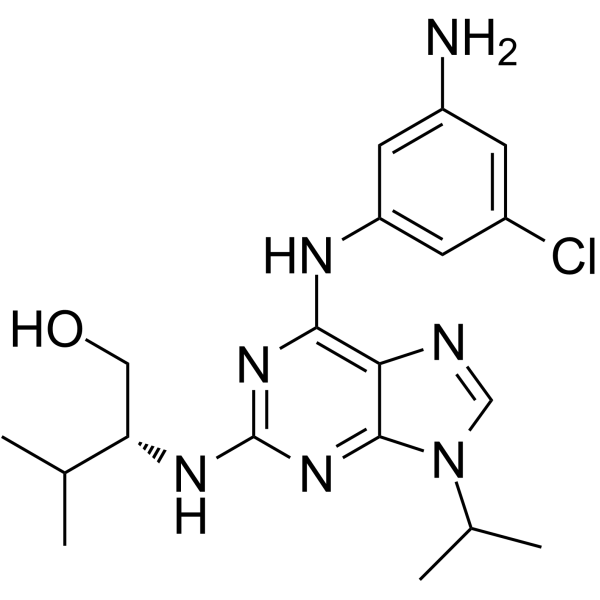
Aminopurvalanol A is a potent, selective, and cell permeable inhibitor of Cyclins/Cdk complexes. Aminopurvalanol A preferentially targets the G2/M-phase transition inhibiting cancer cell differentiation. Aminopurvalanol A causes the inhibition of sperm fertilizing ability via the inhibition of physiological capacitation-dependent actin polymerization.
For research use only. We do not sell to patients.
- Purity : 98.03%
- CAS No.: 220792-57-4
- Formula: C19H26ClN7O
- Molecular Weight:403.91
-
Storage:Powder -20°C, 3 years , 4°C, 2 years ; In solvent -80°C, 6 months , -20°C, 1 month

To place orders, for customer services and technical support, please contact: MedChemExpress USA
Tel: 609-228-6898 E-mail: [email protected] [email protected]


-
 Biological Activity
-
Chemical Information
-
Solvent & Solubility
- Protocols
- Purity & Documentation
- References
-
 Help & FAQs


-
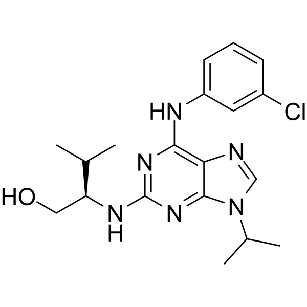
HY-18299A
Purvalanol ACDK Inhibitor -
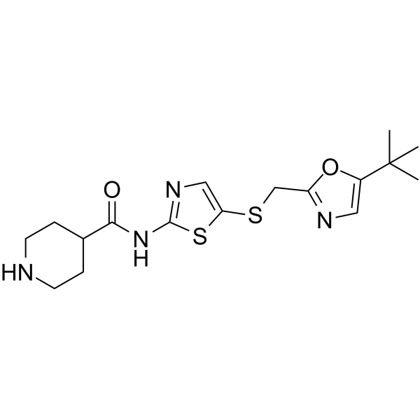
HY-10008
SNS-032CDK Inhibitor -
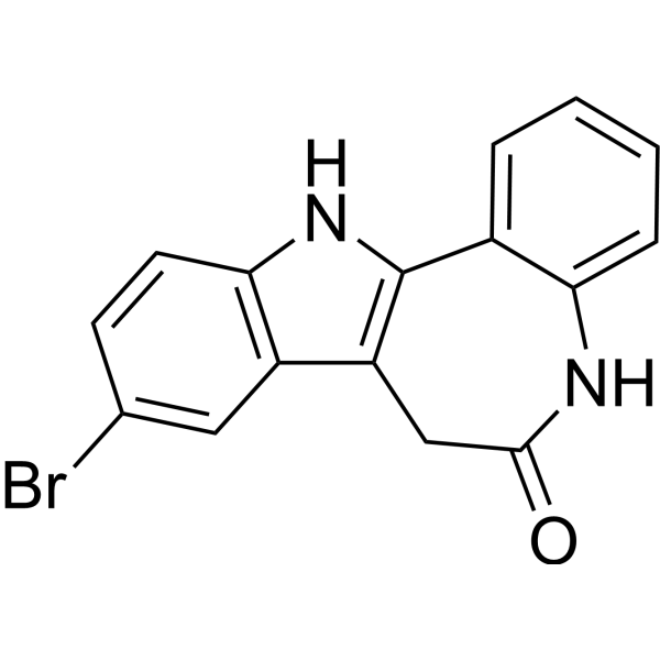
HY-12302
KenpaulloneCDK1/GSK-3β Inhibitor -
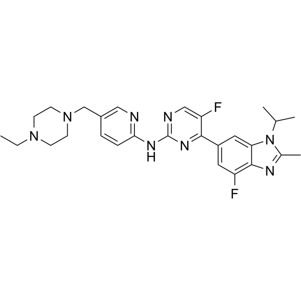
HY-16297A
AbemaciclibCDK4/6 Inhibitor -
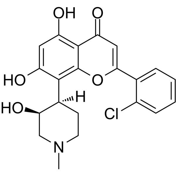
HY-10005
FlavopiridolCDK Inhibitor -
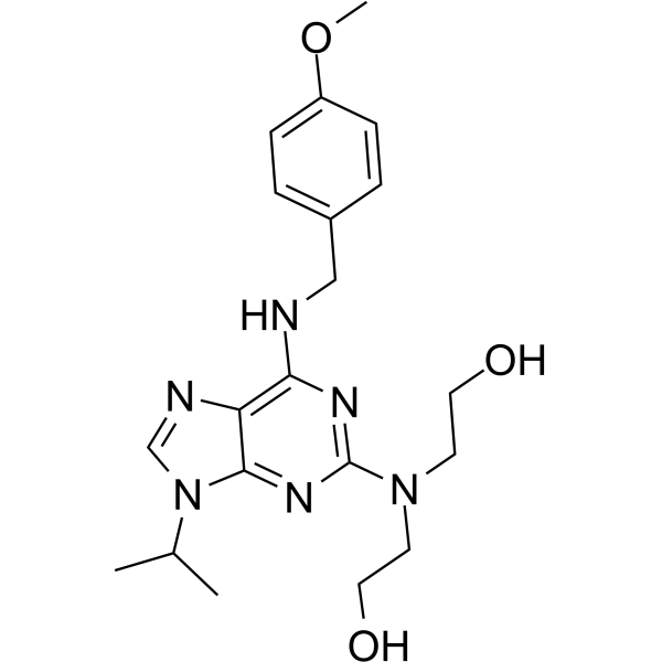
HY-15339
CVT-313CDK2 Inhibitor -
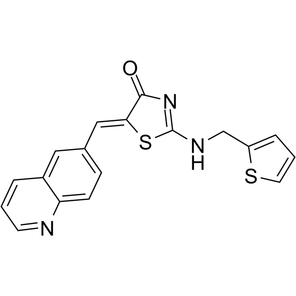
HY-12529
Ro-3306CDK1 Inhibitor -
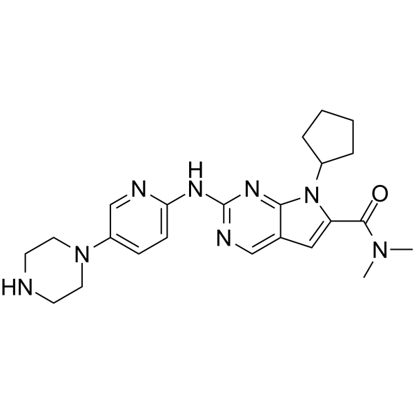
HY-15777
RibociclibCDK4/6 Inhibitor -
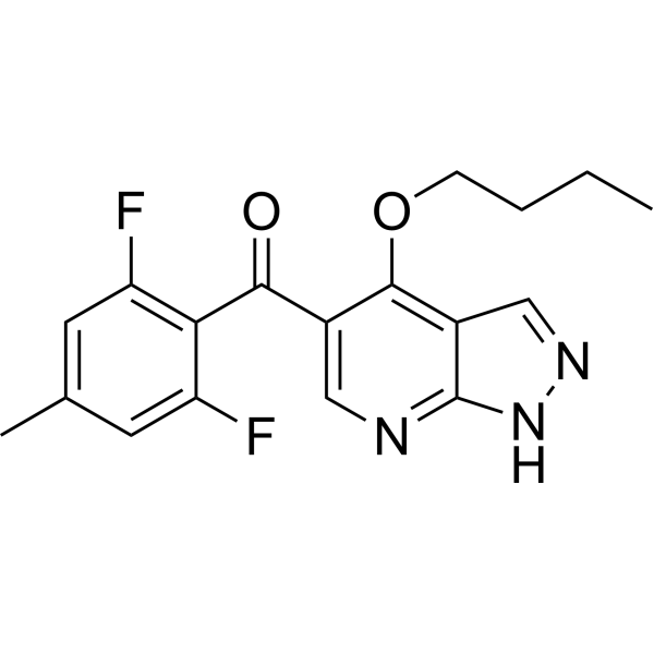
HY-15275
BMS-265246CDK1/2 Inhibitor -
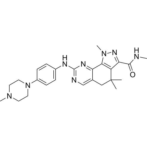
HY-10424
MilciclibCDK/TRK Inhibitor -

HY-16297AR
Abemaciclib (Standard)CDK4/6 Inhibitor -
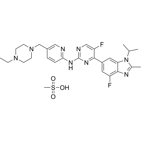
HY-16297
Abemaciclib methanesulfonateCDK4/6 Inhibitor
Publications Citing Use of MedChemExpress (MCE) Aminopurvalanol A
More

Biological Activity
Description
IC50 & Target
Cyclins/Cdk[1]
Cellular Effect
|
Cell Line

|
Type | Value | Description | References |
|---|---|---|---|---|
| KM12 | GI50 |
30 nM
Compound: K00613a, Aminopurvalanol
|
Inhibition of human KM12 cells
Inhibition of human KM12 cells
|
[PMID: 18077363] |
In Vitro
Aminopurvalanol A (5 and 40 μM; 8 hours) inhibits cell growth primarily by arresting the cells in the G2 phase of the cell cycle and, at higher concentration, triggering apoptosis[2].
MedChemExpress (MCE) has not independently confirmed the accuracy of these methods. They are for reference only. Further protocols information, click here.
-
Cell Line:Human U937 leukemic cells
-
Concentration:5 and 40 μM
-
Incubation Time:8 hours
-
Result:Increased the number of cells with a 4N DNA content as early as 8 h after the beginning of treatment at 5 μM. 40 μM led to cellular fragmentation and cells with an irregular DNA distribution, characteristic of apoptotic cell populations.
-
Cell Line:Human U937 leukemic cells
-
Concentration:5 and 40 μM
-
Incubation Time:8 hours
-
Result:40 μM Aminopurvalanol A led to apoptosis rather than after the beginning of treatment at 5 μM.


Chemical Information
-
CAS No. 220792-57-4
-
Appearance Solid
-
Molecular Weight 403.91
-
Formula C19H26ClN7O
-
Color Off-white to light yellow
-
SMILES
CC(C)N1C2=NC(N[C@@H](CO)C(C)C)=NC(NC3=CC(Cl)=CC(N)=C3)=C2N=C1
-
Shipping
Room temperature in continental US; may vary elsewhere.
-
Storage
Powder -20°C 3 years 4°C 2 years In solvent -80°C 6 months -20°C 1 month


Publications (1)
-
Journal Impact Factor
-
Most Recent
-
Biomed Pharmacother
Predicting potential therapeutic targets and small molecule drugs for early-stage lung adenocarcinoma. [Abstract]2024 May:174:116528. PMID: 38555814


Solvent & Solubility
In Vitro:
DMSO : 100 mg/mL (247.58 mM; Need ultrasonic; Hygroscopic DMSO has a significant impact on the solubility of product, please use newly opened DMSO)
Please refer to the solubility information to select the appropriate solvent. Once prepared, please aliquot and store the solution to prevent product inactivation from repeated freeze-thaw cycles.
Storage method and period of stock solution: -80°C, 6 months; -20°C, 1 month. When stored at -80°C, please use it within 6 months. When stored at -20°C, please use it within 1 month.
Please refer to the solubility information to select the appropriate solvent. Once prepared, please aliquot and store the solution to prevent product inactivation from repeated freeze-thaw cycles.
Storage method and period of stock solution: -80°C, 6 months; -20°C, 1 month. When stored at -80°C, please use it within 6 months. When stored at -20°C, please use it within 1 month.
Concentration (start) × Volume (start) = Concentration (final) × Volume (final)
In Vivo:
Select the appropriate dissolution method based on your experimental animal and administration route.
- For the following dissolution methods, please ensure to first prepare a clear stock solution using an In Vitro approach and then sequentially add co-solvents:
- To ensure reliable experimental results, the clarified stock solution can be appropriately stored based on storage conditions. As for the working solution for In Vivo experiments, it is recommended to prepare freshly and use it on the same day.
- The percentages shown for the solvents indicate their volumetric ratio in the final prepared solution. If precipitation or phase separation occurs during preparation, heat and/or sonication can be used to aid dissolution.
Add each solvent one by one: 10% DMSO 40% PEG300 5% Tween-80 45% Saline
Solubility: ≥ 2.5 mg/mL (6.19 mM); Clear solution
This protocol yields a clear solution of ≥ 2.5 mg/mL (saturation unknown).
Taking 1 mL working solution as an example, add 100 μL DMSO stock solution (25.0 mg/mL) to 400 μL PEG300, and mix evenly; then add 50 μL Tween-80 and mix evenly; then add 450 μL Saline to adjust the volume to 1 mL.
Preparation of Saline: Dissolve 0.9 g sodium chloride in ddH₂O and dilute to 100 mL to obtain a clear Saline solution.
In Vivo Dissolution Calculator
Please enter the basic information of animal experiments:
-
-
-
-
Recommended: Prepare an additional quantity of animals to account for potential losses during experiments.
Please enter your animal formula composition:
-
%DMSO +
Recommended: Keep the proportion of DMSO in working solution below 2% if your animal is weak.
-
%+
-
+%Tween-80 + +
-
%Saline +
The co-solvents required include: DMSO, . All of co-solvents are available by MedChemExpress (MCE). , Tween 80. All of co-solvents are available by MedChemExpress (MCE).
Working solution concentration: 0.22 mg/mL
Method for preparing stock solution: mg drug dissolved in μL DMSO. Stock solution concentration: mg/mL.
1. Take μL DMSO stock solution;
2. Add μL .
μL , mix evenly;
3. Then add μL Tween 80, mix evenly;
4. Then add μL
Please ensure that the stock solution in the first step is dissolved to a clear state, and add co-solvents in sequence. You can use ultrasonic heating (ultrasonic cleaner, recommended frequency 20-40 kHz), vortexing, etc. to assist dissolution.

Protocols
-
Flow cytometric DNA-content cell-cycle staining
Flow cytometric DNA-content cell-cycle staining measures the fluorescence intensity of DNA-bound fluorochromes in single cells or nuclei to estimate DNA content distributions, allowing assignment of populations to G0/G1, S, and G2/M phases by DNA histogram deconvolution. Propidium iodide (PI) intercalates into DNA, and PI fluorescence is proportional to cellular DNA content when staining is performed under conditions that make DNA accessible and minimize non-DNA signal. Cells with G2/M DNA content are expected to show approximately twice the fluorescence intensity of G0/G1 cells, while S-phase cells occupy intermediate fluorescence values. PI-based DNA-content analysis can also detect cells with fractional DNA content, often reported as sub-G1, when DNA fragmentation and extraction during staining reduce retained DNA signal in apoptotic cells. DAPI is an alternative DNA fluorochrome for univariate DNA-content analysis, while bivariate approaches combining DNA content with proliferation
-
Protocol for Cell Cycle
Cell-cycle analysis by flow cytometry measures DNA content in single cells to estimate the fraction of cells in G0/G1, S, and G2/M phases. Propidium iodide intercalates into DNA, and after RNA removal with RNase, fluorescence intensity reflects cellular DNA content: 2N cells are assigned to G0/G1, cells between 2N and 4N to S phase, and 4N cells to G2/M. DNA-content analysis alone cannot reliably separate G0 from G1 or G2 from M. Ki-67 can distinguish quiescent G0 cells from cycling cells, EdU or BrdU incorporation marks active DNA synthesis in S phase, and phospho-histone H3 staining identifies mitotic cells within the 4N population.
-
iPSC cell differentiation
Induced pluripotent stem cells (iPSCs) are a type of cell that has similar properties to embryonic stem cells through somatic cell reprogramming.
-
Phalloidin F-actin cytoskeleton staining
Phalloidin F-actin staining detects polymerized filamentous actin in fixed and permeabilized specimens by using fluorescent phalloidin or phalloidin-derived phallotoxins that bind actin filaments and generate a fluorescence microscopy readout corresponding to F-actin organization, including stress fibers, cortical actin, filament bundles, and tissue-specific actin networks. Phalloidin stabilizes F-actin by reducing actin subunit dissociation from filament ends, and fluorescent phallotoxins were established as tools for visualizing actin-containing structures in eukaryotic cells.


Purity & Documentation
-
Data Sheet (278 KB)
-
SDS (393 KB)
- English - EN (393 KB)
- Français - FR (393 KB)
- Deutsch - DE (393 KB)
- Norwegian - NO (393 KB)
- Español - ES (393 KB)
- Swedish - SV (393 KB)
- Italian - IT (393 KB)
- Korean - KR (393 KB)
- Portuguese - PT (393 KB)
-
Handling Instructions (2659 KB)
References
[1]. Bernabò N, et al. Aminopurvalanol A, a Potent, Selective, and Cell Permeable Inhibitor of Cyclins/Cdk Complexes, Causes the Reduction of in Vitro Fertilizing Ability of Boar Spermatozoa, by Negatively Affecting the Capacitation-Dependent Actin Polymerization. Front Physiol. 2017;8:1097. [Content Brief]
[2]. Rosania GR, et al. A cyclin-dependent kinase inhibitor inducing cancer cell differentiation: biochemical identification using Xenopus egg extracts. Proc Natl Acad Sci U S A. 1999;96(9):4797-4802. [Content Brief]


Complete Stock Solution Preparation Table
Please refer to the solubility information to select the appropriate solvent. Once prepared, please aliquot and store the solution to prevent product inactivation from repeated freeze-thaw cycles.
Storage method and period of stock solution: -80°C, 6 months; -20°C, 1 month. When stored at -80°C, please use it within 6 months. When stored at -20°C, please use it within 1 month.
| Optional Solvent | Concentration Solvent Mass | 1 mg | 5 mg | 10 mg | 25 mg |
|---|---|---|---|---|---|
| DMSO | 1 mM | 2.4758 mL | 12.3790 mL | 24.7580 mL | 61.8950 mL |
| 5 mM | 0.4952 mL | 2.4758 mL | 4.9516 mL | 12.3790 mL | |
| 10 mM | 0.2476 mL | 1.2379 mL | 2.4758 mL | 6.1895 mL | |
| 15 mM | 0.1651 mL | 0.8253 mL | 1.6505 mL | 4.1263 mL | |
| 20 mM | 0.1238 mL | 0.6189 mL | 1.2379 mL | 3.0947 mL | |
| 25 mM | 0.0990 mL | 0.4952 mL | 0.9903 mL | 2.4758 mL | |
| 30 mM | 0.0825 mL | 0.4126 mL | 0.8253 mL | 2.0632 mL | |
| 40 mM | 0.0619 mL | 0.3095 mL | 0.6189 mL | 1.5474 mL | |
| 50 mM | 0.0495 mL | 0.2476 mL | 0.4952 mL | 1.2379 mL | |
| 60 mM | 0.0413 mL | 0.2063 mL | 0.4126 mL | 1.0316 mL | |
| 80 mM | 0.0309 mL | 0.1547 mL | 0.3095 mL | 0.7737 mL | |
| 100 mM | 0.0248 mL | 0.1238 mL | 0.2476 mL | 0.6189 mL |


Aminopurvalanol A Related Classifications
HY-104013 Related Classifications
 Powered by Bioz