



Physcion
Based on 11 publication(s) in Google Scholar
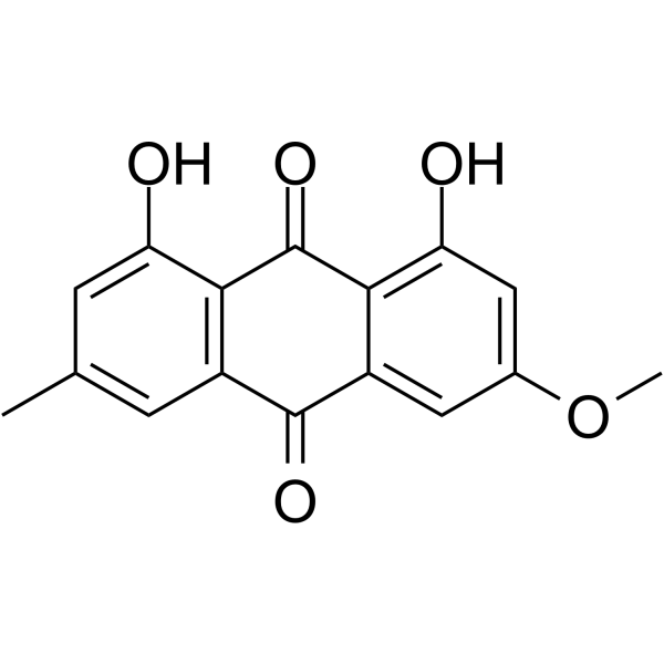
Physcion (Parietin), an anthraquinone derivative derived from the traditional Chinese medicine rhubarb, is an effective oral active 6-phosphogluconate dehydrogenase inhibitor with blood-brain barrier permeability, with IC50 and Kd values of 38.5 μM and 26.0 μM, respectively. Additionally, Physcion is an inhibitor of the <>bTLR4/NF-κB signaling pathway, exhibiting anti-inflammatory, antibacterial, and anticancer effects, and can induce Apoptosis and Autophagy in cancer cells.
Nur für Forschungszwecke. Wir verkaufen nicht an Patienten.
- Reinheit : 99.40%
- CAS. Nr.: 521-61-9
- Formel: C16H12O5
- Molecular Weight:284.26
-
Speicherung:Powder -20°C, 3 years , 4°C, 2 years ; In solvent -80°C, 6 months , -20°C, 1 month

To place orders, for customer services and technical support, please contact: MedChemExpress USA
Tel: 609-228-6898 E-mail: [email protected] [email protected]


-
 Biologische Aktivität
-
Chemical Information
-
Lösungsmittel & Löslichkeit
- Protokoll
- Reinheit & Dokumentation
- Verweise
-
 Help & FAQs


-
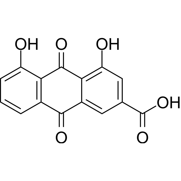
HY-N0105
RheinAntioxidant -
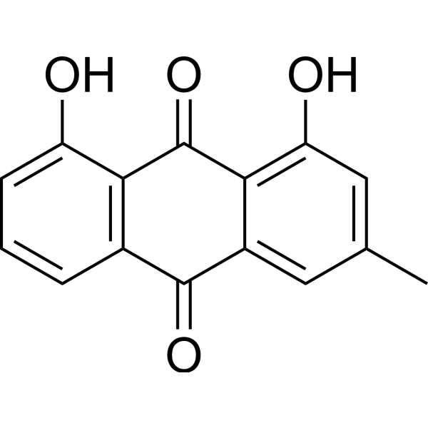
HY-13595
ChrysophanolEGFR Inhibitor -
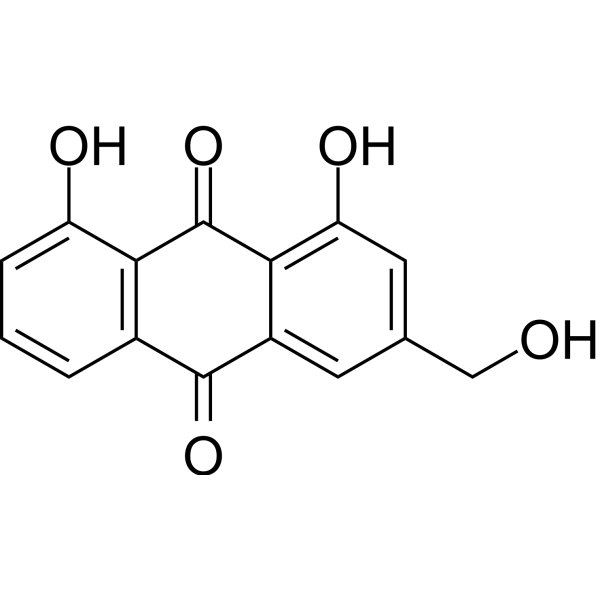
HY-N0189
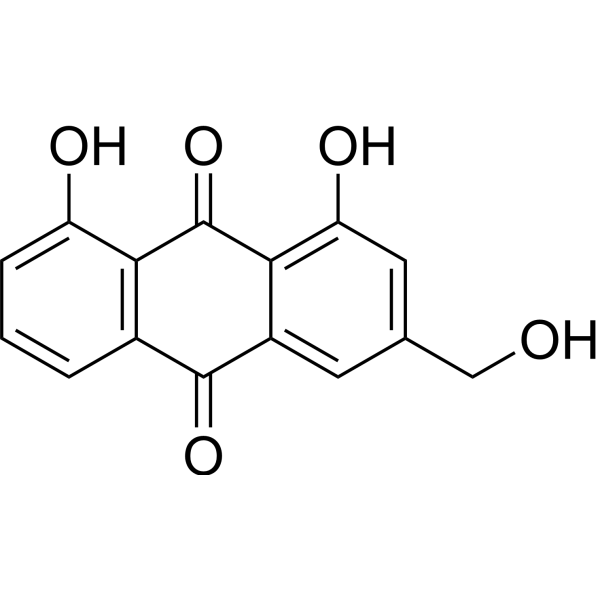
Aloe emodinmTORC2 Inhibitor -
HY-W010342
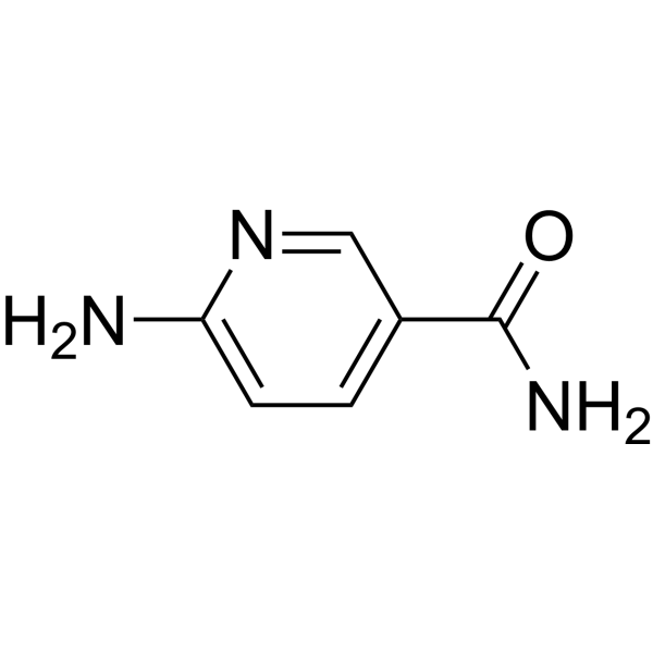
6-AminonicotinamideG6PD Inhibitor -
HY-14393
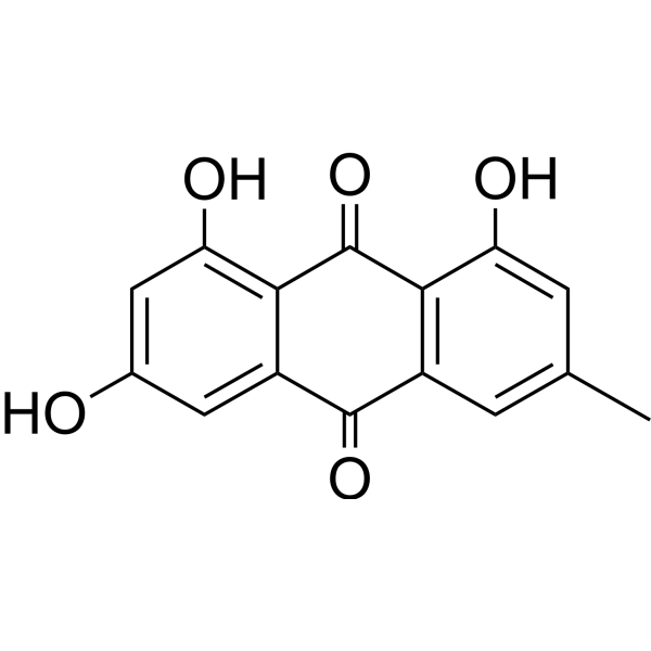
EmodinNatural Anthraquinone -
HY-N0189R
Aloe emodin (Standard) -
HY-N0005
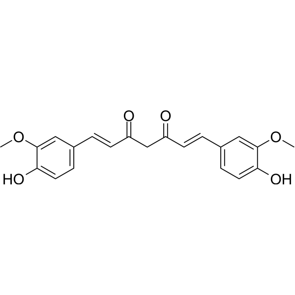
Curcuminp300 Histone Acetylatransferase Inhibitor -
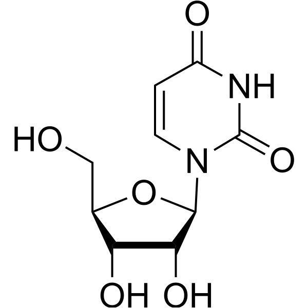
HY-B1449
UridineNucleoside -
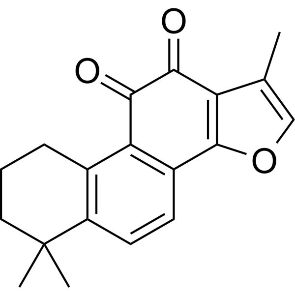
HY-N0135
Tanshinone IIAVEGFR Inhibitor -
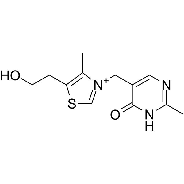
HY-107430
OxythiamineTransketolase Inhibitor -

HY-B0015
PaclitaxelAntitumor Agent -
HY-K0301
Cell Counting Kit-8
Publications Citing Use of MedChemExpress (MCE) Physcion
More- Mol Cancer. 2025 Mar 26;24(1):97. [Abstract]
- Cancer Lett. 2024 Oct 1:601:217177. [Abstract]
- Chin Herb Med. 2026 Jun 19.
- J Ethnopharmacol. 2026 May 27:121893. [Abstract]
- Drug Des Devel Ther. 2021 Jan 25:15:277-287. [Abstract]
- Exp Eye Res. 2026 Jul 28:271:111186.
- Biomed Res Int. 2021 Sep 9;2021:9066938. [Abstract]
- World J Gastrointest Oncol. 2023 Aug 15;15(8):1400-1411. [Abstract]
- Drug Chem Toxicol. 2025 Oct 14:1-9. [Abstract]
- Curr Eye Res. 2023 Mar;48(3):238-250. [Abstract]
- Authorea. September 07, 2022.
 Customer Validation & Images

-
Cell Proliferation/Viability Assay
-
Flow Cytometry
-
WB
-
IF
-
In Vivo Efficacy Study
Alle Antibiotic Isoform-spezifische Produkte anzeigen
More

Biologische Aktivität
Beschreibung
IC50 & Target
|
TLR4 |
Cellular Effect
|
Cell Line

|
Type | Value | Description | References |
|---|---|---|---|---|
| A549 | IC50 |
>100 μg/mL
Compound: 6
|
Antiproliferative activity against human A549 cells assessed as growth inhibition after 48 hrs by MTT assay
Antiproliferative activity against human A549 cells assessed as growth inhibition after 48 hrs by MTT assay
|
[PMID: 26995526] |
| A549 | IC50 |
1.5 μM
Compound: 80
|
Antiproliferative activity against human A549 cells assessed as reduction in cell viability measured after 72 hrs by sulforhodamine B assay
Antiproliferative activity against human A549 cells assessed as reduction in cell viability measured after 72 hrs by sulforhodamine B assay
|
[PMID: 33691166] |
| HCT-116 | IC50 |
19 μM
Compound: 2
|
Cytotoxicity against MDR1 Pgp under-expressing human HCT116 cells after 24 hrs by MTT assay
Cytotoxicity against MDR1 Pgp under-expressing human HCT116 cells after 24 hrs by MTT assay
|
[PMID: 17949858] |
| HepG2 | IC50 |
77.3 μM
Compound: 2
|
Cytotoxicity against MDR1 Pgp overexpressing human HepG2 cells after 24 hrs by MTT assay
Cytotoxicity against MDR1 Pgp overexpressing human HepG2 cells after 24 hrs by MTT assay
|
[PMID: 17949858] |
| HepG2 2.2.15 | IC50 |
300 μg/mL
Compound: 53
|
Antiviral activity against Hepatitis B virus infected in human HepG2.2.15 cells assessed as decrease in HBV DNA production
Antiviral activity against Hepatitis B virus infected in human HepG2.2.15 cells assessed as decrease in HBV DNA production
|
[PMID: 24549242] |
| HepG2 2.2.15 | IC50 |
300 μg/mL
Compound: 53
|
Antiviral activity against Hepatitis B virus infected in human HepG2.2.15 cells assessed as decrease in HBV e antigen secretion
Antiviral activity against Hepatitis B virus infected in human HepG2.2.15 cells assessed as decrease in HBV e antigen secretion
|
[PMID: 24549242] |
| HepG2 2.2.15 | IC50 |
300 μg/mL
Compound: 53
|
Antiviral activity against Hepatitis B virus infected in human HepG2.2.15 cells assessed as decrease in HBV surface antigen secretion
Antiviral activity against Hepatitis B virus infected in human HepG2.2.15 cells assessed as decrease in HBV surface antigen secretion
|
[PMID: 24549242] |
| HT-29 | IC50 |
>100 μg/mL
Compound: 6
|
Antiproliferative activity against human HT-29 cells assessed as growth inhibition after 48 hrs by MTT assay
Antiproliferative activity against human HT-29 cells assessed as growth inhibition after 48 hrs by MTT assay
|
[PMID: 26995526] |
| HUVEC | IC50 |
>100 μg/mL
Compound: 6
|
Antiproliferative activity against human HUVEC cells assessed as growth inhibition after 48 hrs by MTT assay
Antiproliferative activity against human HUVEC cells assessed as growth inhibition after 48 hrs by MTT assay
|
[PMID: 26995526] |
| KB | ED50 |
1.55 μg/mL
Compound: 2
|
Cytotoxicity against human KB cells
Cytotoxicity against human KB cells
|
10.1021/np50085a021 |
| MCF7 | IC50 |
>100 μg/mL
Compound: 6
|
Antiproliferative activity against human MCF7 cells assessed as growth inhibition after 48 hrs by MTT assay
Antiproliferative activity against human MCF7 cells assessed as growth inhibition after 48 hrs by MTT assay
|
[PMID: 26995526] |
| NCI-H157 | IC50 |
3 μM
Compound: 80
|
Antiproliferative activity against human NCI-H157 cells assessed as reduction in cell viability measured after 72 hrs by sulforhodamine B assay
Antiproliferative activity against human NCI-H157 cells assessed as reduction in cell viability measured after 72 hrs by sulforhodamine B assay
|
[PMID: 33691166] |
| PLC-PRF-5 | ED50 |
1.44 μg/mL
Compound: 2
|
Cytotoxicity against human PLC/PRF/5 cells
Cytotoxicity against human PLC/PRF/5 cells
|
10.1021/np50085a021 |
In Vitro
Physcion (10 or 20 μM, 48 h) induces apoptosis and autophagy in human nasopharyngeal carcinoma (CNE2 cells)[2]. Physcion (10-40 μM, 24 h) attenuates OGD/R (oxygen-glucose deprivation/reperfusion)-induced neuronal damage and inflammatory response in SH-SY5Y cells[3].
MedChemExpress (MCE) has not independently confirmed the accuracy of these methods. They are for reference only.
-
Cell Line:The nasopharyngeal carcinoma cell line CNE2
-
Concentration:10 or 20 μM
-
Incubation Time:48 h
-
Result:Induced apoptosis in CNE2 cells in a dose-dependent manner.
-
Cell Line:The nasopharyngeal carcinoma cell line CNE2
-
Concentration:10 or 20 μM
-
Incubation Time:48 h
-
Result:Significantly increased the level of LC3B-II, which means physcion induced autophagy in CNE2 cells.
-
Cell Line:Oxygen-glucose deprivation/reperfusion (OGD/R)-induced SH-SY5Y cells
-
Concentration:40 μM
-
Incubation Time:24 h
-
Result:Significantly reversed the OGD/R-induced upregulation of TLR4, p-p65, and p-IκB expression.
In Vivo
MedChemExpress (MCE) has not independently confirmed the accuracy of these methods. They are for reference only.
-
Animal Model:Ischemia-reperfusion (I/R)-induced focal cerebral ischemia model in SD rats[3]
-
Dosage:20 or 40 mg/kg
-
Administration:Oral gavage (p.o.), once daily for 7 days
-
Result:Significantly reduced the brain water content and the NSS (Neurological Severity Score) in I/R rats, compared to I/R group. Obviously decreased areas of infarction compared to I/R group.
-
Animal Model:SPF chickens (21 days old) infected intra-laryngeally with Chlamydia psittaci 6 BCE[4]
-
Dosage:4 or 9 mg/kg
-
Administration:Oral gavage (p.o.), once daily for 6 days
-
Result:Dose-dependently alleviated respiratory distress, air sac and lung lesions, and bacterial load in the spleen in infected chickens.
- Research Protocol for Infectious Diseases
- Detection of Apoptosis
- TUNEL staining for apoptotic DNA fragmentation
- Annexin V plus membrane-impermeant dye apoptosis staining
- Autophagy
- Transepithelial/transendothelial electrical resistance assay
- Apoptosis Solutions
- Research Protocol for Inflammation-related Diseases
- Lysosome and acidic-vesicle live-cell staining
- Macroautophagy Solutions


Chemical Information
-
CAS. Nr. 521-61-9
-
Appearance Solid
-
Molecular Weight 284.26
-
Formel C16H12O5
-
SMILES
O=C1C2=C(C=C(C)C=C2O)C(C3=CC(OC)=CC(O)=C13)=O
-
Synonyms
Parietin; Rheochrysidin
-
Structure Classification
-
Initial Source
Microsporum sp;Teloschistes flavicans
-
Versand
Room temperature in continental US; may vary elsewhere.
-
Speicherung
Powder -20°C 3 years 4°C 2 years In solvent -80°C 6 months -20°C 1 month


Publications (11)
-
Journal Impact Factor
-
Most Recent
-
Mol Cancer
Inhibition of 6-phosphogluconate dehydrogenase suppresses esophageal squamous cell carcinoma growth and enhances the anti-tumor effects of metformin via the AMPK/mTOR pathway. [Abstract]2025 Mar 26;24(1):97. PMID: 40140842
Physcion purchased from MedChemExpress. Usage Cited in: Mol Cancer. 2025 Mar 26;24(1):97. [Abstract]
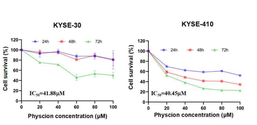
The cell viability of KYSE-30 and KYSE-410 cells was evaluated after treatment with various concentrations of Physcion (20, 40, 60, 80 μM) for 24 to 72 h.
Physcion purchased from MedChemExpress. Usage Cited in: Mol Cancer. 2025 Mar 26;24(1):97. [Abstract]
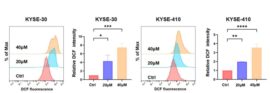
Intracellular ROS levels in ESCC cells fluctuated after 48 h of Physcion (20, 40 μM) treatment.
Physcion purchased from MedChemExpress. Usage Cited in: Mol Cancer. 2025 Mar 26;24(1):97. [Abstract]
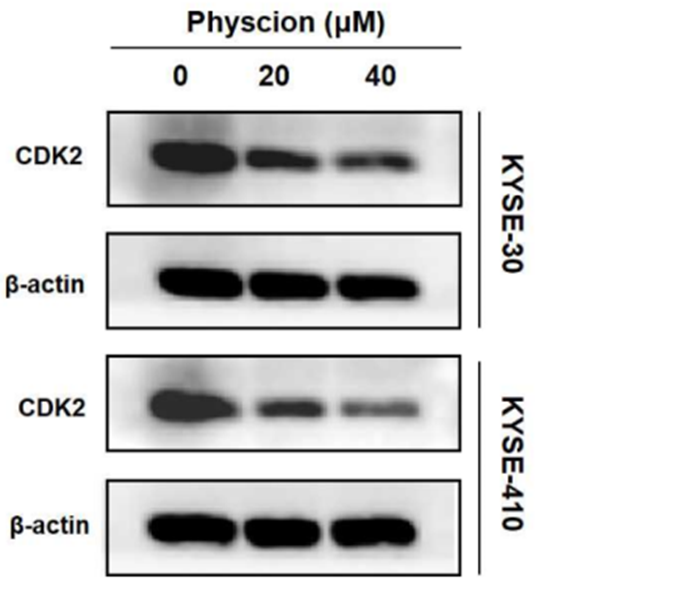
Western blot analysis revealed a reduction in CDK2 protein expression in ESCC cells following Physcion (20, 40 μM) treatment.
Physcion purchased from MedChemExpress. Usage Cited in: Mol Cancer. 2025 Mar 26;24(1):97. [Abstract]
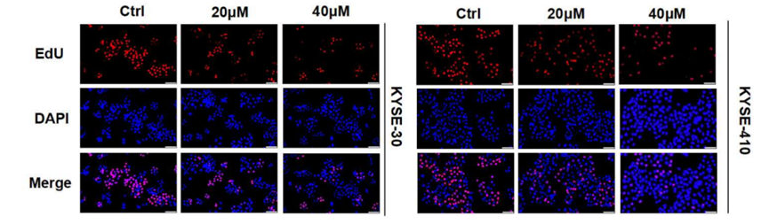
The effect of Physcion (20, 40 μM) on DNA synthesis in ESCC cells was detected using an EdU staining assay.
Physcion purchased from MedChemExpress. Usage Cited in: Mol Cancer. 2025 Mar 26;24(1):97. [Abstract]
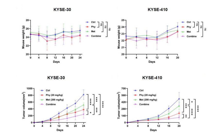
Effects of physcion combined with metformin on the growth of ESCC in vivo. The changes in body weight and tumor volume in nude mice after administration of Physcion (20 mg/kg) and Metformin (200 mg/kg) as single drugs or in combination.
-
Cancer Lett
6-Phosphogluconate dehydrogenase promotes glycolysis and fatty acid synthesis by inhibiting the AMPK pathway in lung adenocarcinoma cells. [Abstract]2024 Oct 1:601:217177. PMID: 39179096 -
-
J Ethnopharmacol
Physcion alleviates ulcerative colitis by upregulating SIRT3 to suppress PANoptosis via inhibition of the cGAS-STING pathway. [Abstract]2026 May 27:121893. PMID: 42202913 -
Drug Des Devel Ther
Physcion Protects Rats Against Cerebral Ischemia-Reperfusion Injury via Inhibition of TLR4/NF-kB Signaling Pathway. [Abstract]2021 Jan 25:15:277-287. PMID: 33536742 -
-
Biomed Res Int
Anti-influenza A Virus Effects and Mechanisms of Emodin and Its Analogs via Regulating PPAR α/ γ-AMPK-SIRT1 Pathway and Fatty Acid Metabolism. [Abstract]2021 Sep 9;2021:9066938. PMID: 34540999 -
World J Gastrointest Oncol
Physcion increases the sensitivity of hepatocellular carcinoma to sorafenib through miRNA-370/PIM1 axis-regulated glycolysis. [Abstract]2023 Aug 15;15(8):1400-1411. PMID: 37663938 -
Drug Chem Toxicol
Physcion alleviates cisplatin-induced apo in HK-2 cells by activating the apo signaling for treating acute kidney injury: Integrating network pharmacology and informatics. [Abstract]2025 Oct 14:1-9. PMID: 41084754 -
Curr Eye Res
Hyperosmolar potassium inhibits corneal myofibroblast transformation and prevent corneal scar. [Abstract]2023 Mar;48(3):238-250. PMID: 36149345 -


Lösungsmittel & Löslichkeit
In Vitro:
THF : 2.5 mg/mL (8.79 mM; ultrasonic and warming and heat to 60°C)
DMF : 1 mg/mL (3.52 mM; Need ultrasonic)
Ethanol : < 1 mg/mL (insoluble)
Please refer to the solubility information to select the appropriate solvent. Once prepared, please aliquot and store the solution to prevent product inactivation from repeated freeze-thaw cycles.
Storage method and period of stock solution: -80°C, 6 months; -20°C, 1 month. When stored at -80°C, please use it within 6 months. When stored at -20°C, please use it within 1 month.
Please refer to the solubility information to select the appropriate solvent. Once prepared, please aliquot and store the solution to prevent product inactivation from repeated freeze-thaw cycles.
Storage method and period of stock solution: -80°C, 6 months; -20°C, 1 month. When stored at -80°C, please use it within 6 months. When stored at -20°C, please use it within 1 month.
Konzentration (Stammlösung) × Volumen (Stammlösung) = Konzentration (Ziellösung) × Volumen (Ziellösung)


Reinheit & Dokumentation
-
Data Sheet (281 KB)
-
SDS (396 KB)
- English - EN (396 KB)
- Français - FR (396 KB)
- Deutsch - DE (396 KB)
- Norwegian - NO (396 KB)
- Español - ES (396 KB)
- Swedish - SV (396 KB)
- Italian - IT (396 KB)
- Korean - KR (396 KB)
- Portuguese - PT (396 KB)
-
Handling Instructions (2659 KB)
Verweise
[1]. Lin R, et al. 6-Phosphogluconate dehydrogenase links oxidative PPP, lipogenesis and tumour growth by inhibiting LKB1-AMPK signalling. Nat Cell Biol. 2015 Nov;17(11):1484-96. [Content Brief]
[2]. Pang MJ, et al. Physcion, a naturally occurring anthraquinone derivative, induces apoptosis and autophagy in human nasopharyngeal carcinoma. Acta Pharmacol Sin. 2016 Dec;37(12):1623-1640. [Content Brief]
[3]. Dong X, et al. Physcion Protects Rats Against Cerebral Ischemia-Reperfusion Injury via Inhibition of TLR4/NF-kB Signaling Pathway. Drug Des Devel Ther. 2021 Jan 25;15:277-287. [Content Brief]
[4]. Liu X, et al. Physcion, a novel anthraquinone derivative against Chlamydia psittaci infection. Vet Microbiol. 2023 Apr;279:109664. doi: 10.1016/j.vetmic.2023.109664. [Content Brief]


Complete Stock Solution Preparation Table
Please refer to the solubility information to select the appropriate solvent. Once prepared, please aliquot and store the solution to prevent product inactivation from repeated freeze-thaw cycles.
Storage method and period of stock solution: -80°C, 6 months; -20°C, 1 month. When stored at -80°C, please use it within 6 months. When stored at -20°C, please use it within 1 month.
| Optional Solvent | Concentration Solvent Mass | 1 mg | 5 mg | 10 mg | 25 mg |
|---|---|---|---|---|---|
| DMF / THF | 1 mM | 3.5179 mL | 17.5895 mL | 35.1791 mL | 87.9477 mL |
| THF | 5 mM | 0.7036 mL | 3.5179 mL | 7.0358 mL | 17.5895 mL |


Physcion Related Classifications
HY-N0108 Related Classifications
 Powered by Bioz