



Hypocrellin B
Based on 1 publication(s) in Google Scholar
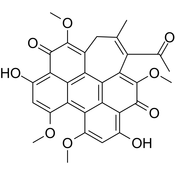
Hypocrellin B, a pigment isolated from the fungi Hypocrella bambusae and Shiraia bambusicola, is an apoptosis inducer. Hypocrellin B can be used as a photosensitizer for photodynamic therapy of cancer. Hypocrellin B also has antimicrobial and antileishmanial activities.
Nur für Forschungszwecke. Wir verkaufen nicht an Patienten.
- Reinheit : 99.63%
- CAS. Nr.: 123940-54-5
- Formel: C30H24O9
- Molecular Weight:528.51
-
Speicherung:
4°C, protect from light
* In solvent : -80°C, 6 months; -20°C, 1 month (protect from light)

To place orders, for customer services and technical support, please contact: MedChemExpress USA
Tel: 609-228-6898 E-mail: [email protected] [email protected]


-
 Biologische Aktivität
-
Chemical Information
-
Lösungsmittel & Löslichkeit
- Protokoll
- Reinheit & Dokumentation
- Verweise
-
 Help & FAQs


-
HY-14596
GenisteinAutophagy Inducer -
HY-B1328
PyridoxinePyridine Derivative -
HY-N0035
ArctigeninAntitumor Agent -
HY-114245
Se-MethylselenocysteineChemopreventive Agent -
HY-D0803
ThymoquinoneNatural Product -
HY-13425
DeguelinChemopreventive Agent -
HY-N6074
Soyasapogenol BAutophagy Inducer -
HY-N2369
ChelidonineIsoquinoline Alkaloid -
HY-N2193
HirsutineApoptosis Inducer -
HY-N2575
Hypocrellin APhotosensitizer -

HY-N1453R
Hypocrellin B (Standard)Anti-Fungal Agent -
HY-N0453
HypericinPKC Inhibitor
Publications Citing Use of MedChemExpress (MCE) Hypocrellin B
MoreAlle Parasite Isoform-spezifische Produkte anzeigen
More

Biologische Aktivität
Beschreibung
IC50 & Target
|
Leishmania |
Cellular Effect
|
Cell Line

|
Type | Value | Description | References |
|---|---|---|---|---|
| BGC-823 | IC50 |
40 nM
Compound: 2
|
Cytotoxicity against human BGC823 cells assessed as cell death preincubated for 5 hrs in dark condition followed by irradiation with a 532 nm laser of 20 J/cm2 for 1000 seconds measured after 24 hrs by MTT assay
Cytotoxicity against human BGC823 cells assessed as cell death preincubated for 5 hrs in dark condition followed by irradiation with a 532 nm laser of 20 J/cm2 for 1000 seconds measured after 24 hrs by MTT assay
|
[PMID: 22352922] |
In Vitro
Hypocrellin B (2.5 μM, 4 h) induces cell cytotoxicity 24 h after sonodynamic therapy (SDT) in HepG2 cells, and induces induce the early apoptosis[2].
Hypocrellin B shows antifungal and anti-parasite activity against C. albicans and Leishmania donovani respectively (IC50: 5 and 12.7 μg/mL)[3].
MedChemExpress (MCE) has not independently confirmed the accuracy of these methods. They are for reference only.
In Vivo
MedChemExpress (MCE) has not independently confirmed the accuracy of these methods. They are for reference only.


Chemical Information
-
CAS. Nr. 123940-54-5
-
Appearance Solid
-
Molecular Weight 528.51
-
Formel C30H24O9
-
Color Brown to black
-
SMILES
CC(C1)=C(C(C)=O)C(C2=C(C3=C(O)C=C4OC)C4=C5C(OC)=CC(O)=C6C5=C2C1=C(OC)C6=O)=C(OC)C3=O
-
Structure Classification
-
Initial Source
-
Versand
Room temperature in continental US; may vary elsewhere.
-
Speicherung
4°C, protect from light
* In solvent : -80°C, 6 months; -20°C, 1 month (protect from light)


Publications (1)
-
Journal Impact Factor
-
Most Recent


Lösungsmittel & Löslichkeit
In Vitro:
DMSO : < 1 mg/mL (insoluble or slightly soluble)
* Hypocrellin B is usually formulated as a suspension.


Reinheit & Dokumentation
-
Data Sheet (282 KB)
-
SDS (394 KB)
- English - EN (394 KB)
- Français - FR (394 KB)
- Deutsch - DE (394 KB)
- Norwegian - NO (394 KB)
- Español - ES (394 KB)
- Swedish - SV (394 KB)
- Italian - IT (394 KB)
- Korean - KR (394 KB)
- Portuguese - PT (394 KB)
-
Handling Instructions (2659 KB)
Verweise
[1]. Miller GG, et al. Preclinical assessment of hypocrellin B and hypocrellin B derivatives as sensitizers for photodynamic therapy of cancer: progress update. Photochem Photobiol. 1997 Apr;65(4):714-22. [Content Brief]
[2]. Wang X, et al. Hypocrellin B-mediated sonodynamic action induces apoptosis of hepatocellular carcinoma cells. Ultrasonics. 2012 Apr;52(4):543-6. [Content Brief]
[3]. Ma G, et al. Antimicrobial and antileishmanial activities of hypocrellins A and B. Antimicrob Agents Chemother. 2004 Nov;48(11):4450-2. [Content Brief]
[4]. Chang JE, et al. Hypocrellin B and paclitaxel-encapsulated hyaluronic acid-ceramide nanoparticles for targeted photodynamic therapy in lung cancer. J Photochem Photobiol B. 2016 May;158:113-21. [Content Brief]

Calculators
Konzentration (Stammlösung) × Volumen (Stammlösung) = Konzentration (Ziellösung) × Volumen (Ziellösung)


Hypocrellin B Related Classifications
HY-N1453 Related Classifications
 Powered by Bioz