



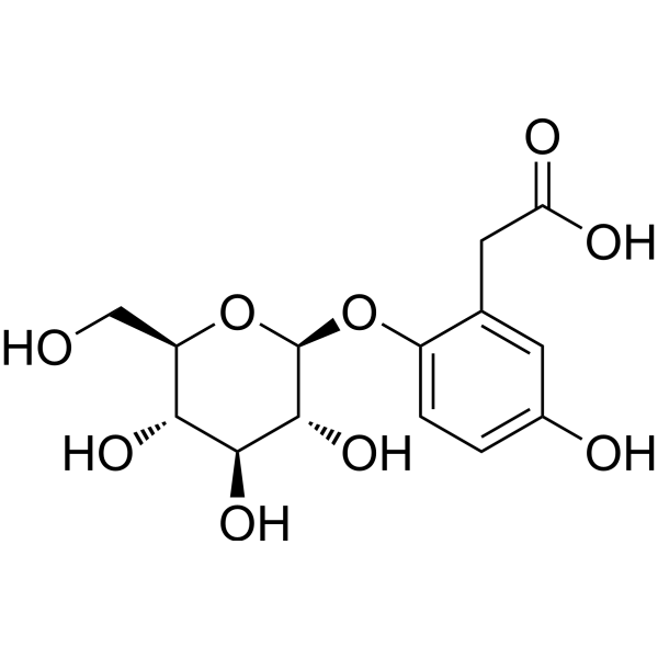
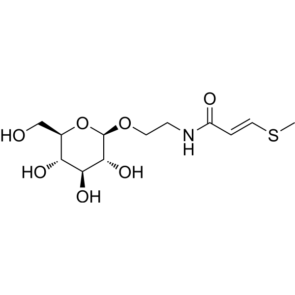
Phaseoloidin

Based on 1 Customer Validation
Phaseoloidin is an orally active multi-target inhibitor. Phaseoloidin inhibits the activation of the NLRP3 inflammasome and blocks the caspase-11-GSDMD pyroptosis axis. Phaseoloidin reduces the expression of collagen-degrading enzymes to maintain the integrity of cartilage matrix. Phaseoloidin activates the AMPK/mTOR pathway to enhance autophagy function and reverse apoptosis resistance. Phaseoloidin inhibits the growth and development of Manduca sexta and Spodoptera littoralis larvae, thereby helping Nicotiana attenuata defend against lepidopteran herbivorous insects. Phaseoloidin can be used in research related to acute gouty arthritis and pulmonary fibrosis.
For research use only. We do not sell to patients.
- Purity : 99.92%
- CAS No.: 118555-82-1
- Formula: C14H18O9
- Molecular Weight:330.29
-
Storage:
4°C, protect from light
* In solvent : -80°C, 6 months; -20°C, 1 month (protect from light)

To place orders, for customer services and technical support, please contact: MedChemExpress USA
Tel: 609-228-6898 E-mail: [email protected] [email protected]


-
 Biological Activity
-
Chemical Information
-
Solvent & Solubility
- Protocols
- Purity & Documentation
- References
-
 Help & FAQs




Biological Activity
Description
In Vitro
Phaseoloidin (6.25-200 μM; 24 h) is non-cytotoxic to mouse peritoneal macrophages at concentrations up to 200 μM following 24 h incubation[1].
Phaseoloidin (6.25-200 μM; 24 h) is non-cytotoxic to rat articular chondrocytes at concentrations up to 100 μM following 24 h incubation, with only mild cytotoxicity at 200 μM[1].
Phaseoloidin (25-100 μM; 1 h pre-incubation, followed by 22 h total incubation with LPS and sodium urate crystals) dose-dependently inhibits NLRP3 inflammasome activation and downstream inflammatory mediator release in LPS/sodium urate-stimulated mouse peritoneal macrophages, with maximal effects at 100 μM[1].
Phaseoloidin (10-100 μM; 48 h) activates the AMPK/mTOR pathway to promote autophagy in TGF-β1-induced primary mouse lung myofibroblasts[2].
Phaseoloidin (3-300 μM) does not reduce the viability of TGF-β1-induced primary mouse lung myofibroblasts[2].
Phaseoloidin (10-100 μM; 48 h) promotes autophagy-mediated collagen degradation in TGF-β1-induced primary mouse lung myofibroblasts via the AMPK pathway[2].
Phaseoloidin (10-100 μM; 48 h co-treatment with FasL) reverses FasL-induced apoptosis resistance in TGF-β1-induced primary mouse lung myofibroblasts via the AMPK pathway[2].
Phaseoloidin (10-100 μM; 48 h co-treatment with FasL) promotes autophagy-mediated degradation of PTPN13 to restore FasL-induced apoptosis sensitivity in TGF-β1-induced primary mouse lung myofibroblasts[2].
MedChemExpress (MCE) has not independently confirmed the accuracy of these methods. They are for reference only.
-
Cell Line:Rat articular chondrocytes
-
Concentration:6.25-200 μM
-
Incubation Time:24 h
-
Result:Showed no significant cytotoxicity at concentrations ≤100 μM relative to untreated cells.
Caused a minor but significant reduction in viability at 200 μM relative to untreated cells.
In Vivo
Phaseoloidin (25-100 mg/kg; i.g.; single dose 1 hour pre-challenge) dose-dependently suppresses sodium urate-induced acute gout inflammation in mouse air pouches by inhibiting NLRP3 inflammasome activation, pyroptosis, and NETosis, with high-dose treatment normalizing lavage fluid IL-1β levels[1].
Phaseoloidin (1-25 mg/kg; i.p.; daily; days 14-28) exerts dose-dependent antifibrotic effects in Bleomycin (HY-108345)-induced pulmonary fibrosis in male C57/BL mice via AMPK-mediated autophagy activation[2].
Phaseoloidin (0.16 μg/mg of diet; oral; continuous feeding; 12 days) reduces Manduca sexta larval growth by 50%[3].
Phaseoloidin (0.16 μg/mg of diet; oral; continuous feeding; 8 days) reduces Spodoptera littoralis larval growth by 55%[3].
MedChemExpress (MCE) has not independently confirmed the accuracy of these methods. They are for reference only.
-
Animal Model:Kunming mice (male, 6-8 weeks old, acute gouty arthritis induced by sodium urate crystal paw injection)[1]
-
Dosage:25-100 mg/kg
-
Administration:i.g.; single dose 1 hour pre-challenge
-
Result:Attenuated paw swelling dose-dependently at 1 hour post-sodium urate injection.
Reduced paw erythema and swelling.
Decreased immune cell infiltration in articular cavity and periarticular muscle tissues.
Downregulated expression of macrophage marker F4/80 dose-dependently.
Inhibited expression of NLRP3 inflammasome components (NLRP3, ASC, pro-caspase-1, cleaved caspase-1, pro-IL-1β, mature IL-1β, HMGB1, Cathepsin B) dose-dependently.
Reduced levels of caspase-11 pathway proteins (pro-caspase-11, cleaved caspase-11, GSDMD, GSDMD-N) dose-dependently.
Suppressed sodium urate-induced expression of NETosis-associated proteins (Cathepsin G, CitH3, PAD4, PR3, NE) dose-dependently.
Reduced SYTOX Green-labeled extracellular DNA release.
Downregulated sodium urate-induced expression of cartilage-degrading enzymes MMP-3 and MMP-13 dose-dependently.
Restored paw glycosaminoglycan content (toluidine blue staining intensity).
Reduced cartilage erosion.
-
Animal Model:Kunming mice (male, 6-8 weeks old, acute gout induced by sodium urate crystal subcutaneous air pouch injection)[1]
-
Dosage:25-100 mg/kg
-
Administration:i.g.; single dose 1 hour pre-challenge
-
Result:Reduced inflammatory cell infiltration in air pouch subcutaneous tissue dose-dependently.
Inhibited NLRP3 inflammasome activation (downregulated HMGB1, Cathepsin B, NLRP3, ASC, pro-caspase-1, cleaved caspase-1, pro-IL-1β, mature IL-1β) dose-dependently.
Suppressed caspase-11-GSDMD pathway activity (downregulated pro-caspase-11, cleaved caspase-11, GSDMD, GSDMD-N) dose-dependently.
Reduced IL-1β levels in air pouch lavage fluid from ~1800 pg/mL (model group) to near-normal levels (~300 pg/mL) at 100 mg/kg.
Inhibited sodium urate-induced NETosis dose-dependently, reducing expression of Cathepsin G, CitH3, PAD4, PR3, and NE.
Decreased co-localization of MPO and SYTOX Green signals.
-
Animal Model:C57/BL (male)[2]
-
Dosage:1 mg/kg; 5 mg/kg; 25 mg/kg
-
Administration:i.p.; daily; days 14-28
-
Result:Reduced alveolar wall thickening and extracellular matrix deposition .
Dose-dependently reduced mortality, partially reversed bleomycin-induced weight loss, and lowered lung hydroxyproline content relative to untreated fibrotic mice.
Decreased bleomycin-induced upregulation of Collagen I and α-SMA mRNA and protein levels.
Activated AMPK phosphorylation in lung tissue, increased autophagic activity, and promoted myofibroblast sensitivity to FasL-induced apoptosis by reducing PTPN13 protein abundance.
Completely reversed antifibrotic, survival-improving, autophagy-activating, and apoptosis-sensitizing effects when co-administered with AMPK inhibitor Compound C .
- RNA extraction experimental
- Collagen: Sirius Red Staining
- Annexin V plus membrane-impermeant dye apoptosis staining
- Connective Tissue: Masson's Trichrome/Collagen Trichrome Staining
- Fibrosis/Collagen Morphometry
- Collagen-Induced Arthritis
- Detection of Apoptosis
- TUNEL staining for apoptotic DNA fragmentation
- Autophagy
- Apoptosis Solutions
- Lysosome and acidic-vesicle live-cell staining
- Macroautophagy Solutions
- Pyroptosis Solutions


Chemical Information
-
CAS No. 118555-82-1
-
Appearance Solid
-
Molecular Weight 330.29
-
Formula C14H18O9
-
Color White to off-white
-
SMILES
O=C(O)CC1=CC(O)=CC=C1O[C@H]2[C@@H]([C@H]([C@@H]([C@@H](CO)O2)O)O)O
-
Structure Classification
-
Initial Source
-
Shipping
Room temperature in continental US; may vary elsewhere.
-
Storage
4°C, protect from light
* In solvent : -80°C, 6 months; -20°C, 1 month (protect from light)


Solvent & Solubility
In Vitro:
DMSO : 100 mg/mL (302.76 mM; Need ultrasonic; Hygroscopic DMSO has a significant impact on the solubility of product, please use newly opened DMSO)
Please refer to the solubility information to select the appropriate solvent. Once prepared, please aliquot and store the solution to prevent product inactivation from repeated freeze-thaw cycles.
Storage method and period of stock solution: -80°C, 6 months; -20°C, 1 month (protect from light). When stored at -80°C, please use it within 6 months. When stored at -20°C, please use it within 1 month.
Please refer to the solubility information to select the appropriate solvent. Once prepared, please aliquot and store the solution to prevent product inactivation from repeated freeze-thaw cycles.
Storage method and period of stock solution: -80°C, 6 months; -20°C, 1 month (protect from light). When stored at -80°C, please use it within 6 months. When stored at -20°C, please use it within 1 month.
Concentration (start) × Volume (start) = Concentration (final) × Volume (final)
In Vivo:
Select the appropriate dissolution method based on your experimental animal and administration route.
- For the following dissolution methods, please ensure to first prepare a clear stock solution using an In Vitro approach and then sequentially add co-solvents:
- To ensure reliable experimental results, the clarified stock solution can be appropriately stored based on storage conditions. As for the working solution for In Vivo experiments, it is recommended to prepare freshly and use it on the same day.
- The percentages shown for the solvents indicate their volumetric ratio in the final prepared solution. If precipitation or phase separation occurs during preparation, heat and/or sonication can be used to aid dissolution.
Add each solvent one by one: 10% DMSO 40% PEG300 5% Tween-80 45% Saline
Solubility: ≥ 2.5 mg/mL (7.57 mM); Clear solution
This protocol yields a clear solution of ≥ 2.5 mg/mL (saturation unknown).
Taking 1 mL working solution as an example, add 100 μL DMSO stock solution (25.0 mg/mL) to 400 μL PEG300, and mix evenly; then add 50 μL Tween-80 and mix evenly; then add 450 μL Saline to adjust the volume to 1 mL.
Preparation of Saline: Dissolve 0.9 g sodium chloride in ddH₂O and dilute to 100 mL to obtain a clear Saline solution.
Add each solvent one by one: 10% DMSO 90% (20% SBE-β-CD in Saline)
Solubility: ≥ 2.5 mg/mL (7.57 mM); Clear solution
This protocol yields a clear solution of ≥ 2.5 mg/mL (saturation unknown).
Taking 1 mL working solution as an example, add 100 μL DMSO stock solution (25.0 mg/mL) to 900 μL 20% SBE-β-CD in Saline, and mix evenly.
Preparation of 20% SBE-β-CD in Saline (4°C, storage for one week): 2 g SBE-β-CD powder is dissolved in 10 mL Saline, completely dissolve until clear.
In Vivo Dissolution Calculator
Please enter the basic information of animal experiments:
-
-
-
-
Recommended: Prepare an additional quantity of animals to account for potential losses during experiments.
Please enter your animal formula composition:
-
%DMSO +
Recommended: Keep the proportion of DMSO in working solution below 2% if your animal is weak.
-
%+
-
+%Tween-80 + +
-
%Saline +
The co-solvents required include: DMSO, . All of co-solvents are available by MedChemExpress (MCE). , Tween 80. All of co-solvents are available by MedChemExpress (MCE).
Working solution concentration: 0.22 mg/mL
Method for preparing stock solution: mg drug dissolved in μL DMSO. Stock solution concentration: mg/mL. * In solvent : -80°C, 6 months; -20°C, 1 month (protect from light)
1. Take μL DMSO stock solution;
2. Add μL .
μL , mix evenly;
3. Then add μL Tween 80, mix evenly;
4. Then add μL
Please ensure that the stock solution in the first step is dissolved to a clear state, and add co-solvents in sequence. You can use ultrasonic heating (ultrasonic cleaner, recommended frequency 20-40 kHz), vortexing, etc. to assist dissolution.


Purity & Documentation
-
Data Sheet (286 KB)
-
SDS (393 KB)
- English - EN (393 KB)
- Français - FR (393 KB)
- Deutsch - DE (393 KB)
- Norwegian - NO (393 KB)
- Español - ES (393 KB)
- Swedish - SV (393 KB)
- Italian - IT (393 KB)
- Korean - KR (393 KB)
- Portuguese - PT (393 KB)
-
Handling Instructions (2659 KB)
References
[1]. Jiang YJ, et al. Phaseoloidin, a homogentisic acid glucoside from Entada phaseoloides, suppresses gout inflammation via NLRP3 inflammasome. Phytomedicine. 2026;152:157848. [Content Brief]
[2]. Li S, et al. Reversal of Myofibroblast Apoptosis Resistance and Collagen Deposition by Phaseoloidin-Induced Autophagy Attenuates Pulmonary Fibrosis. Biomedicines. 2025;13(11):2679. Published 2025 Oct 31. [Content Brief]
[3]. Weinhold A, et al. Phaseoloidin, a homogentisic acid glucoside from Nicotiana attenuata trichomes, contributes to the plant's resistance against lepidopteran herbivores. J Chem Ecol. 2011;37(10):1091-1098. [Content Brief]


Complete Stock Solution Preparation Table
Please refer to the solubility information to select the appropriate solvent. Once prepared, please aliquot and store the solution to prevent product inactivation from repeated freeze-thaw cycles.
Storage method and period of stock solution: -80°C, 6 months; -20°C, 1 month (protect from light). When stored at -80°C, please use it within 6 months. When stored at -20°C, please use it within 1 month.
| Optional Solvent | Concentration Solvent Mass | 1 mg | 5 mg | 10 mg | 25 mg |
|---|---|---|---|---|---|
| DMSO | 1 mM | 3.0276 mL | 15.1382 mL | 30.2764 mL | 75.6911 mL |
| 5 mM | 0.6055 mL | 3.0276 mL | 6.0553 mL | 15.1382 mL | |
| 10 mM | 0.3028 mL | 1.5138 mL | 3.0276 mL | 7.5691 mL | |
| 15 mM | 0.2018 mL | 1.0092 mL | 2.0184 mL | 5.0461 mL | |
| 20 mM | 0.1514 mL | 0.7569 mL | 1.5138 mL | 3.7846 mL | |
| 25 mM | 0.1211 mL | 0.6055 mL | 1.2111 mL | 3.0276 mL | |
| 30 mM | 0.1009 mL | 0.5046 mL | 1.0092 mL | 2.5230 mL | |
| 40 mM | 0.0757 mL | 0.3785 mL | 0.7569 mL | 1.8923 mL | |
| 50 mM | 0.0606 mL | 0.3028 mL | 0.6055 mL | 1.5138 mL | |
| 60 mM | 0.0505 mL | 0.2523 mL | 0.5046 mL | 1.2615 mL | |
| 80 mM | 0.0378 mL | 0.1892 mL | 0.3785 mL | 0.9461 mL | |
| 100 mM | 0.0303 mL | 0.1514 mL | 0.3028 mL | 0.7569 mL |


Phaseoloidin Related Classifications
HY-N7400 Related Classifications
 Powered by Bioz