



ERD-3111

Based on 1 Customer Validation
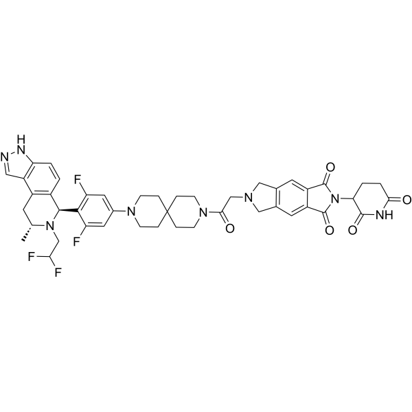
ERD-3111 is an orally active PROTAC degrader targeting estrogen receptor α (ERα), with a DC50 of 0.5 nM. ERD-3111 induces ERα degradation in breast cancer cells, and triggers tumor regression or complete inhibition of tumor growth in ER-positive breast cancer xenograft models. ERD-3111 can be used in breast cancer-related research.
(Pink: ER ligand (HY-175927); Blue: Cereblon ligand (HY-W248665); Black: linker).
Nos produits utilisent uniquement pour la recherche. Nous ne vendons pas aux patients.
- Pureté : 98.72%
- CAS No.: 2832865-25-3
- Formule: C45H46F4N8O5
- Masse moléculaire:854.89
-
Stockage:
-20°C, protect from light, stored under nitrogen
* In solvent : -80°C, 6 months; -20°C, 1 month (protect from light, stored under nitrogen)

To place orders, for customer services and technical support, please contact: MedChemExpress USA
Tel: 609-228-6898 E-mail: [email protected] [email protected]


-
 Activité biologique
-
Chemical Information
-
Solvant et solubilité
- Protocole
- Pureté et documentation
- Références
-
 Help & FAQs


Voir tous les produits spécifiques à Isoform PROTACs
More

Activité biologique
Description
IC50 & Target
[1]|
ERα 0.5 nM (DC50) |
In Vitro
ERD-3111 (compound 44) potently degrades ERα in MCF-7 breast cancer cells, with a DC50 of 0.5 nM and a maximum degradation efficiency of 91%[1].
ERD-3111 (0.1-30 nM) induces dose-dependent ERα degradation in MCF-7 breast cancer cells[1].
ERD-3111 (0.1-100 nM) induces dose-dependent ERα degradation in T47D breast cancer cells[1].
ERD-3111 does not significantly inhibit hERG channels in HEK293 cells at concentrations up to 30 μM, with an IC50 greater than 30 μM; it does not significantly inhibit any tested human CYP enzyme subtypes at concentrations up to 10 μM, with an IC50 value greater than 10 μM[1].
MedChemExpress (MCE) has not independently confirmed the accuracy of these methods. They are for reference only. Further protocols information, click here.
Parmacokinetics
| Species | Dose | Route | Vss | CL | T1/2 | Cmax | AUC | F |
|---|---|---|---|---|---|---|---|---|
| Rat[1] | 1 mg/kg | i.v. | 1.3 L/kg | 7.4 mL/min/kg | 4.0 h | 141.1 ng/mL | 1317 ng·h/mL | / |
| Mice[1] | 1 mg/kg | i.v. | 3.2 L/kg | 5.7 mL/min/kg | 6.4 h | 260 ng/mL | 3366 ng·h/mL | / |
| Dog[1] | 0.5 mg/kg | i.v. | 5.2 L/kg | 11.0 mL/min/kg | 7.9 h | 87 ng/mL | 937 ng·h/mL | / |
| Rat[1] | 3 mg/kg | p.o. | / | / | / | / | / | 20 % |
| Mice[1] | 3 mg/kg | p.o. | / | / | / | / | / | 42 % |
| Dog[1] | 1 mg/kg | p.o. | / | / | / | / | / | 66 % |
In Vivo
ERD-3111 (10 mg/kg; p.o.; once daily; for 3 consecutive days) reduces the protein level of ESR1D538G-mutant ERα in MCF-7 xenograft tumors[1].
ERD-3111 (10-30 mg/kg; p.o.; once daily; for 35 consecutive days) induces sustained tumor regression in wild-type ERα MCF-7 xenografts, and the therapeutic effect is maintained after drug withdrawal[1].
ERD-3111 (10-30 mg/kg; p.o.; once daily; for 28 consecutive days) induces sustained tumor growth inhibition in ESR1Y537S-mutant and ESR1D538G-mutant MCF-7 xenografts, with enhanced efficacy observed even after discontinuation of treatment[1].
MedChemExpress (MCE) has not independently confirmed the accuracy of these methods. They are for reference only.
-
Animal Model:CB17 SCID (female, wild-type ERα MCF-7 xenograft and ESR1Y537S mutant MCF-7 xenograft, maintained on 17β-estradiol-supplemented drinking water)[1]
-
Dosage:10 mg/kg; 30 mg/kg
-
Administration:p.o.; daily; 3 days
-
Result:Reduced ERα protein levels after 3-day daily oral administration in female CB17 SCID mice bearing wild-type or ESR1Y537S mutant MCF-7 xenografts.
Achieved dose-dependent depletion of wild-type ERα, reaching maximum reduction of 70% (10 mg/kg) and 78% (30 mg/kg) at 24 h, with detectable compound exposure in plasma and tumor tissues at both doses.
Induced dose-dependent downregulation of ESR1Y537S mutant ERα, achieving 70% suppression at 10 mg/kg and 85% suppression at 30 mg/kg, accompanied by measurable plasma and intratumoral drug concentrations.
-
Animal Model:CB17 SCID (female, wild-type ERα MCF-7 xenograft, maintained on 17β-estradiol-supplemented drinking water)[1]
-
Dosage:10 mg/kg; 30 mg/kg
-
Administration:p.o.; daily; 35 days
-
Result:Caused 24% tumor regression at the end of treatment (day 73) at 10 mg/kg.
Caused 42% tumor regression at the end of treatment (day 73) at 30 mg/kg.
Maintained 13% tumor regression three weeks post-treatment (day 93) at 10 mg/kg.
Maintained 42% tumor regression three weeks post-treatment (day 93) at 30 mg/kg.
Exhibited significantly better antitumor activity than 10 mg/kg ARV-471 at day 93 at 10 mg/kg.
-
Animal Model:CB17 SCID (female, ESR1D538G mutant MCF-7 xenograft)[1]
-
Dosage:10 mg/kg
-
Administration:p.o.; daily; 3 days
-
Result:Reduced ESR1D538G mutant ERα protein levels in tumors by 68%.
Achieved plasma concentration of 609 ng/mL and tumor concentration of 1052 ng/mL.
-
Animal Model:CB17 SCID (female, ESR1Y537S mutant MCF-7 xenograft and ESR1D538G mutant MCF-7 xenograft)[1]
-
Dosage:10 mg/kg; 30 mg/kg
-
Administration:p.o.; daily; 28 days
-
Result:Exhibited dose-dependent antitumor activity after 28-day daily oral dosing in female CB17 SCID mice bearing ESR1-mutant MCF-7 xenografts.
Achieved 72% (10 mg/kg) and 86% (30 mg/kg) tumor growth inhibition at treatment endpoint in ESR1Y537S mutant tumors; antitumor effects persisted until day 82 with 99% and 101% sustained inhibition.
Induced 38% (10 mg/kg) and 51% (30 mg/kg) tumor regression at treatment completion in ESR1D538G mutant tumors, with tumor growth suppression maintained for one week post-treatment cessation.


Chemical Information
-
CAS No. 2832865-25-3
-
Appearance Solid
-
Masse moléculaire 854.89
-
Formule C45H46F4N8O5
-
Color Off-white to light yellow
-
SMILES
FC(CN1[C@@H](CC2=C([C@H]1C3=C(C=C(C=C3F)N4CCC5(CC4)CCN(C(CN6CC(C=C(C7=C8)C(N(C7=O)C9CCC(NC9=O)=O)=O)=C8C6)=O)CC5)F)C=CC%10=C2C=NN%10)C)F
-
Livraison
Room temperature in continental US; may vary elsewhere.
-
Stockage
-20°C, protect from light, stored under nitrogen
* In solvent : -80°C, 6 months; -20°C, 1 month (protect from light, stored under nitrogen)


Solvant et solubilité
In Vitro:
DMSO : 100 mg/mL (116.97 mM; Need ultrasonic; Hygroscopic DMSO has a significant impact on the solubility of product, please use newly opened DMSO)
Please refer to the solubility information to select the appropriate solvent. Once prepared, please aliquot and store the solution to prevent product inactivation from repeated freeze-thaw cycles.
Storage method and period of stock solution: -80°C, 6 months; -20°C, 1 month (protect from light, stored under nitrogen). When stored at -80°C, please use it within 6 months. When stored at -20°C, please use it within 1 month.
Please refer to the solubility information to select the appropriate solvent. Once prepared, please aliquot and store the solution to prevent product inactivation from repeated freeze-thaw cycles.
Storage method and period of stock solution: -80°C, 6 months; -20°C, 1 month (protect from light, stored under nitrogen). When stored at -80°C, please use it within 6 months. When stored at -20°C, please use it within 1 month.
Concentration (start) × Volume (start) = Concentration (final) × Volume (final)

Protocole
-
Research Protocol for Endocrine Diseases
Endocrine diseases often arise from disrupted hormone production, hormone signaling, or target-tissue responsiveness; for diabetes-focused endocrine disease models, insulin signaling regulates glucose uptake, hepatic glucose output, lipid metabolism, and β-cell compensation. Type 2 diabetes develops through interacting defects in insulin resistance, β-cell dysfunction, adipose inflammation, hepatic glucose overproduction, altered incretin signaling, and ectopic lipid metabolism. A major unresolved question is whether endocrine dysfunction is driven primarily by target-tissue insulin resistance, intrinsic β-cell failure, immune/inflammatory stress, or combined multi-organ failure that differs by disease stage.
-
Patient-Derived Xenograft (PDX)
Patient-derived xenograft (PDX) models are generated by engrafting primary human tumor tissue directly into immunodeficient mice, allowing in vivo propagation of patient tumor biology without initial in vitro adaptation. These models are used to preserve key histopathological and molecular characteristics of the original tumor and enable assessment of tumor growth dynamics and therapeutic response in a living organism. The biological readout is tumor engraftment and subsequent growth in the murine host, which reflects the ability of human tumor cells to survive, vascularize, and expand in an immunocompromised microenvironment.
-
Patient-Derived Orthotopic Xenograft (PDOX)
Patient-derived orthotopic xenograft (PDOX) modeling implants fresh patient tumor tissue or patient-derived tumor cells into the anatomically corresponding organ or tissue site of immunodeficient mice, usually by surgical orthotopic implantation, to preserve patient tumor histology, local microenvironmental context, invasion, metastatic behavior, and treatment-response features better than subcutaneous implantation. PDOX readouts include tumor engraftment, orthotopic tumor growth, local invasion, metastasis, recurrence after resection, histologic similarity to the donor tumor, biomarker retention, molecular concordance, survival, and response or resistance to therapy. PDOX models are used for preclinical drug testing and individualized therapy evaluation, but engraftment success varies by tumor type and specimen quality.
-
Splenic/Portal-Vein Liver Metastasis Xenograft
Splenic and portal-vein liver metastasis xenograft models deliver tumor cells into the portal circulation so that cells reach the liver first and form hepatic metastatic lesions; splenic injection uses the spleen as an access route to the portal system, while direct portal-vein injection introduces cells into the portal vein without requiring splenectomy. The assay detects liver colonization, intrahepatic tumor growth, tumor distribution, treatment response, survival, and liver-metastasis microenvironment changes; readouts include bioluminescence or fluorescence imaging, gross liver nodule counts, liver weight or tumor burden, histology, and survival.
-
Subcutaneous Cell-Line-Derived Xenograft
Subcutaneous cell-line-derived xenograft (CDX) models are established by implanting cultured human cancer cell lines into immunodeficient mice, where the injected cells form localized tumors that can be monitored in vivo as a measure of tumorigenic potential, growth kinetics, and treatment response. These models are widely used in oncology research because they allow reproducible tumor formation and enable comparative assessment of tumor growth between different cell lines or genetic manipulations in a controlled in vivo microenvironment. Subcutaneous implantation of cancer cells in immunodeficient mice is a standard approach for evaluating tumor growth behavior and therapeutic response across multiple cancer types, including prostate, esophageal, pancreatic, and colon cancer models.
-
Breast Cancer Modeling
Breast cancer is a heterogeneous cancer, and it has been distinguished into four subtypes: luminal A, luminal B, HER2-positive and basal-like. Molecular mutations, epigenetic alterations, hormone exposure and immune microenvironment are related to the progression of breast cancer.
-
Orthotopic Cell-Line Xenograft
Orthotopic cell-line xenograft models involve implantation of human cancer cell lines into the anatomically corresponding organ of immunodeficient mice to reproduce tumor growth within a native microenvironment, enabling more clinically relevant tumor behavior compared with subcutaneous models. These models are widely used because orthotopic placement better recapitulates tumor progression, including invasion and metastatic spread, which are often underrepresented in heterotopic implantation systems. Compared with conventional xenografts, orthotopic implantation is described as more technically complex but provides improved simulation of tumor-microenvironment interactions and metastatic behavior, making it particularly valuable for translational oncology research. Surgical orthotopic implantation approaches have been emphasized as enabling faithful reproduction of clinical cancer features, including metastasis and disease progression patterns that align with the tumor’s organ of origi
-
Intraperitoneal/Peritoneal Dissemination Xenograft
Intraperitoneal (IP) or peritoneal dissemination xenograft models are based on the introduction of human cancer cells into the peritoneal cavity of immunodeficient mice, where they attach to peritoneal surfaces, form multicellular aggregates or spheroids, and progressively generate disseminated tumor nodules that mimic advanced peritoneal metastatic disease. These models are widely used to study ovarian cancer progression, tumor-microenvironment interactions, and intraperitoneal therapeutic responses, often incorporating bioluminescence or fluorescence imaging to longitudinally monitor tumor burden in vivo. The biological principle relies on the capacity of tumor cells such as SKOV3 or related ovarian carcinoma lines to survive in suspension, aggregate within ascites-like fluid, adhere to mesothelial surfaces, and invade peritoneal organs, thereby recapitulating human peritoneal carcinomatosis patterns observed in advanced disease.


Pureté et documentation
-
Fiche technique (285 KB)
-
SDS (252 KB)
- English - EN (252 KB)
- Français - FR (252 KB)
- Deutsch - DE (252 KB)
- Norwegian - NO (252 KB)
- Español - ES (252 KB)
- Swedish - SV (252 KB)
- Italian - IT (252 KB)
- Korean - KR (252 KB)
- Portuguese - PT (252 KB)
-
Instruction de manipulation (2659 KB)
Références


Complete Stock Solution Preparation Table
Please refer to the solubility information to select the appropriate solvent. Once prepared, please aliquot and store the solution to prevent product inactivation from repeated freeze-thaw cycles.
Storage method and period of stock solution: -80°C, 6 months; -20°C, 1 month (protect from light, stored under nitrogen). When stored at -80°C, please use it within 6 months. When stored at -20°C, please use it within 1 month.
| Optional Solvent | Concentration Solvent Mass | 1 mg | 5 mg | 10 mg | 25 mg |
|---|---|---|---|---|---|
| DMSO | 1 mM | 1.1697 mL | 5.8487 mL | 11.6974 mL | 29.2435 mL |
| 5 mM | 0.2339 mL | 1.1697 mL | 2.3395 mL | 5.8487 mL | |
| 10 mM | 0.1170 mL | 0.5849 mL | 1.1697 mL | 2.9244 mL | |
| 15 mM | 0.0780 mL | 0.3899 mL | 0.7798 mL | 1.9496 mL | |
| 20 mM | 0.0585 mL | 0.2924 mL | 0.5849 mL | 1.4622 mL | |
| 25 mM | 0.0468 mL | 0.2339 mL | 0.4679 mL | 1.1697 mL | |
| 30 mM | 0.0390 mL | 0.1950 mL | 0.3899 mL | 0.9748 mL | |
| 40 mM | 0.0292 mL | 0.1462 mL | 0.2924 mL | 0.7311 mL | |
| 50 mM | 0.0234 mL | 0.1170 mL | 0.2339 mL | 0.5849 mL | |
| 60 mM | 0.0195 mL | 0.0975 mL | 0.1950 mL | 0.4874 mL | |
| 80 mM | 0.0146 mL | 0.0731 mL | 0.1462 mL | 0.3655 mL | |
| 100 mM | 0.0117 mL | 0.0585 mL | 0.1170 mL | 0.2924 mL |


ERD-3111 Related Classifications
HY-149480 Related Classifications
 Powered by Bioz