



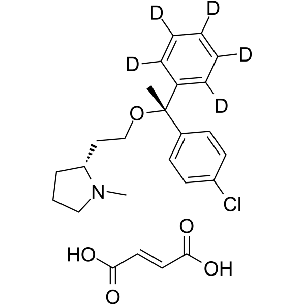
Clemastine-d5 fumarate
Based on 1 publication(s) in Google Scholar
Clemastine (HS-592; Meclastine)-d5 fumarate is the deuterium labeled Clemastine fumarate. Clemastine fumarate is an orally active, blood-brain barrier-permeable H1 histamine receptor (H1 histamine receptor) antagonist with potent antiallergic effects. Clemastine fumarate also antagonizes muscarinic acetylcholine receptors (mAChR), particularly the M1 and M4 subtypes. In addition to antihistamine effects, Clemastine fumarate exhibits multiple pharmacological activities, especially in promoting central nervous system remyelination, activating autophagy and pyroptosis, exerting anti-apoptotic and neuroprotective effects, and suppressing inflammation .
For research use only. We do not sell to patients.
- Purity : 99.79%
- Formula: C25H25D5ClNO5
- Molecular Weight:464.99
-
Storage:
4°C, sealed storage, away from moisture
* In solvent : -80°C, 6 months; -20°C, 1 month (sealed storage, away from moisture)

To place orders, for customer services and technical support, please contact: MedChemExpress USA
Tel: 609-228-6898 E-mail: [email protected] [email protected]


-
 Biological Activity
-
Chemical Information
-
Solvent & Solubility
- Protocols
- Purity & Documentation
- References
-
 Help & FAQs


Publications Citing Use of MedChemExpress (MCE) Clemastine-d5 fumarate
MoreAll Histamine Receptor Isoforms
More

Biological Activity
Description
IC50 & Target
|
H1 Receptor |
In Vitro
Stable heavy isotopes of hydrogen, carbon, and other elements have been incorporated into drug molecules, largely as tracers for quantitation during the drug development process. Deuteration has gained attention because of its potential to affect the pharmacokinetic and metabolic profiles of drugs[1].
MedChemExpress (MCE) has not independently confirmed the accuracy of these methods. They are for reference only. Further protocols information, click here.
Application
1. This compound can be used as a tracer
2. This compound can be used as an internal standard for quantitative analysis by NMR, GC-MS, or LC-MS.


Chemical Information
-
Unlabeled CAS 14976-57-9
-
Appearance Solid
-
Molecular Weight 464.99
-
Formula C25H25D5ClNO5
-
Color White to off-white
-
SMILES
CN1[C@@H](CCO[C@](C2=C([2H])C([2H])=C([2H])C([2H])=C2[2H])(C3=CC=C(Cl)C=C3)C)CCC1.O=C(O)/C=C/C(O)=O
-
Synonyms
HS-592-d5 fumarate; Meclastine-d5 fumarate
-
Shipping
Room temperature in continental US; may vary elsewhere.
-
Storage
4°C, sealed storage, away from moisture
* In solvent : -80°C, 6 months; -20°C, 1 month (sealed storage, away from moisture)


Publications (1)
-
Journal Impact Factor
-
Most Recent
-
Biochem Biophys Res Commun
Spatial and temporal alterations of developing oligodendrocytes induced by repeated sevoflurane exposure in neonatal mice. [Abstract]2023 Jan 15:640:12-20. PMID: 36495605


Solvent & Solubility
In Vitro:
DMSO : 100 mg/mL (215.06 mM; Need ultrasonic; Hygroscopic DMSO has a significant impact on the solubility of product, please use newly opened DMSO)
Please refer to the solubility information to select the appropriate solvent. Once prepared, please aliquot and store the solution to prevent product inactivation from repeated freeze-thaw cycles.
Storage method and period of stock solution: -80°C, 6 months; -20°C, 1 month (sealed storage, away from moisture). When stored at -80°C, please use it within 6 months. When stored at -20°C, please use it within 1 month.
Please refer to the solubility information to select the appropriate solvent. Once prepared, please aliquot and store the solution to prevent product inactivation from repeated freeze-thaw cycles.
Storage method and period of stock solution: -80°C, 6 months; -20°C, 1 month (sealed storage, away from moisture). When stored at -80°C, please use it within 6 months. When stored at -20°C, please use it within 1 month.
Concentration (start) × Volume (start) = Concentration (final) × Volume (final)

Protocols
-
Apoptosis
Apoptosis, also called programmed cell death, is generally characterized by distinct morphological characteristics.
-
Autophagy
Autophagy is a process in which eukaryotic cells use lysosomes to degrade their own cytoplasmic proteins and damaged organelles under the regulation of autophagy related gene (Atg). Microtubule-associated proteins light chain 3 (LC3) is recognized as autophagy marker, which transfers from cytoplasmic LC3 (LC3-I) to membrane type (LC3-II). LC3-II/I ratio could be detected by Western Blot and fluorescence microscopy.
-
Apoptosis Solutions
Apoptosis is a regulated, generally non-lytic cell-death pathway that removes unwanted, damaged, infected, or abnormal cells through coordinated morphological changes, caspase activation, DNA fragmentation, and membrane remodeling. The intrinsic apoptosis pathway is controlled mainly by mitochondrial outer membrane permeabilization, BCL-2 family proteins, cytochrome c release, apoptosome formation, caspase-9 activation, and downstream executioner caspase-3/7 activation. The extrinsic apoptosis pathway is initiated by death receptors such as Fas, TNFR, and TRAIL receptors, which recruit adaptor proteins and activate caspase-8 before engaging executioner caspases or mitochondrial amplification through BID cleavage. Apoptosis is linked to many phenotypes, including cancer cell killing, tissue homeostasis, immune regulation, neurodegeneration, infection response, and treatment-induced cytotoxicity; unresolved questions include how apoptosis interacts with necroptosis, pyroptosis, ferroptos
-
Lysosome and acidic-vesicle live-cell staining
Lysosome and acidic-vesicle live-cell staining detects acidic intracellular compartments by using membrane-permeant acidotropic probes that accumulate in low-pH vesicles, including lysosomes, late endosomes, autolysosomes, and acidic phagosomes. LysoTracker staining is commonly used as an intensity-based readout of acidic lysosomal compartment abundance or enlargement, while acridine orange produces green fluorescence in less concentrated compartments and red fluorescence after concentration-dependent accumulation in acidic vesicular organelles. Loss or reduction of acridine-orange red signal can be used as a readout of lysosomal membrane permeabilization or reduced acidic-vesicle integrity. This protocol is designed for live cultured cells and can be adapted for fluorescence microscopy, high-content imaging, plate-reader readout, or flow cytometry when the selected literature supports the readout. Because these dyes report acidotropic accumulation rather than lysosome identity alone,
-
Research Protocol for Inflammation-related Diseases
The NLRP3 inflammasome is a cytosolic innate immune signaling platform that integrates priming signals and danger-signal activation to promote caspase-1 activation, maturation of IL-1β and IL-18, and gasdermin D-mediated pyroptotic cell death. The core experimental logic is to determine whether inflammatory disease phenotypes are driven by increased NLRP3 expression, ASC-containing inflammasome assembly, caspase-1 cleavage, GSDMD cleavage, and extracellular release of IL-1β/IL-18 rather than by nonspecific cell injury alone. The pathway is strongly linked to inflammation-related disease phenotypes because monosodium urate crystals activate NALP3/NLRP3 inflammasome signaling in gout-like crystal inflammation, cholesterol crystals activate NLRP3 inflammasomes in atherogenesis models, and DSS-induced intestinal inflammation has been reported to involve NLRP3 inflammasome activity. However, experimental colitis studies also show context-dependent protective effects of NLRP3 inflammasome co
-
Macroautophagy Solutions
Macroautophagy is a conserved lysosome-dependent degradation pathway in which cytoplasmic material is sequestered into double-membrane autophagosomes and delivered to lysosomes for degradation and recycling. The pathway supports cellular homeostasis during nutrient limitation, organelle stress, protein-aggregate accumulation, infection, differentiation, and tissue remodeling by coupling cargo sequestration, autophagosome maturation, lysosomal fusion, and degradation of cargo-derived macromolecules. The core molecular sequence includes initiation by nutrient- and stress-regulated autophagy machinery, autophagosome nucleation, LC3/ATG8-family conjugation to autophagosomal membranes, cargo selection through receptors such as SQSTM1/p62, autophagosome-lysosome fusion, and lysosomal degradation. LC3 was identified as a mammalian homolog of yeast Atg8 that localizes to autophagosomal membranes after processing, and p62/SQSTM1 was shown to connect ubiquitinated cargo with autophagic degradati
-
Pyroptosis Solutions
Pyroptosis is a lytic inflammatory cell-death pathway executed by gasdermin pores, most classically through inflammasome-mediated activation of caspase-1, cleavage of gasdermin D, membrane pore formation, LDH release, and secretion of IL-1β and IL-18. The canonical pathway is commonly modeled by priming cells with an inflammatory signal such as LPS to induce pro-IL-1β and inflammasome components, followed by an activation signal such as ATP or nigericin to activate NLRP3, ASC speck formation, caspase-1 cleavage, GSDMD cleavage, cytokine release, and pyroptotic membrane rupture. The non-canonical pathway is triggered when cytosolic LPS activates mouse caspase-11 or human caspase-4/5, leading to GSDMD cleavage and pyroptosis, and this can secondarily activate NLRP3-dependent IL-1β release. Pyroptosis is linked to inflammatory injury, infection, cancer, liver disease, ocular disease, placental inflammation, and other disease phenotypes, but unresolved questions include which gasdermin fam


Purity & Documentation
-
Data Sheet (281 KB)
-
SDS (252 KB)
- English - EN (252 KB)
- Français - FR (252 KB)
- Deutsch - DE (252 KB)
- Norwegian - NO (252 KB)
- Español - ES (252 KB)
- Swedish - SV (252 KB)
- Italian - IT (252 KB)
- Korean - KR (252 KB)
- Portuguese - PT (252 KB)
-
Handling Instructions (2659 KB)
References
[1]. Zou Y, et al. Clemastine attenuates subarachnoid haemorrhage pathology in a mouse model via Nrf2/SQSTM1-mediated autophagy. Br J Pharmacol. 2025;182(12):2730-2753. [Content Brief]
[2]. Kocot J, et al. Clemastine fumarate accelerates accumulation of disability in progressive multiple sclerosis by enhancing pyroptosis. J Clin Invest. 2025;135(10):e183941. Published 2025 May 15. [Content Brief]
[3]. Yamazaki R, et al. The potential of repurposing clemastine to promote remyelination. Front Cell Neurosci. 2025;19:1582902. Published 2025 May 7. [Content Brief]
[4]. Myatich A, et al. Clemastine in remyelination and protection of neurons and skeletal muscle after spinal cord injury. Neural Regen Res. 2023;18(5):940-946. [Content Brief]
[5].
Wang X, et al. Clemastine protects against sepsis-induced myocardial injury in vivo and in vitro. Bioengineered. 2022 Mar;13(3):7134-7146.
[Content Brief]
[6]. Liu K, et al. Remyelination-oriented clemastine treatment attenuates neuropathies of optic nerve and retina in glaucoma. Glia. 2024;72(9):1555-1571. [Content Brief]


Complete Stock Solution Preparation Table
Please refer to the solubility information to select the appropriate solvent. Once prepared, please aliquot and store the solution to prevent product inactivation from repeated freeze-thaw cycles.
Storage method and period of stock solution: -80°C, 6 months; -20°C, 1 month (sealed storage, away from moisture). When stored at -80°C, please use it within 6 months. When stored at -20°C, please use it within 1 month.
| Optional Solvent | Concentration Solvent Mass | 1 mg | 5 mg | 10 mg | 25 mg |
|---|---|---|---|---|---|
| DMSO | 1 mM | 2.1506 mL | 10.7529 mL | 21.5058 mL | 53.7646 mL |
| 5 mM | 0.4301 mL | 2.1506 mL | 4.3012 mL | 10.7529 mL | |
| 10 mM | 0.2151 mL | 1.0753 mL | 2.1506 mL | 5.3765 mL | |
| 15 mM | 0.1434 mL | 0.7169 mL | 1.4337 mL | 3.5843 mL | |
| 20 mM | 0.1075 mL | 0.5376 mL | 1.0753 mL | 2.6882 mL | |
| 25 mM | 0.0860 mL | 0.4301 mL | 0.8602 mL | 2.1506 mL | |
| 30 mM | 0.0717 mL | 0.3584 mL | 0.7169 mL | 1.7922 mL | |
| 40 mM | 0.0538 mL | 0.2688 mL | 0.5376 mL | 1.3441 mL | |
| 50 mM | 0.0430 mL | 0.2151 mL | 0.4301 mL | 1.0753 mL | |
| 60 mM | 0.0358 mL | 0.1792 mL | 0.3584 mL | 0.8961 mL | |
| 80 mM | 0.0269 mL | 0.1344 mL | 0.2688 mL | 0.6721 mL | |
| 100 mM | 0.0215 mL | 0.1075 mL | 0.2151 mL | 0.5376 mL |


Clemastine-d5 fumarate Related Classifications
HY-B0298AS Related Classifications
 Powered by Bioz