



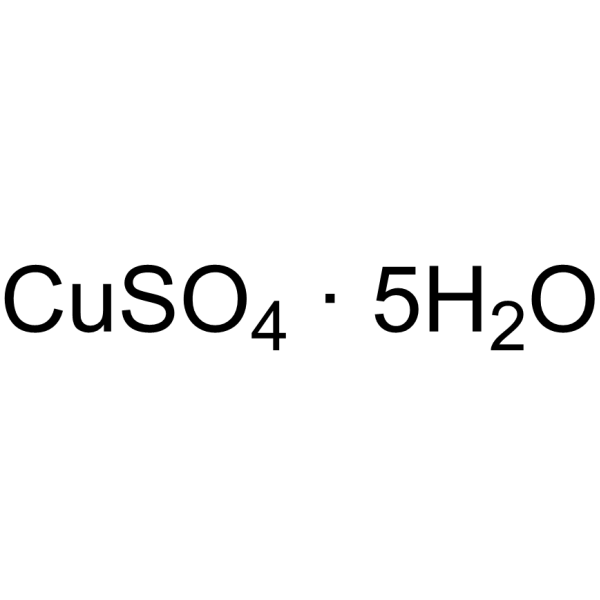
Copper sulfate pentahydrate

Based on 1 Customer Validation
Copper sulfate pentahydrate, 99% is a biochemical reagent. Copper sulfate pentahydrate, 99% reduces the production of ROS and the expression levels of MyD88 as well as c-Rel genes. Copper sulfate pentahydrate, 99% decreases the activities of T-SOD, CAT, and GSH, increases the activities of caspase-3, caspase-8, and caspase-9. Copper sulfate pentahydrate, 99% is cytotoxic to various cells. Copper sulfate pentahydrate, 99% has antioxidant activity. Copper sulfate pentahydrate, 99% can be used in the research of diabetes, Parkinson's disease and DMBA (HY-W011845)-induced tumors.
For research use only. We do not sell to patients.
- CAS No.: 7758-99-8
- Formula: CuSO4·5H2O
- Molecular Weight:249.69
-
Storage:
Store at room temperature, keep dry and cool.
In solvent -80°C, 1 year , -20°C, 6 months

To place orders, for customer services and technical support, please contact: MedChemExpress USA
Tel: 609-228-6898 E-mail: [email protected] [email protected]


-
 Biological Activity
-
Chemical Information
-
Solvent & Solubility
- Protocols
- Purity & Documentation
- References
-
 Help & FAQs


-
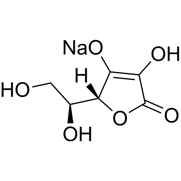
HY-B0166A
L-Ascorbic acid sodium saltCalcium Channel Inhibitor -
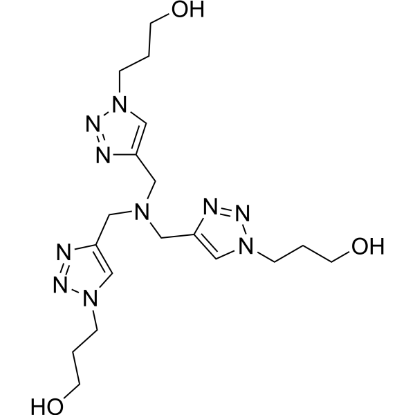
HY-W021042
Tris(3-hydroxypropyltriazolylmethyl)amineCuAAC Ligand -
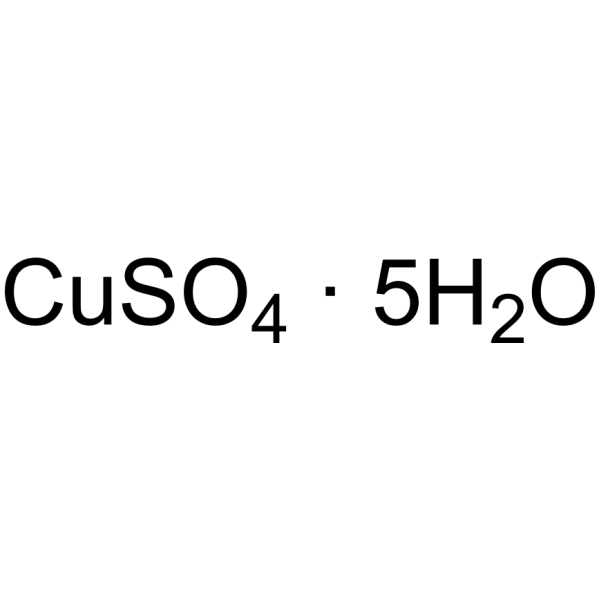
HY-Y1881B
Copper sulfate pentahydrate, for cell culture, 98%Biochemical Reagent -

HY-Y1103
Iron(II) sulfate heptahydrate, 99%Iron Salt -
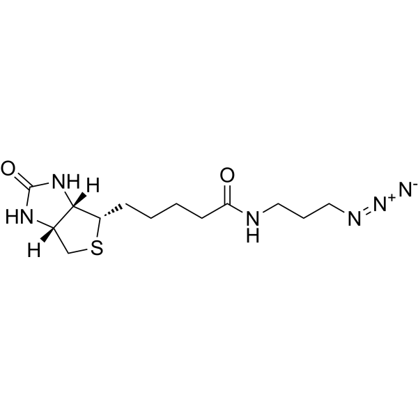
HY-129832
Biotin-azideClick Chemical Reagent -
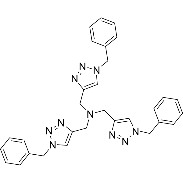
HY-116677
Tris(benzyltriazolylmethyl)amineBiochemical Tool -

HY-Y1103A
Iron(II) sulfate heptahydrate, for cell culture, 99%Iron Salt -

HY-N3025
Zinc sulfate heptahydrateDietary Supplement -

HY-Y1267
Magnesium sulfateAnti-Inflammatory Agent -
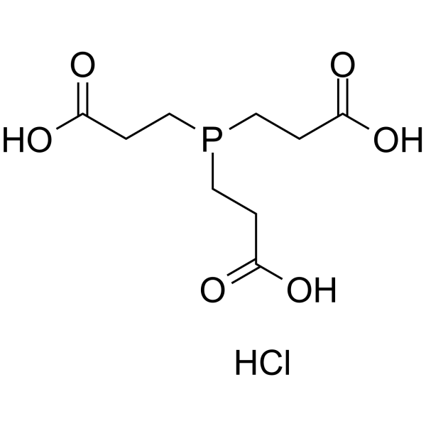
HY-W011500
TCEP hydrochlorideNon-Thiol Reducing Agent -

HY-D0852A
Sodium metavanadate, 99%PTPases Inhibitor -

HY-Y1878
Copper(Ⅱ) sulfateBiochemical Reagent
All Caspase Isoforms
More

Biological Activity
Description
|
Caspase-3 |
Caspase-9 |
Caspase-8 |
In Vitro
Copper sulfate pentahydrate (6.25-200 mg/mL; 24 h), 99.9% significantly decreases the cell viability of three human intestinal epithelial cell lines (HCT116, HT-29 and SW480)[1].
Copper sulfate (2.4 mg Cu/L; 24 h) pentahydrate, 99.9% significantly decreases the viability of primary hepatocytes of Epinephelus coioides[2].
Copper sulfate (5-100 µM) pentahydrate, 99.9% has no effect on CHO cell growth or antibody production, with a slight dose-dependent decrease in culture viability[3].
MedChemExpress (MCE) has not independently confirmed the accuracy of these methods. They are for reference only. Further protocols information, click here.
-
Cell Line:primary hepatocytes of Epinephelus coioides
-
Concentration:2.4 mg Cu/L
-
Incubation Time:24 h
-
Result:Increased the percentages of apoptosis and necrosis.
Increased the activities of caspase-3, caspase-8, and caspase-9.
In Vivo
Copper sulfate (2.5 mg/kg; i.p.; 16/24 h before MPP+) pentahydrate, 99% blocks MPP+-induced striatal lipid peroxidation and dopamine depletion in mice[6].
Copper sulfate (approximately 50 mg Cu2+/liter in drinking water) pentahydrate, 99% delays DMBA (HY-W011845)-induced ovarian granulosa cell tumor development in C57BL/6J female mice[7].
MedChemExpress (MCE) has not independently confirmed the accuracy of these methods. They are for reference only.


Chemical Information
-
CAS No. 7758-99-8
-
Appearance Solid
-
Molecular Weight 249.69
-
Formula CuSO4·5H2O
-
Color Light blue to blue
-
SMILES
O=S([O-])([O-])=O.[H]O[H].[H]O[H].[H]O[H].[H]O[H].[H]O[H].[Cu+2]
-
Shipping
Room temperature in continental US; may vary elsewhere.
-
Storage
Store at room temperature, keep dry and cool
In solvent -80°C 1 year -20°C 6 months


Solvent & Solubility
In Vitro:
H2O : 100 mg/mL (400.50 mM; Need ultrasonic)
Please refer to the solubility information to select the appropriate solvent. Once prepared, please aliquot and store the solution to prevent product inactivation from repeated freeze-thaw cycles.
Storage method and period of stock solution: -80°C, 1 year; -20°C, 6 months. When stored at -80°C, please use it within 1 year. When stored at -20°C, please use it within 6 months.
* Note: If you choose water as the stock solution, please dilute it to the working solution, then filter and sterilize it with a 0.22 μm filter before use.
Please refer to the solubility information to select the appropriate solvent. Once prepared, please aliquot and store the solution to prevent product inactivation from repeated freeze-thaw cycles.
Storage method and period of stock solution: -80°C, 1 year; -20°C, 6 months. When stored at -80°C, please use it within 1 year. When stored at -20°C, please use it within 6 months.
* Note: If you choose water as the stock solution, please dilute it to the working solution, then filter and sterilize it with a 0.22 μm filter before use.
Concentration (start) × Volume (start) = Concentration (final) × Volume (final)

Protocols
-
RNA extraction experimental
By lysing cells, releasing RNA, and removing impurities such as proteins and DNA, high-purity RNA products are finally obtained. The commonly used traditional method is the guanidine isothiocyanate/phenol/chloroform method (Trizol), which is suitable for a variety of animal materials including animal tissues, microorganisms, cultured cells, etc., and most plant materials.
-
ROS/oxidative-stress fluorescent staining
ROS/oxidative-stress fluorescent staining uses cell-permeant fluorogenic probes that become fluorescent after oxidation inside cells or tissues; commonly used examples include DCFH-DA/DCFDA for broad cellular oxidant detection, DHE for superoxide-related signal detection, MitoSOX for mitochondrial superoxide-related signal detection, and CellROX probes for oxidative-stress-associated fluorescence readouts. The assay detects probe oxidation rather than a single ROS species unless the probe and analysis method have been chemically validated for that species. DCFH-DA enters cells, is deacetylated by intracellular esterases to DCFH, and produces fluorescent DCF after oxidation, so the readout is used as an operational measure of total cellular oxidative stress rather than a species-specific ROS measurement. DHE and MitoSOX can report superoxide-related oxidation, but red fluorescence alone can include non-specific ethidium-like oxidation products; HPLC or optimized spectral approaches are
-
Human pluripotent stem cell midbrain dopaminergic neuron differentiation
Human pluripotent stem cells are directed toward midbrain dopaminergic neurons by first inducing a neural floor-plate-like progenitor state, then patterning cells with ventralizing SHH signaling and midbrain/WNT-FGF cues, and finally maturing progenitors into neurons expressing dopaminergic markers such as TH, NURR1/NR4A2, PITX3, DAT/SLC6A3, VMAT2/SLC18A2, GIRK2/KCNJ6, FOXA2, LMX1A, and EN1. The main readouts are loss of pluripotency, acquisition of FOXA2+/LMX1A+ midbrain floor-plate progenitors, emergence of βIII-tubulin+/MAP2+ neurons, and production of TH+ dopaminergic neurons with molecular, dopamine-release, and electrophysiological features of midbrain dopaminergic identity.
-
Apoptosis Solutions
Apoptosis is a regulated, generally non-lytic cell-death pathway that removes unwanted, damaged, infected, or abnormal cells through coordinated morphological changes, caspase activation, DNA fragmentation, and membrane remodeling. The intrinsic apoptosis pathway is controlled mainly by mitochondrial outer membrane permeabilization, BCL-2 family proteins, cytochrome c release, apoptosome formation, caspase-9 activation, and downstream executioner caspase-3/7 activation. The extrinsic apoptosis pathway is initiated by death receptors such as Fas, TNFR, and TRAIL receptors, which recruit adaptor proteins and activate caspase-8 before engaging executioner caspases or mitochondrial amplification through BID cleavage. Apoptosis is linked to many phenotypes, including cancer cell killing, tissue homeostasis, immune regulation, neurodegeneration, infection response, and treatment-induced cytotoxicity; unresolved questions include how apoptosis interacts with necroptosis, pyroptosis, ferroptos
-
Research Protocol for Metabolic Diseases
AMP-activated protein kinase, AMPK, is a conserved cellular energy sensor that responds to reduced cellular energy status and coordinates metabolism by increasing ATP-generating catabolic pathways while suppressing ATP-consuming anabolic processes. In metabolic disease research, the AMPK pathway is experimentally relevant because it regulates hepatic lipid synthesis, fatty acid oxidation, glucose production, skeletal-muscle glucose disposal, mTORC1-linked biosynthesis, autophagy, mitochondrial homeostasis, and whole-body energy balance. The central pathway logic is that energy stress, metformin, exercise-like stimulation, or direct AMPK activators increase AMPKα Thr172 phosphorylation and downstream substrate phosphorylation, including ACC and RAPTOR. Phosphorylation of ACC suppresses lipogenesis and supports fatty acid oxidation, whereas phosphorylation of RAPTOR suppresses mTORC1 signaling and links cellular energy status to growth and protein synthesis control. The pathway is linked
-
Cell Viability Determination by MTT Colorimetric Assay
The following protocol uses the MTT colorimetric assay as a classic literature-established method for assessing cell viability/metabolic activity in cultured mammalian cells. MTT[3-(4,5-dimethylthiazol-2-yl)-2,5-diphenyltetrazolium bromide] is reduced by metabolically active cells to a colored formazan product; the amount of formazan is quantified spectrophotometrically and provides an indirect measure of metabolically active viable cells. Importantly, MTT reduction reflects cellular oxidoreductase/metabolic activity rather than an absolute direct count of living cells, so changes in cellular metabolism can alter the signal independently of cell number.


Purity & Documentation
-
Data Sheet (280 KB)
-
SDS (758 KB)
- English - EN (758 KB)
- Français - FR (758 KB)
- Deutsch - DE (758 KB)
- Norwegian - NO (758 KB)
- Español - ES (758 KB)
- Swedish - SV (758 KB)
- Italian - IT (758 KB)
- Korean - KR (758 KB)
- Portuguese - PT (758 KB)
-
Handling Instructions (2659 KB)
References
[1]. Feyzi A, et al. Copper sulfate pentahydrate reduced epithelial cytotoxicity induced by lipopolysaccharide from enterogenic bacteria. Biomed Pharmacother. 2017 May;89:454-461. [Content Brief]
[2]. Wang T, et al. Copper Nanoparticles and Copper Sulphate Induced Cytotoxicity in Hepatocyte Primary Cultures of Epinephelus coioides. PLoS One. 2016 Feb 18;11(2):e0149484. [Content Brief]
[3]. Chaderjian WB, et al. Effect of copper sulfate on performance of a serum-free CHO cell culture process and the level of free thiol in the recombinant antibody expressed. Biotechnol Prog. 2005 Mar-Apr;21(2):550-3. [Content Brief]
[5]. Sitasawad S, et al. Beneficial effect of supplementation with copper sulfate on STZ-diabetic mice (IDDM). Diabetes Res Clin Pract. 2001 May;52(2):77-84. [Content Brief]
[6]. Alcaraz-Zubeldia M, et al. Neuroprotective effect of acute and chronic administration of copper (II) sulfate against MPP+ neurotoxicity in mice. Neurochem Res. 2001 Jan;26(1):59-64. [Content Brief]
[7]. Burki HR, et al. Effect of oral copper sulfate on 7,12-dimethylbenz(alpha)anthracene carcinogenesis in mice. Br J Cancer. 1969 Sep;23(3):591-6. [Content Brief]


Complete Stock Solution Preparation Table
Please refer to the solubility information to select the appropriate solvent. Once prepared, please aliquot and store the solution to prevent product inactivation from repeated freeze-thaw cycles.
Storage method and period of stock solution: -80°C, 1 year; -20°C, 6 months. When stored at -80°C, please use it within 1 year. When stored at -20°C, please use it within 6 months.
| Optional Solvent | Concentration Solvent Mass | 1 mg | 5 mg | 10 mg | 25 mg |
|---|---|---|---|---|---|
| H2O | 1 mM | 4.0050 mL | 20.0248 mL | 40.0497 mL | 100.1242 mL |
| 5 mM | 0.8010 mL | 4.0050 mL | 8.0099 mL | 20.0248 mL | |
| 10 mM | 0.4005 mL | 2.0025 mL | 4.0050 mL | 10.0124 mL | |
| 15 mM | 0.2670 mL | 1.3350 mL | 2.6700 mL | 6.6749 mL | |
| 20 mM | 0.2002 mL | 1.0012 mL | 2.0025 mL | 5.0062 mL | |
| 25 mM | 0.1602 mL | 0.8010 mL | 1.6020 mL | 4.0050 mL | |
| 30 mM | 0.1335 mL | 0.6675 mL | 1.3350 mL | 3.3375 mL | |
| 40 mM | 0.1001 mL | 0.5006 mL | 1.0012 mL | 2.5031 mL | |
| 50 mM | 0.0801 mL | 0.4005 mL | 0.8010 mL | 2.0025 mL | |
| 60 mM | 0.0667 mL | 0.3337 mL | 0.6675 mL | 1.6687 mL | |
| 80 mM | 0.0501 mL | 0.2503 mL | 0.5006 mL | 1.2516 mL | |
| 100 mM | 0.0400 mL | 0.2002 mL | 0.4005 mL | 1.0012 mL |
* Note: If you choose water as the stock solution, please dilute it to the working solution, then filter and sterilize it with a 0.22 μm filter before use.


Copper sulfate pentahydrate Related Classifications
HY-Y1881A Related Classifications
 Powered by Bioz